{"translation_price":"50","translation_doc_price":"40","child_coefficient":"1.1","transfer_price":"2.00","transfer_price_vip":"5.00","constant_transfer_price_vip":350,"constant_transfer_price":150,"constant_transfer_distanse":60,"type":"treatment","program_full_story":"<ul>\n\t<li>Initial presentation in the clinic<\/li>\n\t<li>clinical history taking<\/li>\n\t<li>review of medical records<\/li>\n\t<li>physical examination<\/li>\n\t<li>laboratory tests:\n\t<ul>\n\t\t<li>complete blood count<\/li>\n\t\t<li>general urine analysis<\/li>\n\t\t<li>biochemical analysis of blood<\/li>\n\t\t<li>TSH-basal, fT3, fT4<\/li>\n\t\t<li>tumor markers (AFP, CEA, \u0421\u0410-19-9)<\/li>\n\t\t<li>inflammation indicators (CRP, ESR)<\/li>\n\t\t<li>indicators of blood coagulation<\/li>\n\t<\/ul>\n\t<\/li>\n\t<li>abdominal ultrasound\u200b scan<\/li>\n\t<li>CT\/MRI of abdomen<\/li>\n\t<li>preoperative care<\/li>\n\t<li>percutaneous embolization (coiling) or chemoembolization<\/li>\n\t<li>symptomatic treatment<\/li>\n\t<li>cost of essential medicines<\/li>\n\t<li>nursing services<\/li>\n\t<li>elaboration of further recommendations<\/li>\n<\/ul>\n<div class=\"program_how_program_going mt-4\"><h4>How program is carried out<\/h4><p style=\"text-align: justify;\"><strong>During the first visit<\/strong>, the doctor will conduct a clinical examination and go through the results of the available diagnostic tests. After that, you will undergo the necessary additional examination, such as the assessment of liver and kidney function, ultrasound scan, CT scan and MRI. This will allow the doctor to determine which vessels are feeding the tumor and its metastases, as well as determine how well you will tolerate the procedure.<\/p>\n\n<p style=\"text-align: justify;\"><strong>Chemoembolization <\/strong>begins with local anesthesia and catheterization of the femoral artery. The thin catheter is inserted through a few centimeters long incision of the blood vessel. The doctor gradually moves the catheter to the vessel feeding the primary tumor or its metastases. The procedure is carried out under visual control, an angiographic device is used for this. The vascular bed and the position of the catheter in it are displayed on the screen of the angiograph.<\/p>\n\n<p style=\"text-align: justify;\">When the catheter reaches a suspected artery, a contrast agent is injected through it. Due to the introduction of the contrast agent, the doctor clearly sees the smallest vessels of the tumor and the surrounding healthy tissues on the screen of the angiograph. After that, he injects emboli into the tumor vessels through the same catheter.<\/p>\n\n<p style=\"text-align: justify;\">Emboli are the spirals or the liquid microspheres. The type of embolus is selected individually, taking into account the diameter of the target vessel. When carrying out chemoembolization, a solution of a chemotherapy drug is additionally injected into the tumor vessel. Due to the subsequent closure of the vessel lumen with an embolus, the chemotherapy drug influences the tumor for a long time. In addition, the drug does not enter the systemic circulation, which allows doctors to use high doses of chemotherapeutic agents without the development of serious side effects. Chemoembolization leads to the destruction of the tumor or slowing down its progression.<\/p>\n\n<p style=\"text-align: justify;\">After that, the catheter is removed from the artery. The doctor puts a vascular suture on the femoral artery and closes it with a sterile dressing. During chemoembolization, you will be awake. General anesthesia is not used, which significantly reduces the risks of the procedure and allows performing it on an outpatient basis, avoiding long hospital stay.<\/p>\n\n<p style=\"text-align: justify;\"><strong>After the first procedure<\/strong>, you will stay under the supervision of an interventional oncologist and general practitioner. If necessary, you will receive symptomatic treatment. As a rule, a second chemoembolization procedure is performed in 3-5 days after the first one in order to consolidate the therapeutic effect. After that, you will receive recommendations for further follow-up and treatment.<\/p>\n<\/div><div class=\"program_required_documents mt-4\"><h4>Required documents<\/h4><ul>\n\t<li style=\"text-align: justify;\">Medical records<\/li>\n\t<li style=\"text-align: justify;\">MRI\/CT scan (not older than 3 months)<\/li>\n\t<li style=\"text-align: justify;\">Biopsy results (if available)<\/li>\n<\/ul>\n<\/div>","program_full_story_crm":"<ul>\n\t<li>Initial presentation in the clinic<\/li>\n\t<li>clinical history taking<\/li>\n\t<li>review of medical records<\/li>\n\t<li>physical examination<\/li>\n\t<li>laboratory tests:\n\t<ul>\n\t\t<li>complete blood count<\/li>\n\t\t<li>general urine analysis<\/li>\n\t\t<li>biochemical analysis of blood<\/li>\n\t\t<li>TSH-basal, fT3, fT4<\/li>\n\t\t<li>tumor markers (AFP, CEA, \u0421\u0410-19-9)<\/li>\n\t\t<li>inflammation indicators (CRP, ESR)<\/li>\n\t\t<li>indicators of blood coagulation<\/li>\n\t<\/ul>\n\t<\/li>\n\t<li>abdominal ultrasound\u200b scan<\/li>\n\t<li>CT\/MRI of abdomen<\/li>\n\t<li>preoperative care<\/li>\n\t<li>percutaneous embolization (coiling) or chemoembolization<\/li>\n\t<li>symptomatic treatment<\/li>\n\t<li>cost of essential medicines<\/li>\n\t<li>nursing services<\/li>\n\t<li>elaboration of further recommendations<\/li>\n<\/ul>\n<div class=\"program_how_program_going mt-4\"><h4>How program is carried out<\/h4><p style=\"text-align: justify;\"><strong>During the first visit<\/strong>, the doctor will conduct a clinical examination and go through the results of the available diagnostic tests. After that, you will undergo the necessary additional examination, such as the assessment of liver and kidney function, ultrasound scan, CT scan and MRI. This will allow the doctor to determine which vessels are feeding the tumor and its metastases, as well as determine how well you will tolerate the procedure.<\/p>\n\n<p style=\"text-align: justify;\"><strong>Chemoembolization <\/strong>begins with local anesthesia and catheterization of the femoral artery. The thin catheter is inserted through a few centimeters long incision of the blood vessel. The doctor gradually moves the catheter to the vessel feeding the primary tumor or its metastases. The procedure is carried out under visual control, an angiographic device is used for this. The vascular bed and the position of the catheter in it are displayed on the screen of the angiograph.<\/p>\n\n<p style=\"text-align: justify;\">When the catheter reaches a suspected artery, a contrast agent is injected through it. Due to the introduction of the contrast agent, the doctor clearly sees the smallest vessels of the tumor and the surrounding healthy tissues on the screen of the angiograph. After that, he injects emboli into the tumor vessels through the same catheter.<\/p>\n\n<p style=\"text-align: justify;\">Emboli are the spirals or the liquid microspheres. The type of embolus is selected individually, taking into account the diameter of the target vessel. When carrying out chemoembolization, a solution of a chemotherapy drug is additionally injected into the tumor vessel. Due to the subsequent closure of the vessel lumen with an embolus, the chemotherapy drug influences the tumor for a long time. In addition, the drug does not enter the systemic circulation, which allows doctors to use high doses of chemotherapeutic agents without the development of serious side effects. Chemoembolization leads to the destruction of the tumor or slowing down its progression.<\/p>\n\n<p style=\"text-align: justify;\">After that, the catheter is removed from the artery. The doctor puts a vascular suture on the femoral artery and closes it with a sterile dressing. During chemoembolization, you will be awake. General anesthesia is not used, which significantly reduces the risks of the procedure and allows performing it on an outpatient basis, avoiding long hospital stay.<\/p>\n\n<p style=\"text-align: justify;\"><strong>After the first procedure<\/strong>, you will stay under the supervision of an interventional oncologist and general practitioner. If necessary, you will receive symptomatic treatment. As a rule, a second chemoembolization procedure is performed in 3-5 days after the first one in order to consolidate the therapeutic effect. After that, you will receive recommendations for further follow-up and treatment.<\/p>\n<\/div>","is_ambulant":"1","bh_fee":0,"only_for_children":"0","no_service":"0","with_prepayment":"1","show_calculator":"1","paket_type":"1","btn_type":"2","clinic_icon":"600156bf9bd18.jpg","city":"Aarau","clinic_site":"https:\/\/www.hirslanden.ch\/global\/de\/startseite\/kliniken_zentren\/hirslanden_klinikaarau.html","department_recommend":"0","country":"Switzerland","country_id":"2","clinic_name":"Hirslanden Clinic Aarau","cinic_name":"Hirslanden Clinic Aarau","department_id":"3951","duration":"6","direction":"Interventional radiology","min_duration":0,"clinic_id":"305","paketPrice":"0.00","paket":"<ul>\n <li> Interpreter up to 8 hours<\/li>\n <li>Translation of up to 5 pages<\/li>\n <li>Visa support<\/li>\n\n<\/ul>","title":"Treatment of liver cancer with percutaneous embolization (coiling) or chemoembolization","price":{"val":0,"type":"val"},"price_surcharge":0,"price_surcharge_clear":0,"extra_service_clinic":[],"extra_service":[],"translation_hours":"0","translation_doc_count":null,"roads":[{"id":"1","distance":"50","airport_title":"Zurich"},{"id":"18","distance":"62","airport_title":"Basel"},{"id":"17","distance":"235","airport_title":"Geneve"}],"pakets":[],"lang":{"day":"Day","days":"days","ambulatory":"Outpatient","stationaryProgram":"Inpatient"}}
Treatment of liver cancer with percutaneous embolization (coiling) or chemoembolization in Hirslanden Clinic Aarau
percutaneous embolization (coiling) or chemoembolization
symptomatic treatment
cost of essential medicines
nursing services
elaboration of further recommendations
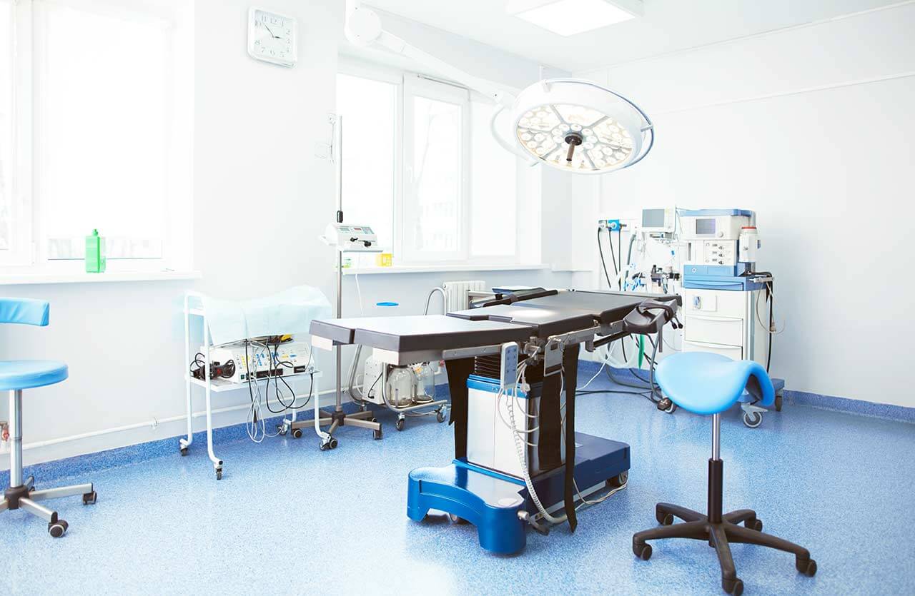
How program is carried out
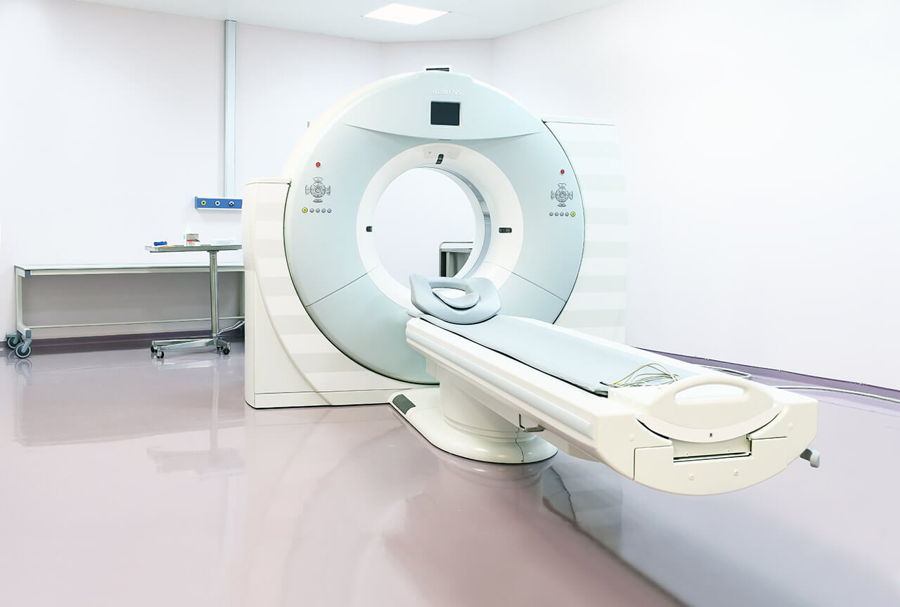
During the first visit, the doctor will conduct a clinical examination and go through the results of the available diagnostic tests. After that, you will undergo the necessary additional examination, such as the assessment of liver and kidney function, ultrasound scan, CT scan and MRI. This will allow the doctor to determine which vessels are feeding the tumor and its metastases, as well as determine how well you will tolerate the procedure.
Chemoembolization begins with local anesthesia and catheterization of the femoral artery. The thin catheter is inserted through a few centimeters long incision of the blood vessel. The doctor gradually moves the catheter to the vessel feeding the primary tumor or its metastases. The procedure is carried out under visual control, an angiographic device is used for this. The vascular bed and the position of the catheter in it are displayed on the screen of the angiograph.
When the catheter reaches a suspected artery, a contrast agent is injected through it. Due to the introduction of the contrast agent, the doctor clearly sees the smallest vessels of the tumor and the surrounding healthy tissues on the screen of the angiograph. After that, he injects emboli into the tumor vessels through the same catheter.
Emboli are the spirals or the liquid microspheres. The type of embolus is selected individually, taking into account the diameter of the target vessel. When carrying out chemoembolization, a solution of a chemotherapy drug is additionally injected into the tumor vessel. Due to the subsequent closure of the vessel lumen with an embolus, the chemotherapy drug influences the tumor for a long time. In addition, the drug does not enter the systemic circulation, which allows doctors to use high doses of chemotherapeutic agents without the development of serious side effects. Chemoembolization leads to the destruction of the tumor or slowing down its progression.
After that, the catheter is removed from the artery. The doctor puts a vascular suture on the femoral artery and closes it with a sterile dressing. During chemoembolization, you will be awake. General anesthesia is not used, which significantly reduces the risks of the procedure and allows performing it on an outpatient basis, avoiding long hospital stay.
After the first procedure, you will stay under the supervision of an interventional oncologist and general practitioner. If necessary, you will receive symptomatic treatment. As a rule, a second chemoembolization procedure is performed in 3-5 days after the first one in order to consolidate the therapeutic effect. After that, you will receive recommendations for further follow-up and treatment.
Required documents
Medical records
MRI/CT scan (not older than 3 months)
Biopsy results (if available)
Service
Price from:
Type of program :
Price for 1 day:
Expected duration of the program:
The minimum duration of the program:
Select
You may also book:
Hospital directly:
Saving:
BookingHealth Price from:
About the department
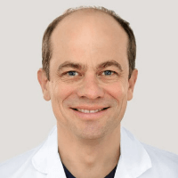
The Department of Diagnostic and Interventional Radiology at the Hirslanden Clinic Aarau offers a wide range of imaging examinations, and also specializes in imaging-guided interventional procedures. The Chief Physician of the department is PD Dr. med. Achim Kaim.
The department has cutting-edge systems for radiography, mammography, sonography, computer and magnetic resonance imaging. The department also has 24-hour emergency service. At the center of all the efforts of a competent team of doctors is high-quality medical care focused on the patient’s high satisfaction. The medical team strictly adheres to radiation protection standards, so the examinations do not harm the health of patients.
The department's range of medical services includes:
Diagnostic radiology
Classic radiography
Lungs
Skeletal system
Gastrointestinal tract
Genitourinary tract
Mammography for the diagnostics of breast pathologies
Prostate and female reproductive organs (ovaries, uterus, etc.)
Vascular system
Breast
Angiography (examination of the carotid arteries, cerebral vessels, peripheral and coronary arteries)
Interventional radiology
Imaging-guided puncture, biopsy and drainage
Imaging-guided catheter placement
Imaging-guided vascular dilation and stent implantation
Other diagnostic and treatment methods
Curriculum vitae
Professional Career
Since 2015 Attending Physician in the Department of Radiology, Hirslanden Clinic Birshof.
Since 2002 Chief Physician of the Department of Diagnostic and Interventional Radiology at the Hirslanden Clinic Aarau.
2002 Anniversary Prize of the Swiss Society of Radiology (SGR).
2002 Lecturer at the Faculty of Medicine, University of Basel.
2000 - 2002 Senior Physician, Department of Nuclear Medicine, University Hospital Zurich.
1995 - 2000 Senior Physician, Department of Diagnostic Radiology, University Hospital Basel.
1991 - 1995 Assistant Physician, Department of Radiology and Nuclear Medicine, University Hospital Basel.
1990 - 1991 Department of Surgery, Stuttgart-Bad Cannstatt.
1989 - 1990 Department of Pathology, University Hospital Ulm.
Higher Education and Postgraduate Training
2007 Specialization in Diagnostic Neuroradiology, Swiss Medical Association (FMH).
2002 Habilitation at the University of Basel.
1996 Board Certification in Diagnostic Radiology.
1993 - 1995 Examination of the Swiss Medical Association (FMH) in Diagnostic Radiology.
1990 Doctoral thesis defense, University of Tuebingen.
1983 - 1989 Study of Human Medicine at the Faculty of Medicine, University of Tuebingen. Internships in Strasbourg (France), St. Louis (USA) and Lausanne (Switzerland).
Memberships in Professional Societies
Swiss Society of Radiology (SRG).
German Radiological Society (DRG).
Swiss Society of Neuroradiology (SGNR).
Swiss Medical Association (FMH).
Medical Association of the Canton of Aargau.
Photo of the doctor: (c) Hirslanden AG
About hospital
The Hirslanden Clinic Aarau enjoys the status of one of the largest and most successful private medical facilities in Bern and Zurich. The clinic is part of the Hirslanden Network known in Europe, which is a provider of first-class medical services in Switzerland.
The main areas of specialization of the clinic in Aarau include cardiology, cardiac surgery, abdominal surgery, urology, oncology, neurosurgery, spinal surgery and orthopedics. The Department of Obstetrics annually delivers more than 700 babies. The high professionalism of the clinic’s medical staff, as well as the excellent technical base, make it possible to successfully cure not only common pathologies, but also very complex clinical cases.
All efforts of doctors and nursing staff of the clinic are focused on meeting the needs of patients and best possible restoration of their health. The work with patients is based on an individual approach to each clinical situation. The clinic has 155 beds for inpatient treatment. The surgical treatment is provided in 7 operating rooms with all the necessary surgical instruments, computer monitoring systems, navigation devices, robot-assisted systems for sparing laparoscopic operations.
Since 2010, the clinic introduced a unified quality management system, which includes all clinics in the Hirslanden Network. The clinical performance indicators and patient reviews are provided in the annual reports available to all those who want to see them. At the moment, the clinic rating is 8.9 out of 10 possible points – based on patient reviews. The clinic is also the first and only health facility in the canton of Aargau, which medical services were certified in accordance with the new ISO 2016 standards.
Photo: (с) depositphotos
Accommodation in hospital
Patients rooms
The patients of the Hirslanden Clinic Aarau live in cozy rooms equipped with everything necessary for a comfortable stay in the hospital. Each patient room has an ensuite bathroom with shower and toilet. The standard patient room includes an automatically adjustable bed, a bedside table, a wardrobe with lockers for storing personal belongings, a hairdryer, a table and chairs for receiving visitors, a TV and a radio. The patient rooms also have Wi-Fi access. If desired, the patients can live in enhanced-comfort patient rooms, which are additionally equipped with a safe, a refrigerator and upholstered furniture.
Meals and Menus
The patient and the accompanying person are offered a daily choice of three menus. If you are on a specific diet for any reason, you will be offered an individual menu. Please inform the medical staff about your dietary preferences prior to the treatment.
Further details
Standard rooms include:
Toilet
Shower
Wi-Fi
TV
Religion
The religious services can be provided upon request.
Accompanying person
During the inpatient program, an accompanying person may stay with the patient in the patient room or at a hotel. Our managers will help you choose the most suitable option.
Hospital accommodation
During the outpatient program, you can live in a hotel at the clinic.
Hotel
During the outpatient program, you can live in a hotel of your choice. Managers will help you choose the most suitable options.
Select your currency:
Your guarantee
We are the only TÜV-certified company with an ISO 9001:2015 certificate
thank you for reaching out! One of our medical advisors is currently reviewing your details. You can expect a call from us within one business day to discuss your request.
Note: The incoming call will appear as a German number or your local area code. This call is entirely free of charge.
Booking Health guarantees:
Expert Placement
We use rigorous statistical analysis to ensure you are treated at the best-rated facility for your specific diagnosis.
Total Cost Protection
No hidden fees or unexpected bills. We provide a fixed price guarantee, backed by insurance that covers any additional complications.
12-Month Extended Care
Your recovery is our priority. Benefit from a full year of direct medical support following your procedure.
 BookingHealth Price from:
