The program includes:
- Initial presentation in the clinic
- clinical history taking
- review of medical records
- physical examination
- laboratory tests:
- complete blood count
- general urine analysis
- biochemical analysis of blood
- TSH-basal, fT3, fT4
- tumor markers (AFP, CEA, СА-19-9)
- inflammation indicators (CRP, ESR)
- indicators of blood coagulation
- abdominal ultrasound scan
- CT/MRI of abdomen
- preoperative care
- percutaneous embolization (coiling) or chemoembolization
- symptomatic treatment
- cost of essential medicines
- nursing services
- elaboration of further recommendations
How program is carried out
During the first visit, the doctor will conduct a clinical examination and go through the results of the available diagnostic tests. After that, you will undergo the necessary additional examination, such as the assessment of liver and kidney function, ultrasound scan, CT scan and MRI. This will allow the doctor to determine which vessels are feeding the tumor and its metastases, as well as determine how well you will tolerate the procedure.
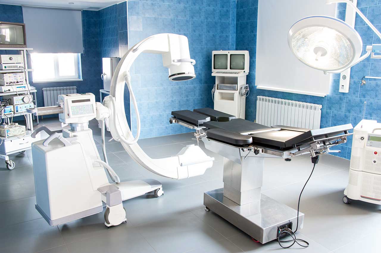
Chemoembolization begins with local anesthesia and catheterization of the femoral artery. The thin catheter is inserted through a few centimeters long incision of the blood vessel. The doctor gradually moves the catheter to the vessel feeding the primary tumor or its metastases. The procedure is carried out under visual control, an angiographic device is used for this. The vascular bed and the position of the catheter in it are displayed on the screen of the angiograph.
When the catheter reaches a suspected artery, a contrast agent is injected through it. Due to the introduction of the contrast agent, the doctor clearly sees the smallest vessels of the tumor and the surrounding healthy tissues on the screen of the angiograph. After that, he injects emboli into the tumor vessels through the same catheter.
Emboli are the spirals or the liquid microspheres. The type of embolus is selected individually, taking into account the diameter of the target vessel. When carrying out chemoembolization, a solution of a chemotherapy drug is additionally injected into the tumor vessel. Due to the subsequent closure of the vessel lumen with an embolus, the chemotherapy drug influences the tumor for a long time. In addition, the drug does not enter the systemic circulation, which allows doctors to use high doses of chemotherapeutic agents without the development of serious side effects. Chemoembolization leads to the destruction of the tumor or slowing down its progression.
After that, the catheter is removed from the artery. The doctor puts a vascular suture on the femoral artery and closes it with a sterile dressing. During chemoembolization, you will be awake. General anesthesia is not used, which significantly reduces the risks of the procedure and allows performing it on an outpatient basis, avoiding long hospital stay.
After the first procedure, you will stay under the supervision of an interventional oncologist and general practitioner. If necessary, you will receive symptomatic treatment. As a rule, a second chemoembolization procedure is performed in 3-5 days after the first one in order to consolidate the therapeutic effect. After that, you will receive recommendations for further follow-up and treatment.
Required documents
- Medical records
- MRI/CT scan (not older than 3 months)
- Biopsy results (if available)
Service
You may also book:
 BookingHealth Price from:
About the department
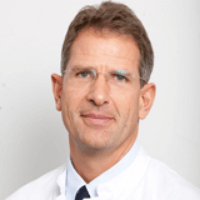
The Department of Interventional Radiology at the Marien Hospital Duesseldorf offers the full range of services in the area of its specialization. The medical facility has an excellent technical base for the full range of imaging tests, including X-ray and ultrasound scanning, angiography, computed tomography, magnetic resonance imaging and others. Since 2004, the department has also been carrying out CT- and MRI-guided angiographic tests without a catheter. Imaging tests are indispensable in the diagnostics of many pathologies. The department also provides patients with image-guided interventional therapeutic procedures. Interventional radiology techniques are an excellent alternative to surgical treatment. Particular attention is paid to the interventional treatment of chronic pain of various localization, oncological diseases and vascular diseases. The department's specialists regularly demonstrate high treatment success rates, so patients can be sure that their health is in the safe hands. The Head Physician of the department is Prof. Dr. med. Stefan Diederich.
Many patients with chronic pain in the back and lower limbs come to the department. Pain is often relieved with painkillers in pill form, but if this treatment does not ensure the desired result, then the patient requires image-guided interventional pain management. Periradicular therapy, facet joint infiltration, sacroiliac joint infiltration, and vertebroplasty are used for the treatment of chronic back pain. The department's doctors also successfully carry out sympathicolysis for the treatment of pain syndrome caused by chronic circulatory disorders of the lower limbs and nerve plexus block for chronic pain caused by oncological diseases. A huge advantage of the above mentioned therapeutic manipulations is the absence of the need for traumatic incisions in the skin and soft tissues. In addition, when performing an interventional procedure, the doctor uses imaging devices to control each action, which guarantees the highest safety of treatment.
The department's team of doctors has a wide range of techniques for the treatment of malignant tumors of various localizations. Therapeutic options in this field include radiofrequency ablation for the treatment of lung and kidney cancer, liver, bone and lung metastases, laser interstitial thermal therapy for the destruction of liver metastases, transarterial chemoembolization for the treatment of liver cancer and liver metastases, percutaneous ethanol injections for the treatment of liver cancer. The department’s specialists also perform uterine fibroid embolization, which is the gold standard for the treatment of this gynecologic pathology in modern medicine.
Another important focus of the department's specialists is on the treatment of vascular stenosis and obstruction using balloon dilatation and stent implantation. The medical facility also carries out lysis therapy for stroke in cooperation with neurologists.
The department's range of diagnostic and therapeutic services includes the following options:
- Diagnostic services
- X-ray scans (also with contrast enhancement)
- Ultrasound scans
- Angiography
- Computed tomography (CT), including cardiac CT
- Magnetic resonance imaging (MRI), including cardiac MRI
- CT scan of the teeth and jaws (orthopantomography)
- Therapeutic services
- Interventional pain management
- Periradicular therapy (PRT) for inflammation and swelling of the nerve roots of the cervical, thoracic and lumbar spinal cord
- Facet joint infiltration for pain in the small intervertebral joints of the cervical, thoracic or lumbar spine
- Sacroiliac joint infiltration for pain in this area
- Infiltration for hip arthrosis
- Sympathicolysis: treatment of pain caused by chronic circulatory disorders of the lower limbs
- Nerve plexus block for chronic pain caused by oncological diseases, for example, in pancreatic cancer
- Vertebroplasty for back pain caused by osteoporosis or spinal metastases
- Local therapy for malignant tumors (ablation)
- Radiofrequency ablation
- Laser interstitial thermal therapy
- Transarterial chemoembolization
- Percutaneous ethanol injections: injection of concentrated alcohol using fine needles directly into the tumor (for example, for liver cancer)
- Uterine artery embolization for the treatment of uterine fibroids
- Interventional catheter-based treatment for vascular diseases
- Balloon dilatation and stent implantation for vascular stenosis or occlusion
- Lysis therapy for stroke
- Interventional pain management
- Other diagnostic tests and treatment methods
Curriculum vitae
University Education
- 1981 - 1987 Study of Medicine at the Universities of Muenster and Heidelberg.
- June 1987 Medical license by the Head of Detmold District Administration.
- July 1987 Doctoral thesis defense, Institute of Clinical Radiology, University of Muenster (note: magna cum laude).
Medical and Scientific Activities
- July 1987 FMGEMS (American Exam).
- 1988 - 1989 Fellow of the Institute of Pathology at the Ruhr University in Bochum.
- 1990 Fellow of the Institute of Clinical Radiology at the University of Muenster.
- 1992 - 1993 Senior Resident of the Department of Radiology, Addenbrooke's Hospital, Cambridge.
- Since 1995 Appointed as an Acting Head of the department, later as a Senior Physician.
- 1997 Board certification in Diagnostic Radiology.
- 1998 Habilitation, Institute of Clinical Radiology, University of Muenster, Venia Legendi in Diagnostic Radiology.
- Since 2002 Head Physician of the Department of Interventional Radiology at the Marien Hospital Duesseldorf.
- 2004 Extraordinary Professorship, Faculty of Medicine, Westphalian Wilhelm University.
Awards
- 2000 Hanns Langendorf Award of the German Association of Radiologists and the Hanns Langendorff Foundation.
- 2006 Eugenie and Felix Award of the German Roentgen Society.
Review Activities in Scientific Journals
- Fortschritte auf dem Gebiet der Röntgenstrahlen.
- Radiologe.
- European Radiology.
- European Journal of Radiology.
- British Journal of Radiology.
- Eur Respiratory Journal.
- Oncologist.
- Cancer.
- Lung Cancer.
Memberships in Professional Societies
- Elected President of the International Cancer Imaging Society.
- Since 2002 Fellow of the International Cancer Imaging Society.
- 2014 President of the 95th German Congress on Radiology.
- 2010 and 2011 President of the Congress on Radiology in the Ruhr region.
- 2009 - 2015 Board Member of the German Roentgen Society.
- 2010 - 2011 President of the Roentgen Society of North Rhine-Westphalia.
- 2011 - 2013 Board Member of the Roentgen Society of North Rhine-Westphalia.
- 2002 - 2011 Chairman of the Working Group on Thoracic Diseases of the German Roentgen Society.
- Since 2011 Board Member of the Working Group on Thoracic Diseases of the German Roentgen Society.
Photo of the doctor: (c) Marien Hospital Düsseldorf
About hospital
The Marien Hospital Duesseldorf is a modern medical facility in the very center of Duesseldorf and a recognized Research Center. The foundation stone of the hospital was laid back in 1864 with the support of the Catholic Church. The Christian principles of helping and supporting patients have not changed at all over the years, but the level of medical care has become fundamentally different: a small hospital has been transformed into a multidisciplinary medical center with 11 internal departments, institutes and 7 interdisciplinary centers. Thus, patients can receive a comprehensive diagnostic examination and treatment under one roof. The medical facility is an academic hospital, so patients have access to innovative achievements in the field of modern medicine and receive the most effective treatment.
The hospital has 437 beds for inpatient treatment. It provides first-class medical services to 63,000 patients every year. The doctors and nursing staff of the hospital strive to provide each patient with customized treatment, taking into account their wishes and needs. In addition to providing top-class medical care, the specialists pay attention to the humane attitude towards patients and their life situation. Doctors devote enough time to personal communication with patients and support them in every possible way on their path to recovery.
The team of the hospital consists of competent doctors who are the best professionals in their medical field. They all work in interdisciplinary cooperation. In their clinical practice, doctors use state-of-the-art equipment, the very latest technologies, as well as innovative treatment methods, which in combination allows achieving optimal treatment outcomes.
Photo: (с) depositphotos
Accommodation in hospital
Patients rooms
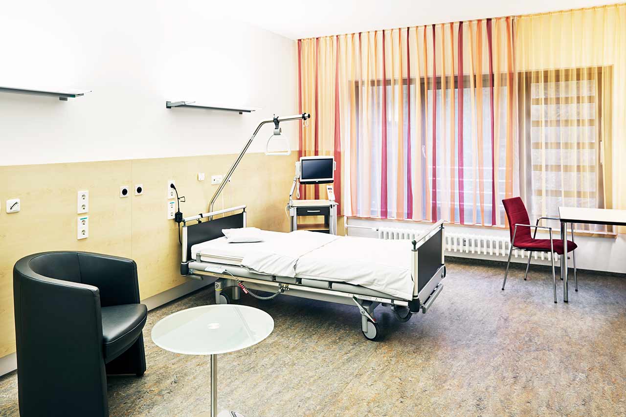
The patients of the Marien Hospital Duesseldorf live in comfortable patient rooms equipped with everything necessary. The room furnishing includes a comfortable bed with an orthopedic mattress, a bedside table, a wardrobe, a table, chairs, and a TV. The rooms are designed in light colors so that patients feel comfortable here. Each patient room also has an ensuite bathroom with shower and toilet.
Meals and Menus
The patients of the hospital are offered a healthy and balanced three meals a day: buffet breakfast, lunch and dinner. The menu also includes dietary and vegetarian dishes.
If for some reason you do not eat all the foods, you will be offered an individual menu. Please inform the medical staff about your food preferences prior to treatment.
Further details
Standard rooms include:
Religion
Religious services are available upon request.
Accompanying person
Your accompanying person may stay with you in your patient room or at the hotel of your choice during the inpatient program.
Hotel
You may stay at the hotel of your choice during the outpatient program. Our managers will support you for selecting the best option.


