About the Department of Interventional Radiology at ATOS Clinic Heidelberg
The Department of Interventional Radiology at the ATOS Clinic Heidelberg offers a wide range of medical services in its area of specialization. The medical facility started its work in 1991 and since then has been successfully admitting patients, providing them with top-notch medical care. One of the key areas of the department's daily clinical practice is computed tomography (CT) and magnetic resonance imaging (MRI). For this purpose, the specialists have at their disposal three 1.5 Tesla MRI scanners, two of which are equipped with the latest SENSE technology for cardiac examinations, an open high-field 1 Tesla MRI scanner, and a 64-slice SOMATOM go.Top CT scanner from Siemens. The department also offers digital mammography and digital X-rays. The medical facility has particular expertise in CT-guided periradicular therapy (PRT) for the treatment of chronic back pain, such as that caused by herniated discs. The department's specialists work with the latest generation of equipment, which allows diagnostic and therapeutic procedures to be carried out with high efficiency and minimal radiation exposure to the body. The Head Physician of the department is Dr. med. Wolfgang Wrazidlo.
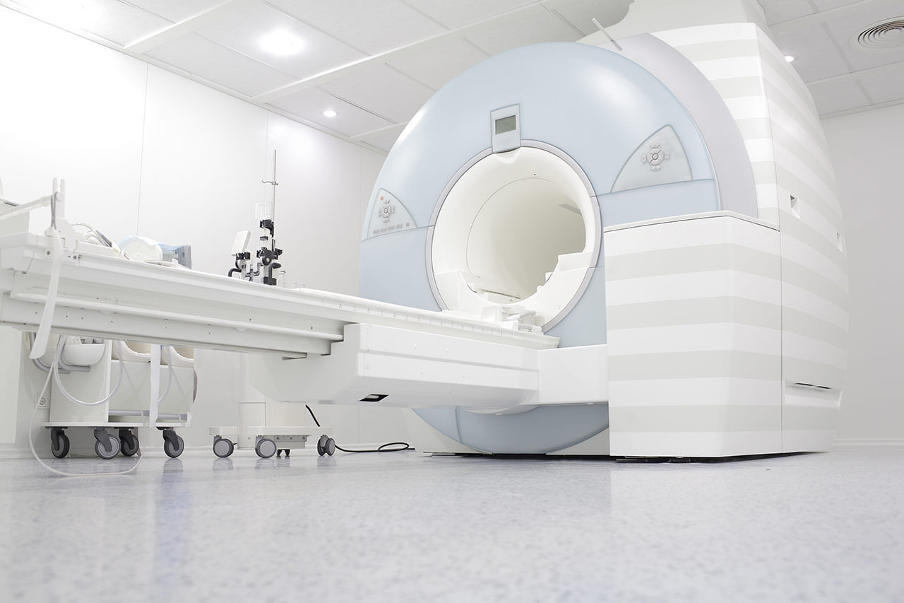
The most commonly requested imaging test in the department is magnetic resonance imaging (MRI). This diagnostic test uses a magnetic field and radio waves, rather than the ionizing radiation used in X-rays or CT scans. Therefore, MRI is a safe method of instrumental diagnosis. It can be performed as often as the patient needs. Moreover, it is a highly informative method, which is indispensable in modern diagnostics of pathologies of many internal organs and anatomical structures. MRI is performed in a special room where a scanner is installed. The doctor places the patient on a comfortable table that moves through a tunnel-shaped magnet. During the procedure, the patient is inside the machine; there is lighting and a fan that provides fresh air. MRI is accompanied by noise from electromagnetic pulse generators; earplugs may be used to reduce discomfort. Painful sensations during MRI are excluded. The procedure can take from 20 minutes to 1 hour. The department also performs contrast-enhanced MRIs. Patients with claustrophobia can undergo an MRI in an open scanner. MRI is used to diagnose diseases of the brain, spinal cord, internal organs, and joints. Imaging allows effective detection of inflammatory processes, tumors, metastases, and other pathological changes.
The department's team of radiologists also performs computed tomography (CT) scans almost daily. This examination uses X-rays. During a CT scan, the scanner head rotates around specific areas of the patient's body, allowing for the creation of multi-layered images in different planes. The department also offers contrast-enhanced CT scans. The patient lies still during the scan. The CT scan is painless. The average duration of the scan is 5-10 minutes. CT scans can be used to examine almost all organs and anatomical structures of the body. The method is especially valuable for diagnosing pathologies of the brain and spinal cord, spine, abdominal organs, retroperitoneal space, chest organs, heart, and pelvic organs.
CT-guided periradicular therapy (PRT), facet joint infiltration, and epidural infiltration are an integral part of the clinical activities of the department's specialists. Such a treatment is indicated for patients with chronic back pain due to intervertebral disc herniation, arthrosis, radiculopathy, facet syndrome, postural disorders, and other pathologies. The essence of infiltration therapy is as follows: a local anesthetic is injected directly into the pathological focus under the guidance of computed tomography; the procedure takes several minutes. Depending on the individual characteristics of the clinical case, the procedure can be performed either once or repeatedly with an interval of several days or weeks. The method is highly effective and in many cases serves as an alternative to spinal surgery when drug treatment and physiotherapy are ineffective.
The department's range of medical services includes the following:
- Diagnostic options
- Magnetic resonance imaging
- Classical magnetic resonance imaging
- Magnetic resonance angiography
- Multiparametric magnetic resonance imaging
- Computed tomography
- Digital X-rays
- Digital mammography
- Magnetic resonance imaging
- Therapeutic options
- Periradicular therapy (PRT)
- Facet joint infiltration
- Epidural infiltration
- Other medical services
Curriculum vitae
Higher Education and Professional Career
- 1972 - 1978 Physics studies, Karlsruhe Institute of Technology.
- 1978 - 198 Medical studies, Ruprecht Karl University of Heidelberg.
- 1984 Admission to medical practice.
- 1984 - 1991 Residency in diagnostic imaging: Department of Radiology, Bruchsal Hospital; Department of Cardiology, Municipal Hospital Karlsruhe; Department of Radiology, University Hospital Heidelberg (research fellow in magnetic resonance imaging); Department of Neuroradiology, University Hospital Mainz.
- 1985 Theoretical foundations of the specialty in emergency medical care.
- 1991 Theoretical foundations of the specialty in nuclear medicine.
- Since 1991 Head Physician, Department of Interventional Radiology, ATOS Clinic Heidelberg.
Memberships in Professional Societies
- German Radiological Society (DRG).
- Working Group on Cardiovascular Imaging and Working Group on Uroradiology of the German Radiological Society (DRG).
- European Society of Cardiology (ESC).
- Association of Southwest German Radiologists and Nuclear Medicine Specialists (VSRN).
- German Cardiac Society (DGK).
Photo of the doctor: (c) ATOS Klinik Heidelberg


