About the Department of Radiology at ATOS Orthoparc Clinic Cologne
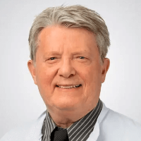
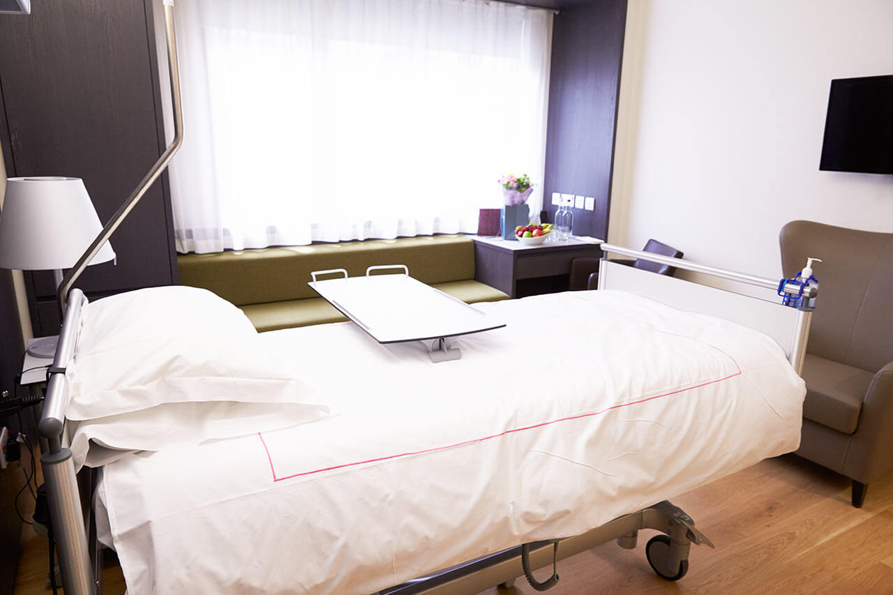
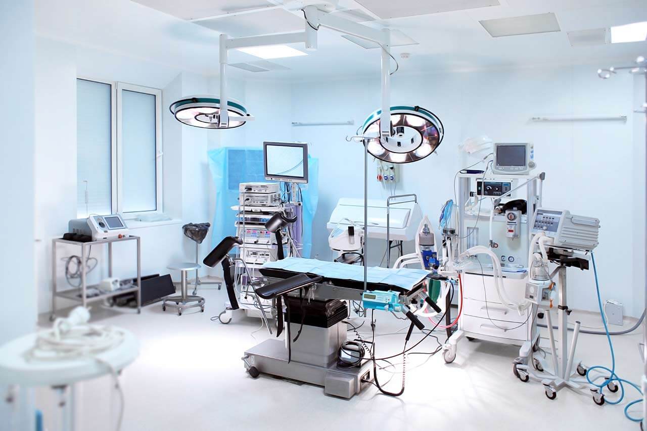
The Department of Radiology at the ATOS Orthoparc Clinic Cologne offers a wide range of modern diagnostic imaging procedures in a pleasant and comfortable environment for the patient. Imaging diagnosis is an integral part of a comprehensive examination for suspected diseases of the musculoskeletal system. The department has state-of-the-art equipment for digital X-ray, sonography, magnetic resonance imaging, and digital volume tomography. A radiologist and an orthopedist are present during the imaging examination. Patients with claustrophobia can undergo MRI in a semi-open machine. A modern picture archiving and communication system is used to store the images, ensuring unhindered access to the images by doctors from different departments of the ATOS Orthoparc Clinic Cologne. It should be noted that the specialists of the department take care of the safety of the examinations, so X-ray and digital volume tomography are performed with a minimum dose of radiation without harming the patient's health. The department is headed by Dr. med. Erwin Dubs.
Magnetic resonance imaging (MRI) is the most popular examination in the department. MRI is a method of imaging diagnostics using magnetic radiation, which allows the creation of a detailed and clear image of internal organs and anatomical structures. The department has both semi-open and closed MRI machines. Patients with claustrophobia find it more comfortable to undergo the examination in a semi-open machine. However, closed MRI machines are more powerful. The department's radiologists use MRI to detect pathological changes in the joints (knee, hip, shoulder, elbow, and ankle), tendons, ligaments, and muscles. In addition, MRI is an indispensable tool in the diagnosis of spinal pathologies. It provides highly accurate images of the intervertebral discs, spinal cord and meninges, cartilaginous structures, blood vessels and nerve endings in the spine, intervertebral joints, and adjacent muscles. For example, MRI allows for highly accurate detection of herniated discs and their size, spinal injuries, inflammatory diseases, malignant neoplasms, and metastases.
MRI is performed without any special preparation. Before the examination, the patient is placed on the table in a supine position. An important requirement for obtaining high-quality images is immobility during the examination, so the patient's extremities are usually fixed with special belts and rollers, or sedatives are recommended. After the patient is immobilized, the examination begins. Characteristic noises are generated during the operation of the device, but they do not cause any particular discomfort. However, the patient may use earplugs or headphones if desired.
The medical facility also offers digital volume tomography, a tomographic examination that uses X-rays to create high-resolution 3D images. This diagnostic method is highly accurate, while the radiation exposure to the body is minimal – 80% lower compared to classic computed tomography. The examination takes only a few seconds, which is also an advantage for the patient.
The department has advanced equipment for digital radiography of the musculoskeletal system. Digital radiography is a modern method of obtaining images of anatomical structures using X-rays with subsequent digital processing. Modern digital radiography equipment allows physicians to quickly obtain highly accurate images of the musculoskeletal system, which speeds up the process of diagnosis and initiation of therapy. Images are generated in digital format, which eliminates the possibility of image distortion on the film. In addition, compared to classical radiography, digital radiography provides almost half the radiation exposure to the body.
The department offers the following imaging tests:
- Magnetic resonance imaging
- Digital volume tomography
- Digital radiography
- Sonography
- Other diagnostic procedures
Curriculum vitae
Higher Education and Professional Career
- Since 2014 Head Physician, Department of Radiology, ATOS Orthoparc Clinic Cologne.
- 1983 - 2013 Physician in private practice specializing in X-ray diagnostics, Cologne.
- 1982 Board certification in diagnostic radiology; medical activities at imaging diagnostic centers in Dueren, Neuss, and Duesseldorf.
- 1997 Admission to medical practice; internship, Institute of Diagnostic Radiology at the University Hospital Cologne.
- 1997 - 1975 Medical activities in the field of pathology, internal medicine, and X-ray diagnostics abroad.
- 1968 Doctorate.
Photo of the doctor: (c) ATOS Orthoparc Klinik Köln


