The program includes:
- Initial presentation in the clinic
- clinical history taking
- review of medical records
- physical examination
- laboratory tests:
- complete blood count
- general urine analysis
- biochemical analysis of blood
- inflammation indicators (CRP, ESR)
- indicators of blood coagulation
- neurological examination
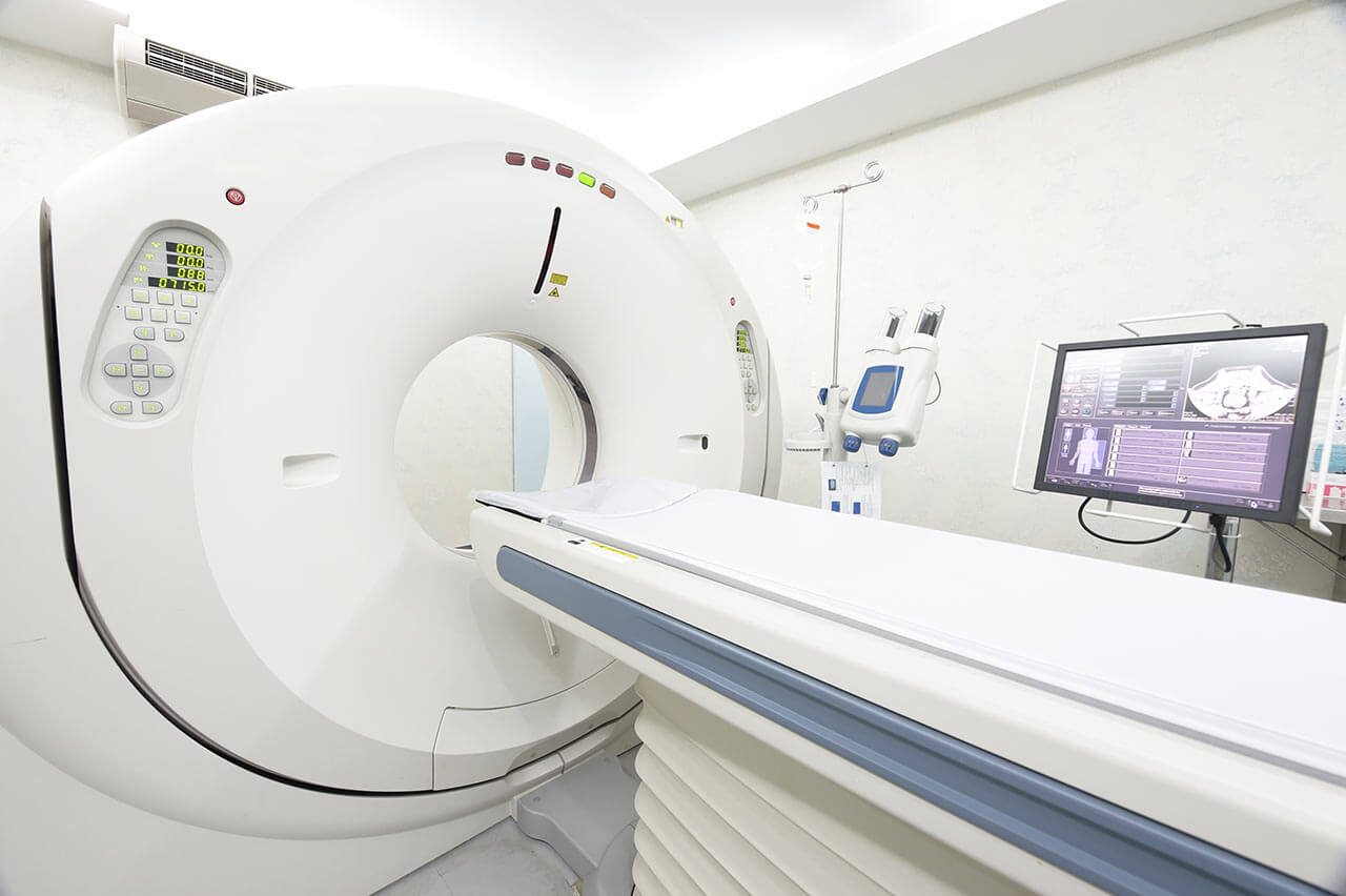
- CT/MRI scan
- neuropsychological tests (if indicated):
- ENMG (electroneuromyography)
- EEG (electroencephalography)
- SEPs (somatosensory evoked potentials)
- VEPs (visually evoked potentials)
- BAEP tests (brainstem auditory evoked potentials)
- preoperative care
- MRI-guided laser ablation of glioblastoma
- postoperative MRI control
- symptomatic treatment
- control examinations
- the cost of essential medicines and materials
- nursing services
- full hospital accommodation
- developing of further guidance by specialists
in oncology and radiotherapy
How program is carried out
During the first visit, the physician will conduct a clinical examination and neurological examination, and go through the results of the available diagnostic tests. After that, you will undergo the necessary additional examination, such as the laboratory blood and urine tests, MRI scan with obtaining 3D images and creating volumetric images of the whole brain. Based on the results of an additional examination, the physician will clarify the localization of glioblastoma. After that, the preparation according to the preoperative standard will begin.
Laser ablation of glioblastoma begins with the formation of access to the brain. When using a catheter with a laser fiber, a hole with a diameter of 3.2 mm is sufficient for this. The miniature size and flexibility of the catheter help preserve healthy brain tissue.
When the catheter reaches glioblastoma, energy is delivered through the laser fiber. Under the influence of high temperature, glioblastoma is destroyed. The procedure is guided by real-time MRI. Surgeons also assess the temperature of healthy brain tissues using thermographic images in order to avoid overheating.
After the completion of the operation, you will be transferred to the ward, under the round-the-clock supervision of physicians and nurses. In several days, the attending physician will evaluate the results of the postoperative MRI scans, schedule the date of discharge from the hospital and give you detailed recommendations for further follow-up and treatment.
Required documents
- Medical records
- MRI/CT scan (not older than 3 months)
- Biopsy results (if available)
Service
You may also book:
 BookingHealth Price from:
About the department
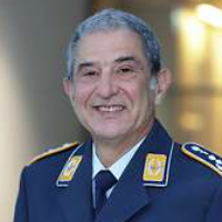
The Department of Neurosurgery and Spinal Surgery at the Bundeswehr Hospital Berlin offers the full range of services for the diagnostics and surgical treatment of diseases of the brain, spinal cord, spine and peripheral nerves. The department uses neuronavigation systems, 3D x-ray, intraoperative dopplerography, stereotaxy, electrophysiology, high-precision surgical microscopes and many other advanced devices that guarantee the effective and safe treatment. The department's surgeons are top-class professionals with rich clinical experience. The doctors always prefer sparing minimally invasive surgical techniques, while classic open surgery is performed only in complex clinical cases. State-of-the-art computer equipment allows the doctors to perform accurate planning of the neurosurgical intervention based on CT and MRI scans, which practically reduces the risk of developing a neurological deficit after the surgical treatment. The Head Physician of the department is Dr. med. Peter Madjurov.
Of particular interest to the department's neurosurgeons is the treatment of tumors of the brain and spinal cord, pituitary neoplasms, degenerative and traumatic lesions of the spine, including vertebral body replacement, acute injuries of the skull and spine, compression syndromes of the cranial and peripheral nerves. The department's doctors strictly adhere to the concept of a comprehensive approach to treatment, which includes preparation for surgery, the surgical intervention itself and postoperative care. The patient is in the safe hands of competent professionals and can count on the most favorable treatment outcomes.
The department uses the cutting-edge neuronavigation systems, which are the gold standard of modern neurosurgery. Prior to the operation, the doctors conduct magnetic resonance imaging to obtain accurate topographic data. During the operation, the surgeon focuses on these data, and also sees in which part of the brain or spinal cord the manipulation is performed. The benefit of neuronavigation is in the fact that it provides maximum accuracy during the operation, down to millimeters. In addition, neuroendoscopy (especially in the treatment of hydrocephalus and brain tumors), stereotaxy (used for the most accurate tissue sampling for its further histological examination), magnetic hyperthermia for malignant brain tumors, and the minimally invasive Spineoplastie® technique for the treatment of vertebral fractures, facet joint denervation, etc. are widely used in clinical practice.
The team of neurosurgeons of the medical facility regularly admits patients with benign and malignant tumors of the brain, skull base and pituitary gland. The medical facility also provides the effective treatment of spinal cord tumors: meningiomas, neurinomas, ependymomas, astrocytes and lipomas. The main diagnostic methods are imaging tests, such as computed tomography (CT) and magnetic resonance imaging (MRI). Based on the patient's diagnosis and the obtained diagnostic data, the optimal type of surgical intervention is selected. In most cases, the department's neurosurgeons use modern microsurgical techniques that guarantee minimal risks to the patient's health. During operations on the brain and spinal cord, intraoperative monitoring is mandatory. The specialists also often use modern surgical microscopes, neuronavigation systems, stereotaxy and other devices, which ensure not only effective, but also safe removal of the central nervous system tumors.
The department also successfully deals with the treatment of intracranial aneurysms. To eliminate the pathology, the clipping technique is mostly used. The essence of the method is that a special titanium clip is delivered to the pathological focus, with the help of which the surgeon pinches the wall of the damaged vessel. The department's specialists also perform microsurgical interventions to remove vascular neoplasms: angiomas and cavernomas. In cooperation with the Department of Neuroradiology at the Charite University Hospital, minimally invasive surgeries are performed for the treatment of neurovascular diseases.
An integral part of the clinical practice of the medical facility is the surgical treatment of degenerative spinal diseases and herniated discs. The main methods for detecting pathological changes in the spine are CT and MRI scans. A comprehensive examination is followed by the selection of the optimal type of the surgical intervention. The most common surgical procedures in the department are microsurgical operations for intervertebral cervical and lumbar disc herniations, microsurgical interventions for spinal canal stenosis dilatation and spinal stabilization, and implantation of artificial intervertebral discs. The recovery period after spinal interventions can vary significantly. For example, after the microsurgical treatment of herniated intervertebral discs or the replacement of intervertebral discs, the patients usually leave the department in 7 days, and in the case of stabilizing spinal surgeries, a full recovery may take several months.
The department's main clinical activities include:
- Surgery for brain, skull base and pituitary tumors
- Surgery for spinal cord tumors
- Surgery for neurovascular diseases: brain aneurysms, angiomas, caverns, vascular malformations
- Surgery for spinal disc herniations
- Intervertebral disc replacement surgery in the cervical and lumbar spine
- Surgery for spinal stenosis
- Spinal stabilization surgery for spondylolisthesis (vertebrae slipping)
- Sclerotherapy for small vertebral joints (facet joints)
- Treatment of chronic back pain (for example, compression radiculopathy)
- Treatment of traumatic brain injuries of varying severity
- Treatment of spinal injuries
- Treatment of pathologies that cause an impaired cerebrospinal fluid outflow (for example, hydrocephalus)
- Treatment of cranial and peripheral nerves compression syndromes
- Other medical services
Photo of the doctor: (c) Bundeswehrkrankenhaus Berlin
About hospital
The Bundeswehr Hospital Berlin enjoys a reputation as one of the leading medical facilities in Germany, where patients receive top-class medical care corresponding to the high standards of European university medicine. The medical center is an academic hospital of the Charite University Hospital Berlin – the largest and the most reputable in all of Europe. The main clinical focuses of the medical facility include emergency care, traumatology, urology, internal medicine, surgery, neurosurgery, neurology, nuclear medicine and others. The hospital offers 15 specialized departments, whose medical teams guarantee excellent treatment results, safety and an individual approach to their patients.
The hospital's staff consists of more than 1,100 competent employees. They work hand in hand to provide their patients with comprehensive and the most effective treatment. Of paramount importance is an individual approach to each patient and his clinical case. The specialists are always open to a dialogue with their patient, tell about the specificities of the upcoming treatment and its expected results. Whenever required, it is possible to involve experienced psychologists in the therapeutic process, who will help the patients to get rid of depressive states and set themselves up for a favorable treatment outcome.
The hospital has at its command state-of-the-art technical resources: equipment for minimally invasive interventions, diagnostic and therapeutic endoscopic procedures, navigation systems, advanced surgical microscopes, various laser devices, imaging systems for the accurate examinations and percutaneous interventional procedures.
The hospital has a quality management system, thanks to which it can strictly control the quality of patient care and other internal processes. In addition, the healthcare facility became the first Bundeswehr hospital, which was awarded the KTQ® certificate (06.02.2006). The KTQ® certification is carried out every three years and the hospital always passes it successfully. And then in 2000, the hospital was certified in accordance with the DIN EN ISO 9001:2000 standards. These achievements testify to the outstanding quality of medical care and the leading position of the hospital in the healthcare sector.
Photo: (с) depositphotos
Accommodation in hospital
Patients rooms
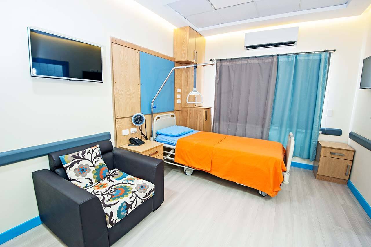
The patients of the Bundeswehr Hospital Berlin live in comfortable single and double patient rooms. Each patient room is equipped with an ensuite bathroom with a shower and a toilet. The patient room furnishing includes a comfortable bed, a bedside table, a TV and a telephone. To ensure a pleasant hospital stay, here is a library with an excellent choice of audiobooks, magazines and games. There is also a beautiful park on the territory of the hospital where one can enjoy peace, clean air and beautiful nature.
Meals and Menus
The hospital offers delicious and healthy three meals a day: a buffet breakfast, a hearty lunch and a light dinner. Every day, patients have a choice of three menus. Vegetarian dishes are also available.
If for some reason you do not eat all the foods, you will be offered an individual menu. Please inform the medical staff about your dietary preferences prior to the treatment.
Further details
Standard rooms include:
Accompanying person
Your accompanying person may stay with you in your patient room or at the hotel of your choice during the inpatient program.
Hotel
You may stay at the hotel of your choice during the outpatient program. Our managers will support you for selecting the best option.


