Retinal detachment
Due to the difficulties associated with the organization of treatment in Turkey, Switzerland, South Korea and India, we are not currently processing requests to these regions.
If you are interested in treatment in Germany, please leave a request and our specialists will contact you as soon as possible.
Retinal detachment is a separation of the retina from the choroid. This illness often leads to blindness within some time. Blindness develops as a result of a retinal dystrophy and optic nerve atrophy. The incidence of the illness in different countries is from 10 to 25 cases per 100,000 population a year. Retinal detachment is more often diagnosed among the people who suffer from myopia (nearsightedness).
The Booking Health portal presents 45 German clinics specializing in retinal detachment treatment
Show all clinics
Retinal detachment – Diagnostics
At the beginning stage of diagnostics doctors examine the vision acuity, perform a perimetry (estimate the peripheral vision), and measure the intraocular pressure.
Ophthalmoscopy, which is fundus examination with the help of a special device, is the main diagnostic method. If the field of view is insufficient, doctors perform the ultrasound examination. Low informativeness of ophthalmoscopy can be caused by opacity of cornea, vitreous humor or the eye lens.
If the detachment is old, specialists will also examine the stage of retinal dystrophy and state of the optic nerve. An electrophysiological study is carried out for this purpose.
Best clinics for the retinal detachment diagnostics in Germany:
Retinal detachment – Treatment
Retinal detachment can only be treated with the help of surgical intervention. The operation should be performed as soon as possible. It is desirable to perform it immediately after the diagnosis establishment, because early detachment responds to treatment much better.
Extrascleral buckling is carried out as an independent surgery as well as a part of vitreo-retinal surgical intervention. Surgeon uses a fine-meshed silicone sponge as a material for buckling. Sponge is sewed on the eyeball from the external side and thus pressing the eye wall and moving the vascular membrane closer to the detached retina. A break is blocked under the dimple.
Buckling can be local or circumferential. Local scleral buckling can be radial or sectoral. Radial buckling is performed for the single retinal break with the early detachment. Sectoral buckling is applied for patients who have several closely located breaks and also, for the large-sized single breaks with separation from the ora serrata.
Cerclage is used in complex cases. It is a circular buckle of sclera. Procedure is performed when there are numerous areas of retina degeneration, big number of breaks in different areas. Cerclage is performed with the help of either silicone tape or porous bandage.
Vitreoretinal surgery is applied in the most severe clinical cases. The purpose of the surgery is to move the retina closer to the vascular membrane and fix it.
Vitreoretinal surgery is performed in the following cases:
- Large-sized retinal detachment
- Wrapped edges of the break
- Expressed traction syndrome
- Central location of the breaks
- Total retinal detachment
- Retinal separation from the ora serrata
- Traumatic retinal detachment
- Inefficiency of the retinal buckling
Cryopexy can be performed as an independent surgery as well as combined with the buckling. The purpose of this method is to freeze a posterior wall of the eye right after the place of retinal break. This will stimulate the process of the scarring and will block the further detachment.
Laser photocoagulation is applied more often as an addition to the extrascleral buckling for the better control of retinal detachment. Laser photocoagulation provides its moderate adjacency right after the procedure and doesn’t cause any inflammatory reactions. One of the benefits of this method is a short rehabilitation period.
Pneumatic retinopexy. Retina is being pressed towards the eye wall with the help of the air bubble which is inserted into the vitreous humor. Afterwards, the fluid flows out from underneath. Air bubble stays in the eye until the retina is healed (up to several weeks). The use of method is restricted to cases when the single break is located in the upper part of the retina.
Retinal transplantation. In case of retinal dystrophy development its transplantation can be done. An artificial prosthesis is used for this purpose. A perspective aim is the transplantation of the retina cultured from the patients’ stem cells.
Best clinics for the retinal detachment treatment in Germany:
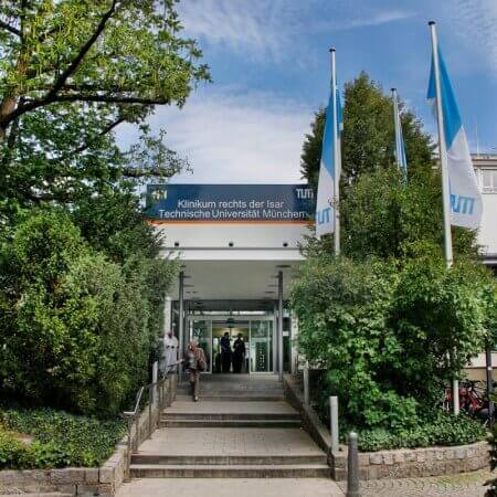
University Hospital Rechts der Isar Munich
2214921299
1942317873
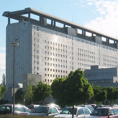
University Hospital of Ludwig Maximilian University of Munich
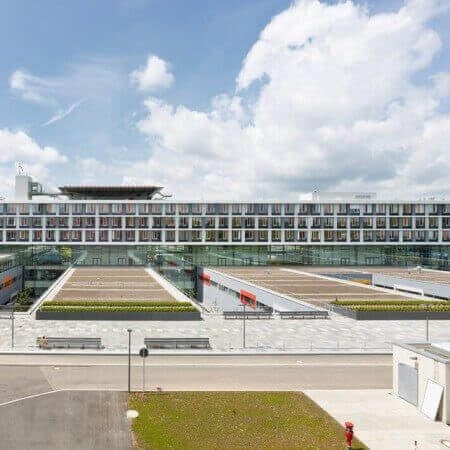
University Hospital Ulm
Retinal detachment – Rehabilitation
Rehabilitation measures during the first 1-3 days after treatment of retinal detachment include using special protective dressing, as well as administration of analgesics and antibacterial drugs.
In the early postoperative period (up to 1.5 months after the treatment) it is recommended to follow such recommendations:
- Limit weight lifting to 3-5 kg
- Exclude intensive physical training, give preference to walking or swimming
- Avoid positions with a tilted head (including housework, washing your head, and so on)
- Avoid visiting the sauna, baths, solarium
- Avoid staying under the active sun and wear sunglasses
- Give preference to foods rich in selenium and zinc, vitamins A, E, C and B
Rehabilitation includes medication support. Antioxidants and drugs for improving condition of retinal vessels are used. Drugs are prescribed in the form of drops, for direct administration to the eye, or in the tablet form. Also, the doctor prescribes medicines for improving the general health condition, for example, for normalization of blood pressure.
Rehabilitation programs in Germany are designed for 2 weeks. If necessary, they can last much longer. In this country, the patient is provided with qualitative care, accommodation in comfortable rooms and individually selected meals.
Rehabilitation programs in Germany show one of the best results in the world. Most patients successfully restore their employability and excellent health there. They remain physically active, return to the full social and family life.
Best clinics for general therapeutic rehabilitation in Germany:

Authors:
The article was edited by medical expert, board certified Dr. Nadezhda Ivanisova. For the treatment of the conditions referred to in the article you must consult a doctor; the information in the article is not intended for self-medication!
Sources:
International Council of Ophthalmology
The cost of services includes
Here you can find the cost of treatment for this disease at the German University Hospitals. Leave a request and we will provide a free consultation with a doctor and will start organizing the whole treatment process.
The program includes the following:
- Issuing of an invitation for getting a visa for treatment as quick as possible
- Fixing an appointment at a time convenient for you
- Preliminary organization of a comprehensive examination and discussion of the forthcoming treatment plan
- Arranging transfer from the airport to the hospital and back to the airport
- Provision of interpreting services and services of a personal medical coordinator
- If necessary, assistance in the organization of further surgical treatment
- Provision of a medical insurance against treatment complications covering up to 200,000 euro
- Preparation and translation of medical records and recommendations from the hospital
- Assistance in the subsequent communication with your attending physician, including consultations on repeated X-ray images through the unique medical document management system E-doc
