{"translation_price":"50","translation_doc_price":"40","child_coefficient":"1.1","transfer_price":"2.00","transfer_price_vip":"5.00","constant_transfer_price_vip":350,"constant_transfer_price":150,"constant_transfer_distanse":60,"type":"treatment","program_full_story":"<ul>\n\t<li>Initial presentation in the clinic<\/li>\n\t<li>clinical history taking<\/li>\n\t<li>review of medical records<\/li>\n\t<li>physical examination<\/li>\n\t<li>laboratory tests:\n\t<ul>\n\t\t<li>complete blood count<\/li>\n\t\t<li>general urine analysis<\/li>\n\t\t<li>biochemical analysis of blood<\/li>\n\t\t<li>TSH-basal, fT3, fT4<\/li>\n\t\t<li>tumor markers (AFP, CEA, \u0421\u0410-19-9)<\/li>\n\t\t<li>inflammation indicators (CRP, ESR)<\/li>\n\t\t<li>indicators of blood coagulation<\/li>\n\t<\/ul>\n\t<\/li>\n\t<li>abdominal ultrasound\u200b scan<\/li>\n\t<li>CT\/MRI of abdomen<\/li>\n\t<li>preoperative care<\/li>\n\t<li>percutaneous embolization (coiling) or chemoembolization<\/li>\n\t<li>symptomatic treatment<\/li>\n\t<li>cost of essential medicines<\/li>\n\t<li>nursing services<\/li>\n\t<li>elaboration of further recommendations<\/li>\n<\/ul>\n<div class=\"program_how_program_going mt-4\"><h4>How program is carried out<\/h4><p style=\"text-align: justify;\"><strong>During the first visit<\/strong>, the doctor will conduct a clinical examination and go through the results of the available diagnostic tests. After that, you will undergo the necessary additional examination, such as the assessment of liver and kidney function, ultrasound scan, CT scan and MRI. This will allow the doctor to determine which vessels are feeding the tumor and its metastases, as well as determine how well you will tolerate the procedure.<\/p>\n\n<p style=\"text-align: justify;\"><strong>Chemoembolization <\/strong>begins with local anesthesia and catheterization of the femoral artery. The thin catheter is inserted through a few centimeters long incision of the blood vessel. The doctor gradually moves the catheter to the vessel feeding the primary tumor or its metastases. The procedure is carried out under visual control, an angiographic device is used for this. The vascular bed and the position of the catheter in it are displayed on the screen of the angiograph.<\/p>\n\n<p style=\"text-align: justify;\">When the catheter reaches a suspected artery, a contrast agent is injected through it. Due to the introduction of the contrast agent, the doctor clearly sees the smallest vessels of the tumor and the surrounding healthy tissues on the screen of the angiograph. After that, he injects emboli into the tumor vessels through the same catheter.<\/p>\n\n<p style=\"text-align: justify;\">Emboli are the spirals or the liquid microspheres. The type of embolus is selected individually, taking into account the diameter of the target vessel. When carrying out chemoembolization, a solution of a chemotherapy drug is additionally injected into the tumor vessel. Due to the subsequent closure of the vessel lumen with an embolus, the chemotherapy drug influences the tumor for a long time. In addition, the drug does not enter the systemic circulation, which allows doctors to use high doses of chemotherapeutic agents without the development of serious side effects. Chemoembolization leads to the destruction of the tumor or slowing down its progression.<\/p>\n\n<p style=\"text-align: justify;\">After that, the catheter is removed from the artery. The doctor puts a vascular suture on the femoral artery and closes it with a sterile dressing. During chemoembolization, you will be awake. General anesthesia is not used, which significantly reduces the risks of the procedure and allows performing it on an outpatient basis, avoiding long hospital stay.<\/p>\n\n<p style=\"text-align: justify;\"><strong>After the first procedure<\/strong>, you will stay under the supervision of an interventional oncologist and general practitioner. If necessary, you will receive symptomatic treatment. As a rule, a second chemoembolization procedure is performed in 3-5 days after the first one in order to consolidate the therapeutic effect. After that, you will receive recommendations for further follow-up and treatment.<\/p>\n<\/div><div class=\"program_required_documents mt-4\"><h4>Required documents<\/h4><ul>\n\t<li style=\"text-align: justify;\">Medical records<\/li>\n\t<li style=\"text-align: justify;\">MRI\/CT scan (not older than 3 months)<\/li>\n\t<li style=\"text-align: justify;\">Biopsy results (if available)<\/li>\n<\/ul>\n<\/div>","program_full_story_crm":"<ul>\n\t<li>Initial presentation in the clinic<\/li>\n\t<li>clinical history taking<\/li>\n\t<li>review of medical records<\/li>\n\t<li>physical examination<\/li>\n\t<li>laboratory tests:\n\t<ul>\n\t\t<li>complete blood count<\/li>\n\t\t<li>general urine analysis<\/li>\n\t\t<li>biochemical analysis of blood<\/li>\n\t\t<li>TSH-basal, fT3, fT4<\/li>\n\t\t<li>tumor markers (AFP, CEA, \u0421\u0410-19-9)<\/li>\n\t\t<li>inflammation indicators (CRP, ESR)<\/li>\n\t\t<li>indicators of blood coagulation<\/li>\n\t<\/ul>\n\t<\/li>\n\t<li>abdominal ultrasound\u200b scan<\/li>\n\t<li>CT\/MRI of abdomen<\/li>\n\t<li>preoperative care<\/li>\n\t<li>percutaneous embolization (coiling) or chemoembolization<\/li>\n\t<li>symptomatic treatment<\/li>\n\t<li>cost of essential medicines<\/li>\n\t<li>nursing services<\/li>\n\t<li>elaboration of further recommendations<\/li>\n<\/ul>\n<div class=\"program_how_program_going mt-4\"><h4>How program is carried out<\/h4><p style=\"text-align: justify;\"><strong>During the first visit<\/strong>, the doctor will conduct a clinical examination and go through the results of the available diagnostic tests. After that, you will undergo the necessary additional examination, such as the assessment of liver and kidney function, ultrasound scan, CT scan and MRI. This will allow the doctor to determine which vessels are feeding the tumor and its metastases, as well as determine how well you will tolerate the procedure.<\/p>\n\n<p style=\"text-align: justify;\"><strong>Chemoembolization <\/strong>begins with local anesthesia and catheterization of the femoral artery. The thin catheter is inserted through a few centimeters long incision of the blood vessel. The doctor gradually moves the catheter to the vessel feeding the primary tumor or its metastases. The procedure is carried out under visual control, an angiographic device is used for this. The vascular bed and the position of the catheter in it are displayed on the screen of the angiograph.<\/p>\n\n<p style=\"text-align: justify;\">When the catheter reaches a suspected artery, a contrast agent is injected through it. Due to the introduction of the contrast agent, the doctor clearly sees the smallest vessels of the tumor and the surrounding healthy tissues on the screen of the angiograph. After that, he injects emboli into the tumor vessels through the same catheter.<\/p>\n\n<p style=\"text-align: justify;\">Emboli are the spirals or the liquid microspheres. The type of embolus is selected individually, taking into account the diameter of the target vessel. When carrying out chemoembolization, a solution of a chemotherapy drug is additionally injected into the tumor vessel. Due to the subsequent closure of the vessel lumen with an embolus, the chemotherapy drug influences the tumor for a long time. In addition, the drug does not enter the systemic circulation, which allows doctors to use high doses of chemotherapeutic agents without the development of serious side effects. Chemoembolization leads to the destruction of the tumor or slowing down its progression.<\/p>\n\n<p style=\"text-align: justify;\">After that, the catheter is removed from the artery. The doctor puts a vascular suture on the femoral artery and closes it with a sterile dressing. During chemoembolization, you will be awake. General anesthesia is not used, which significantly reduces the risks of the procedure and allows performing it on an outpatient basis, avoiding long hospital stay.<\/p>\n\n<p style=\"text-align: justify;\"><strong>After the first procedure<\/strong>, you will stay under the supervision of an interventional oncologist and general practitioner. If necessary, you will receive symptomatic treatment. As a rule, a second chemoembolization procedure is performed in 3-5 days after the first one in order to consolidate the therapeutic effect. After that, you will receive recommendations for further follow-up and treatment.<\/p>\n<\/div>","is_ambulant":"1","bh_fee":"0","only_for_children":"0","no_service":"0","with_prepayment":"1","show_calculator":"1","paket_type":"1","btn_type":"2","clinic_icon":"5ffc4fb11d090.jpg","city":"Krefeld","clinic_site":"https:\/\/www.helios-gesundheit.de\/kliniken\/krefeld\/","department_recommend":"0","country":"Germany","country_id":"1","clinic_name":"HELIOS Clinic Krefeld","cinic_name":"HELIOS Clinic Krefeld","department_id":"2264","duration":"6","direction":"Interventional radiology","min_duration":0,"clinic_id":"1666","paketPrice":3200,"paket":"<ul>\n <li> Interpreter up to 8 hours<\/li>\n <li>Translation of up to 5 pages<\/li>\n <li>Visa support<\/li>\n\n<\/ul>","title":"Treatment of liver cancer with percutaneous embolization (coiling) or chemoembolization","price":{"val":8482.64,"type":"val"},"price_surcharge":0,"price_surcharge_clear":0,"extra_service_clinic":[],"extra_service":[],"translation_hours":"0","translation_doc_count":null,"roads":[{"id":"4","distance":"20","airport_title":"Duesseldorf"},{"id":"7","distance":"83","airport_title":"Cologne-Bonn"},{"id":"6","distance":"244","airport_title":"Frankfurt a.M."}],"pakets":[],"lang":{"day":"Day","days":"days","ambulatory":"Outpatient","stationaryProgram":"Inpatient"}}
Treatment of liver cancer with percutaneous embolization (coiling) or chemoembolization in HELIOS Clinic Krefeld
percutaneous embolization (coiling) or chemoembolization
symptomatic treatment
cost of essential medicines
nursing services
elaboration of further recommendations
How program is carried out
During the first visit, the doctor will conduct a clinical examination and go through the results of the available diagnostic tests. After that, you will undergo the necessary additional examination, such as the assessment of liver and kidney function, ultrasound scan, CT scan and MRI. This will allow the doctor to determine which vessels are feeding the tumor and its metastases, as well as determine how well you will tolerate the procedure.
Chemoembolization begins with local anesthesia and catheterization of the femoral artery. The thin catheter is inserted through a few centimeters long incision of the blood vessel. The doctor gradually moves the catheter to the vessel feeding the primary tumor or its metastases. The procedure is carried out under visual control, an angiographic device is used for this. The vascular bed and the position of the catheter in it are displayed on the screen of the angiograph.
When the catheter reaches a suspected artery, a contrast agent is injected through it. Due to the introduction of the contrast agent, the doctor clearly sees the smallest vessels of the tumor and the surrounding healthy tissues on the screen of the angiograph. After that, he injects emboli into the tumor vessels through the same catheter.
Emboli are the spirals or the liquid microspheres. The type of embolus is selected individually, taking into account the diameter of the target vessel. When carrying out chemoembolization, a solution of a chemotherapy drug is additionally injected into the tumor vessel. Due to the subsequent closure of the vessel lumen with an embolus, the chemotherapy drug influences the tumor for a long time. In addition, the drug does not enter the systemic circulation, which allows doctors to use high doses of chemotherapeutic agents without the development of serious side effects. Chemoembolization leads to the destruction of the tumor or slowing down its progression.
After that, the catheter is removed from the artery. The doctor puts a vascular suture on the femoral artery and closes it with a sterile dressing. During chemoembolization, you will be awake. General anesthesia is not used, which significantly reduces the risks of the procedure and allows performing it on an outpatient basis, avoiding long hospital stay.
After the first procedure, you will stay under the supervision of an interventional oncologist and general practitioner. If necessary, you will receive symptomatic treatment. As a rule, a second chemoembolization procedure is performed in 3-5 days after the first one in order to consolidate the therapeutic effect. After that, you will receive recommendations for further follow-up and treatment.
Required documents
Medical records
MRI/CT scan (not older than 3 months)
Biopsy results (if available)
Service
Price from:
Type of program :
Price for 1 day:
Expected duration of the program:
The minimum duration of the program:
Select
You may also book:
Hospital directly:
Saving:
BookingHealth Price from:
About the department
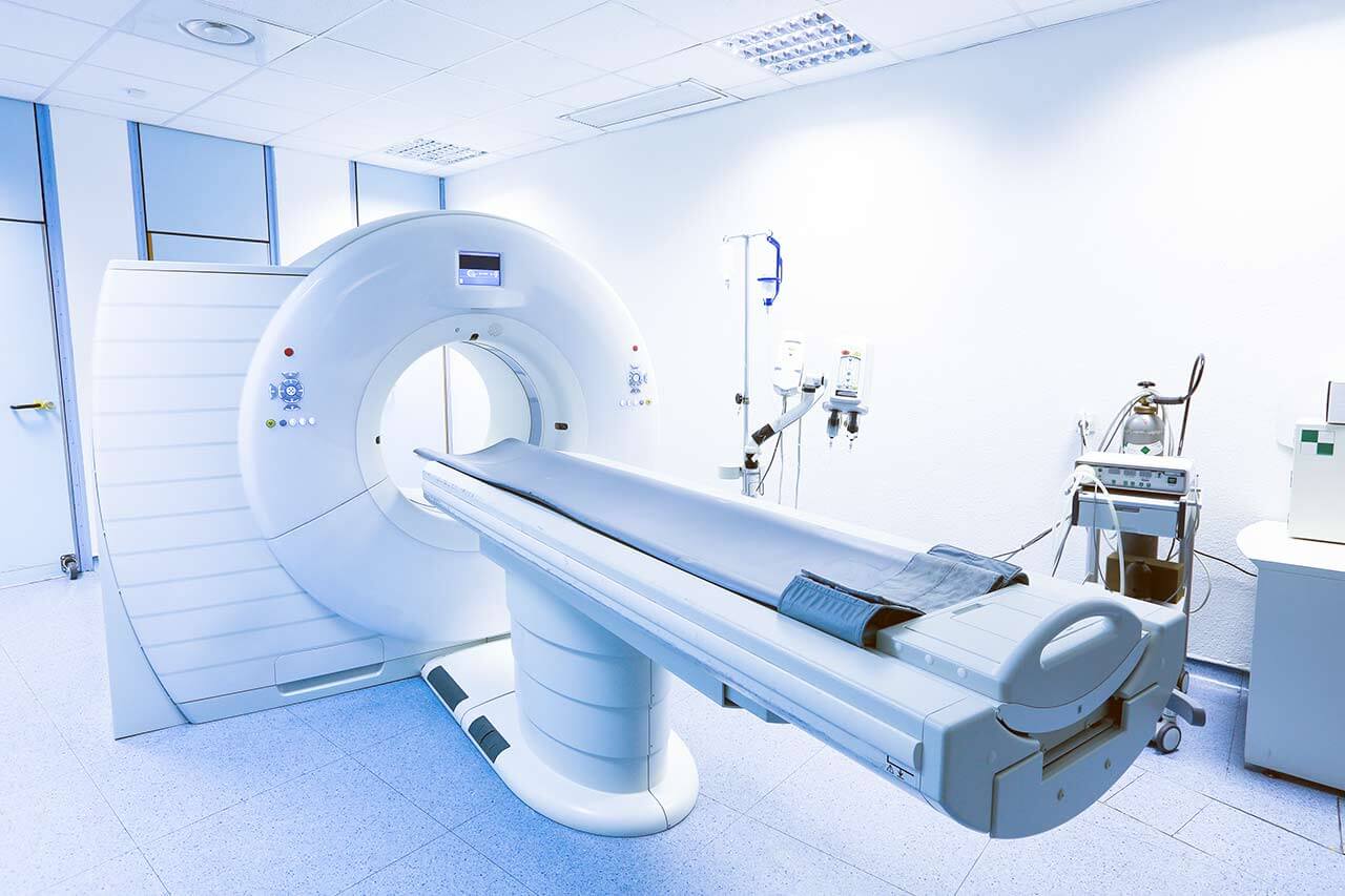
The Department Adult and Pediatric Diagnostic, Interventional Radiology, Neuroradiology at the HELIOS Clinic Krefeld offers the full range of services in the areas of its specialization. The department has state-of-the-art equipment, thanks to which doctors successfully carry out imaging diagnostics allowing them to determine the diagnosis as accurately as possible and develop the most effective treatment regimen. The department performs X-ray studies, ultrasound scanning, computed tomography (CT), magnetic resonance imaging (MRI), angiography, mammography and a combined method of computed tomography with positron emission tomography (PET-CT). Cardiac CT and MRI are also carried out in cooperation with the Department of Cardiology. The department is one of the few medical facilities of this kind in Germany specializing in the imaging diagnostics in children. The range of the department's services is complemented by minimally invasive image-guided therapeutic procedures. In the field of interventional radiology, the focus is on the treatment of pain syndromes, vascular diseases, benign and malignant tumors, as well as metastases. The department's radiologists strictly adhere to the radiation protection standards, so all diagnostic and therapeutic procedures are absolutely safe for patients.The department is headed by Prof. Dr. med. Marcus Katoh.
The advanced diagnostic equipment and high professionalism of doctors allow them to make high-quality diagnostics, detecting the slightest deviations in the work of organs before the onset of clinical symptoms. Ultrasound, CT, MRI, angiography, as well as myelography of the brain and spinal cord are of particular interest to the specialists. In addition, the department focuses on the diagnostics of cancers of various localization and cardiac imaging diagnostics. The specialists of the medical facility use in their work a modern picture archiving and communication system, thanks to which the results of imaging tests are available to doctors in all departments of the clinic.
The diagnostic imaging tests in children are carried out by specially trained doctors with the use of medical equipment adapted to the characteristics of a growing child's body. Pediatricians, pediatric surgeons, orthopedists, neurosurgeons and other specialists are often involved in the diagnostic process. For maximum comfort of young patients, doctors allow the parents to stay with their children during the diagnostic examinations and support them throughout the entire process. The diagnostic procedures in the field of pediatric radiology include ultrasound, radiography, fluoroscopy, CT and MRI. The negative impact of X-rays on the child's body during the diagnostics in the department is completely excluded.
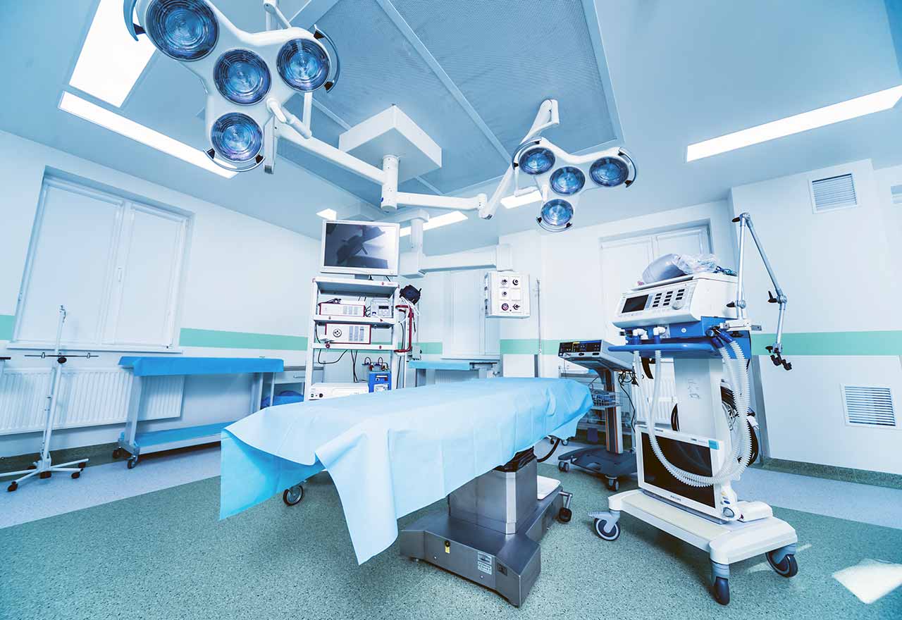
However, the department's work is not limited to diagnostics. It also successfully performs image-guided interventional therapeutic procedures. In many cases, such treatment can effectively eliminate the pathology and save the patient from the need for a full-fledged surgical intervention, which is often associated with risks and long postoperative recovery. The department's interventional radiologists specialize in the treatment of chronic pain, benign neoplasms, stenoses and vascular occlusions caused by atherosclerosis, aneurysms, malignant tumors and their metastases. In addition, the patients can undergo highly competent interventional treatment of diseases of the brain and spinal cord – thrombectomy in the case of stroke, therapeutic procedures for the treatment of cerebral aneurysms, carotid artery stenting, correction of arteriovenous malformations (angiomas).
The department's range of medical services includes:
Diagnostic radiology
Ultrasound scanning
X-ray studies
Angiography
Computed tomography (CT)
Magnetic resonance imaging (MRI)
Cardiac CT and MRI (in collaboration with cardiologists)
Mammography
Combined computed tomography with positron emission tomography (PET-CT)
Radiological diagnostics in children
Ultrasound scanning
X-ray studies
Fluoroscopy
Computed tomography (CT)
Magnetic resonance imaging (MRI)
Interventional radiology
Treatment of benign neoplasms and chronic pain
Prostate artery embolization for benign prostatic hyperplasia
Arterial embolization for chronic joint pain
Hemorrhoidal artery coil embolization
Uterine artery embolization for uterine fibroids
Treatment of stenoses and vascular occlusions due to atherosclerosis
Stent implantation and balloon dilatation of the vessels in the pelvis and lower limbs
Stent implantation and balloon dilatation of the kidney arteries
Treatment of aneurysms in the body, limbs and brain, abdominal and thoracic aortic aneurysms
Treatment of malignant tumors and metastases
Brachytherapy for the treatment of malignant tumors
Transarterial chemoembolization (TACE) and selective internal radiation therapy (SIRT) for the treatment of liver cancer
Percutaneous radiofrequency ablation (for example, for the treatment of lung and liver tumors)
Irreversible electroporation for the treatment of pancreatic tumors
Transjugular intrahepatic portosystemic shunting
Minimally invasive dialysis shunt formation or recanalization
Pain therapy for chronic back pain (injection of anesthetic drugs)
Neuroradiology
Diagnostic tests
X-ray studies
Ultrasound scanning
Computed tomography (CT)
Magnetic resonance imaging (MRI)
Angiography, including contrast-enhanced and digital subtraction angiography
Myelography (examination of the spinal canal with contrast enhancement)
Image-guided interventional therapy
Thrombectomy for the treatment of stroke
Interventional procedures for the treatment of cerebral aneurysms
Carotid artery stenting in the case of their stenosis
Correction of arteriovenous malformations (angiomas) in cooperation with neurosurgeons and radiation surgeons
Other diagnostic and therapeutic options
Curriculum vitae
Higher Education and Professional Career
October 1993 - December 1999 Study of Human Medicine, Heinrich Heine University Duesseldorf. Completed with the third part of the medical licensing examination.
February 2000 - July 2001 Physician in Training, Department of General Surgery, University Hospital Hamburg-Eppendorf.
August 2000 Doctoral thesis defense, Department of General Surgery, University Hospital Duesseldorf. Thesis subject: "Surgical treatment of chronic pancreatitis with special regard to long-term outcomes".
August 2006 Admission to medical practice.
August 2001 - December 2003 Assistant Physician, Department of Diagnostic Radiology, University Hospital RWTH Aachen.
January 2004 - May 2004 Grant, Beth Israel Deaconess Medical Center and Harvard Medical School, Department of Cardiology, Boston, Massachusetts, USA.
June 2004 Visiting Physician, School of Medicine, Johns Hopkins University, Department of Radiology (MRI Division), USA.
June 2004 - Aug 2006 Assistant Physician, Department of Diagnostic Radiology, University Hospital RWTH Aachen.
July 2006 Postdoctoral thesis, Department of Diagnostic Radiology, RWTH Aachen. Postdoctoral thesis subject:
"MR coronary angiography: techniques of imaging the vascular lumen, the vessel wall and the coronary blood flow".
August 2006 Board Certification in Diagnostic Radiology.
August 2006 - October 2006 Senior Physician, Department of Diagnostic and Interventional Radiology, University Hospital RWTH Aachen.
November 2006 - June 2011 Managing Senior Physician, Section for Angiography, Department of Diagnostic and Interventional Radiology, University Hospital Saarland, Homburg.
September 2007 - June 2011 Managing Senior Physician, Department of Diagnostic and Interventional Radiology, University Hospital Saarland, Homburg.
September 2009 3rd place in the appointment proposal while applying for the W2 professorship for cardiopulmonary imaging, Institute for X-Ray Diagnostics, University of Regensburg.
March 2010 Associate Professor, Faculty of Medicine, University of Saarland.
August 2010 Instructor for Interventional Radiology, German Society of Interventional Radiology (DeGIR).
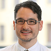
Since July 2011 Head of the Department of Adult and Pediatric Diagnostic, Interventional Radiology, Neuroradiology at the HELIOS Clinic Krefeld.
April 2009 - October 2011 Master of Business Administration (MBA) Program, RheinAhrCampus Remagen.
Photo of the doctor: (c) Helios Klinikum Krefeld
About hospital
Founded in 2014, the HELIOS Clinic Krefeld is one of the most modern medical facilities in Germany today. A team of highly qualified specialists, innovative medical equipment and comfortable accommodation conditions – the clinic has everything to make the treatment run smoothly and efficiently. Having crossed the threshold of the clinic, you will feel the home-like cozy and modern atmosphere. Perhaps you will even forget that you are in the clinic.
As a maximum care medical center, the clinic covers practically all areas of medicine: cardiology, gastroenterology, oncology and hepatology, nephrology, diabetology and rheumatology, general, abdominal and minimally invasive surgery, dermatology and venereology, angiology, cardiac surgery and other medical fields.
Thanks to a continuous fruitful cooperation with the world’s leading universities, research and medical centers, the clinic provides the highest treatment standards.
The doctors of the clinic take up the treatment of the most severe cases, including serious spinal injuries and diseases related to the disorders of the central nervous system. The nursing staff of the clinic regularly undergoes both advanced and special trainings, which contributes to a high-quality, professional patient care at all stages of treatment.
The clinic adheres to a strict quality management system. Not only the patient care, but also the entire treatment process (including monitoring and patient care after the operation) are subject to the quality certification.
Photo: (c) depositphotos
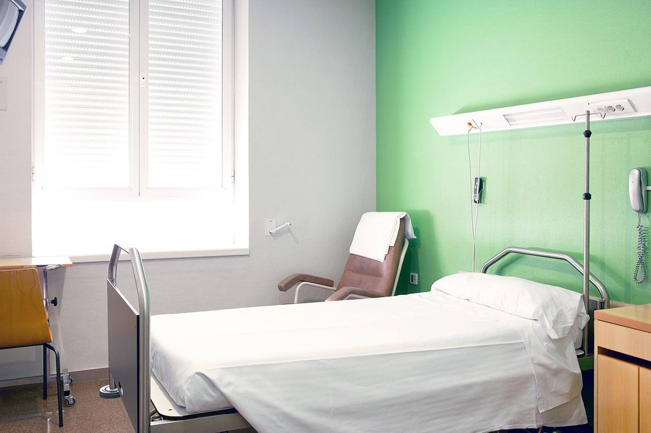
Accommodation in hospital
Patients rooms
The patients of the HELIOS Clinic Krefeld live in comfortable single and double rooms. Each room has a bathroom with a toilet and shower. The room is furnished with a comfortable bed, bedside table, TV, radio and telephone. Each room has access to the Internet, which can be used at extra charge.
The clinic offers an excellent infrastructure: a beautiful park with playgrounds for children, a cafeteria, a chapel, which regularly hosts church services, a prayer room for Muslims, a beauty salon, a shop with a large selection of magazines, drinks and personal hygiene items, an ATM and a large parking lot.
Meals and Menus
The restaurant of the clinic offers three meals a day. For breakfast, lunch and dinner, it serves a large selection of dishes that will please even spoiled gourmets. Every day, the menu features vegetarian and dietary dishes. It is possible to order any dish without leaving the room. In addition, the clinic has a cafeteria where one can always have a cup of aromatic coffee, hot tea or enjoy an exquisite refreshing drink. Also, the cafeteria menu offers many tasty snacks and dishes.
Further details
Standard rooms include:
Shower
Toilet
Wi-Fi
TV
Select your currency:
Your guarantee
One year of support after the treatment
Insurance to cover unforeseen expenses arising from complications during and 48 months after treatment (coverage up to 200,000 €)
Reduced costs by 40-70% (contracts with Hospitals)
In addition, we are the only TÜV-certified company with an ISO 9001:2015 certificate
thank you for reaching out! One of our medical advisors is currently reviewing your details. You can expect a call from us within one business day to discuss your request.
Note: The incoming call will appear as a German number or your local area code. This call is entirely free of charge.
Booking Health guarantees:
Expert Placement
We use rigorous statistical analysis to ensure you are treated at the best-rated facility for your specific diagnosis.
Total Cost Protection
No hidden fees or unexpected bills. We provide a fixed price guarantee, backed by insurance that covers any additional complications.
12-Month Extended Care
Your recovery is our priority. Benefit from a full year of direct medical support following your procedure.
 BookingHealth Price from:
