{"translation_price":"50","translation_doc_price":"40","child_coefficient":"1.1","transfer_price":"2.00","transfer_price_vip":"5.00","constant_transfer_price_vip":350,"constant_transfer_price":150,"constant_transfer_distanse":60,"type":"treatment","program_full_story":"<ul>\n\t<li>Initial presentation in the hospital<\/li>\n\t<li>Clinical history taking<\/li>\n\t<li>Review of available medical records<\/li>\n\t<li>Physical examination<\/li>\n\t<li>Laboratory tests:\n\t<ul>\n\t\t<li>Complete blood count<\/li>\n\t\t<li>General urine analysis<\/li>\n\t\t<li>Biochemical analysis of blood<\/li>\n\t\t<li>Tumor markers<\/li>\n\t\t<li>Inflammation indicators (CRP, ESR)<\/li>\n\t\t<li>Coagulogram<\/li>\n\t<\/ul>\n\t<\/li>\n\t<li>Ultrasound\u200b scan<\/li>\n\t<li>CT scan \/ MRI<\/li>\n\t<li>Preoperative care<\/li>\n\t<li>Embolization or chemoembolization, 2 procedures<\/li>\n\t<li>Symptomatic treatment<\/li>\n\t<li>Cost of essential medicines<\/li>\n\t<li>Nursing services<\/li>\n\t<li>Elaboration of further recommendations<\/li>\n<\/ul>\n<div class=\"program_how_program_going mt-4\"><h4>How program is carried out<\/h4><p style=\"text-align: justify;\"><strong>During the first visit<\/strong>, the doctor will conduct a clinical examination and go through the results of the available diagnostic tests. After that, you will undergo the necessary additional examination, such as the assessment of liver and kidney function, ultrasound scan, CT scan and MRI. This will allow the doctor to determine which vessels are feeding the tumor and its metastases, as well as determine how well you will tolerate the procedure.<\/p>\n\n<p style=\"text-align: justify;\"><strong>Chemoembolization <\/strong>begins with local anesthesia and catheterization of the femoral artery. The thin catheter is inserted through a few centimeters long incision of the blood vessel. The doctor gradually moves the catheter to the vessel feeding the primary tumor or its metastases. The procedure is carried out under visual control, an angiographic device is used for this. The vascular bed and the position of the catheter in it are displayed on the screen of the angiograph.<\/p>\n\n<p style=\"text-align: justify;\">When the catheter reaches a suspected artery, a contrast agent is injected through it. Due to the introduction of the contrast agent, the doctor clearly sees the smallest vessels of the tumor and the surrounding healthy tissues on the screen of the angiograph. After that, he injects emboli into the tumor vessels through the same catheter.<\/p>\n\n<p style=\"text-align: justify;\">Emboli are the spirals or the liquid microspheres. The type of embolus is selected individually, taking into account the diameter of the target vessel. When carrying out chemoembolization, a solution of a chemotherapy drug is additionally injected into the tumor vessel. Due to the subsequent closure of the vessel lumen with an embolus, the chemotherapy drug influences the tumor for a long time. In addition, the drug does not enter the systemic circulation, which allows doctors to use high doses of chemotherapeutic agents without the development of serious side effects. Chemoembolization leads to the destruction of the tumor or slowing down its progression.<\/p>\n\n<p style=\"text-align: justify;\">After that, the catheter is removed from the artery. The doctor puts a vascular suture on the femoral artery and closes it with a sterile dressing. During chemoembolization, you will be awake. General anesthesia is not used, which significantly reduces the risks of the procedure and allows performing it on an outpatient basis, avoiding long hospital stay.<\/p>\n\n<p style=\"text-align: justify;\"><strong>After the first procedure<\/strong>, you will stay under the supervision of an interventional oncologist and general practitioner. If necessary, you will receive symptomatic treatment. As a rule, a second chemoembolization procedure is performed in 3-5 days after the first one in order to consolidate the therapeutic effect. After that, you will receive recommendations for further follow-up and treatment.<\/p>\n<\/div><div class=\"program_required_documents mt-4\"><h4>Required documents<\/h4><ul>\n\t<li style=\"text-align: justify;\">Medical records<\/li>\n\t<li style=\"text-align: justify;\">MRI\/CT scan (not older than 3 months)<\/li>\n\t<li style=\"text-align: justify;\">Biopsy results (if available)<\/li>\n<\/ul>\n<\/div>","program_full_story_crm":"<ul>\n\t<li>Initial presentation in the hospital<\/li>\n\t<li>Clinical history taking<\/li>\n\t<li>Review of available medical records<\/li>\n\t<li>Physical examination<\/li>\n\t<li>Laboratory tests:\n\t<ul>\n\t\t<li>Complete blood count<\/li>\n\t\t<li>General urine analysis<\/li>\n\t\t<li>Biochemical analysis of blood<\/li>\n\t\t<li>Tumor markers<\/li>\n\t\t<li>Inflammation indicators (CRP, ESR)<\/li>\n\t\t<li>Coagulogram<\/li>\n\t<\/ul>\n\t<\/li>\n\t<li>Ultrasound\u200b scan<\/li>\n\t<li>CT scan \/ MRI<\/li>\n\t<li>Preoperative care<\/li>\n\t<li>Embolization or chemoembolization, 2 procedures<\/li>\n\t<li>Symptomatic treatment<\/li>\n\t<li>Cost of essential medicines<\/li>\n\t<li>Nursing services<\/li>\n\t<li>Elaboration of further recommendations<\/li>\n<\/ul>\n<div class=\"program_how_program_going mt-4\"><h4>How program is carried out<\/h4><p style=\"text-align: justify;\"><strong>During the first visit<\/strong>, the doctor will conduct a clinical examination and go through the results of the available diagnostic tests. After that, you will undergo the necessary additional examination, such as the assessment of liver and kidney function, ultrasound scan, CT scan and MRI. This will allow the doctor to determine which vessels are feeding the tumor and its metastases, as well as determine how well you will tolerate the procedure.<\/p>\n\n<p style=\"text-align: justify;\"><strong>Chemoembolization <\/strong>begins with local anesthesia and catheterization of the femoral artery. The thin catheter is inserted through a few centimeters long incision of the blood vessel. The doctor gradually moves the catheter to the vessel feeding the primary tumor or its metastases. The procedure is carried out under visual control, an angiographic device is used for this. The vascular bed and the position of the catheter in it are displayed on the screen of the angiograph.<\/p>\n\n<p style=\"text-align: justify;\">When the catheter reaches a suspected artery, a contrast agent is injected through it. Due to the introduction of the contrast agent, the doctor clearly sees the smallest vessels of the tumor and the surrounding healthy tissues on the screen of the angiograph. After that, he injects emboli into the tumor vessels through the same catheter.<\/p>\n\n<p style=\"text-align: justify;\">Emboli are the spirals or the liquid microspheres. The type of embolus is selected individually, taking into account the diameter of the target vessel. When carrying out chemoembolization, a solution of a chemotherapy drug is additionally injected into the tumor vessel. Due to the subsequent closure of the vessel lumen with an embolus, the chemotherapy drug influences the tumor for a long time. In addition, the drug does not enter the systemic circulation, which allows doctors to use high doses of chemotherapeutic agents without the development of serious side effects. Chemoembolization leads to the destruction of the tumor or slowing down its progression.<\/p>\n\n<p style=\"text-align: justify;\">After that, the catheter is removed from the artery. The doctor puts a vascular suture on the femoral artery and closes it with a sterile dressing. During chemoembolization, you will be awake. General anesthesia is not used, which significantly reduces the risks of the procedure and allows performing it on an outpatient basis, avoiding long hospital stay.<\/p>\n\n<p style=\"text-align: justify;\"><strong>After the first procedure<\/strong>, you will stay under the supervision of an interventional oncologist and general practitioner. If necessary, you will receive symptomatic treatment. As a rule, a second chemoembolization procedure is performed in 3-5 days after the first one in order to consolidate the therapeutic effect. After that, you will receive recommendations for further follow-up and treatment.<\/p>\n<\/div>","is_ambulant":"1","bh_fee":"0","only_for_children":"0","no_service":"0","with_prepayment":"1","show_calculator":"1","paket_type":"1","btn_type":"0","clinic_icon":"6005635c5a6a8.jpg","city":"Munich","clinic_site":"www.mri.tum.de","department_recommend":"1","country":"Germany","country_id":"1","clinic_name":"University Hospital Rechts der Isar Munich","cinic_name":"University Hospital Rechts der Isar Munich","department_id":"2756","duration":"7","direction":"Interventional radiology","min_duration":0,"clinic_id":"28","paketPrice":7800,"paket":"<ul>\n <li>Interpreter up to 17 hours<\/li>\n <li>Translation up to 10 pages<\/li>\n <li>Visa support<\/li>\n\n \n<\/ul>","title":"Treatment of rectal cancer with embolization or chemoembolization","price":{"val":31056.14,"type":"val"},"price_surcharge":0,"price_surcharge_clear":0,"extra_service_clinic":[],"extra_service":[],"translation_hours":"0","translation_doc_count":null,"roads":[{"id":"2","distance":"38","airport_title":"Munich"},{"id":"19","distance":"220","airport_title":"Stuttgart"},{"id":"1","distance":"316","airport_title":"Zurich"}],"pakets":[],"lang":{"day":"Day","days":"days","ambulatory":"Outpatient","stationaryProgram":"Inpatient"}}
Treatment of rectal cancer with embolization or chemoembolization in University Hospital Rechts der Isar Munich
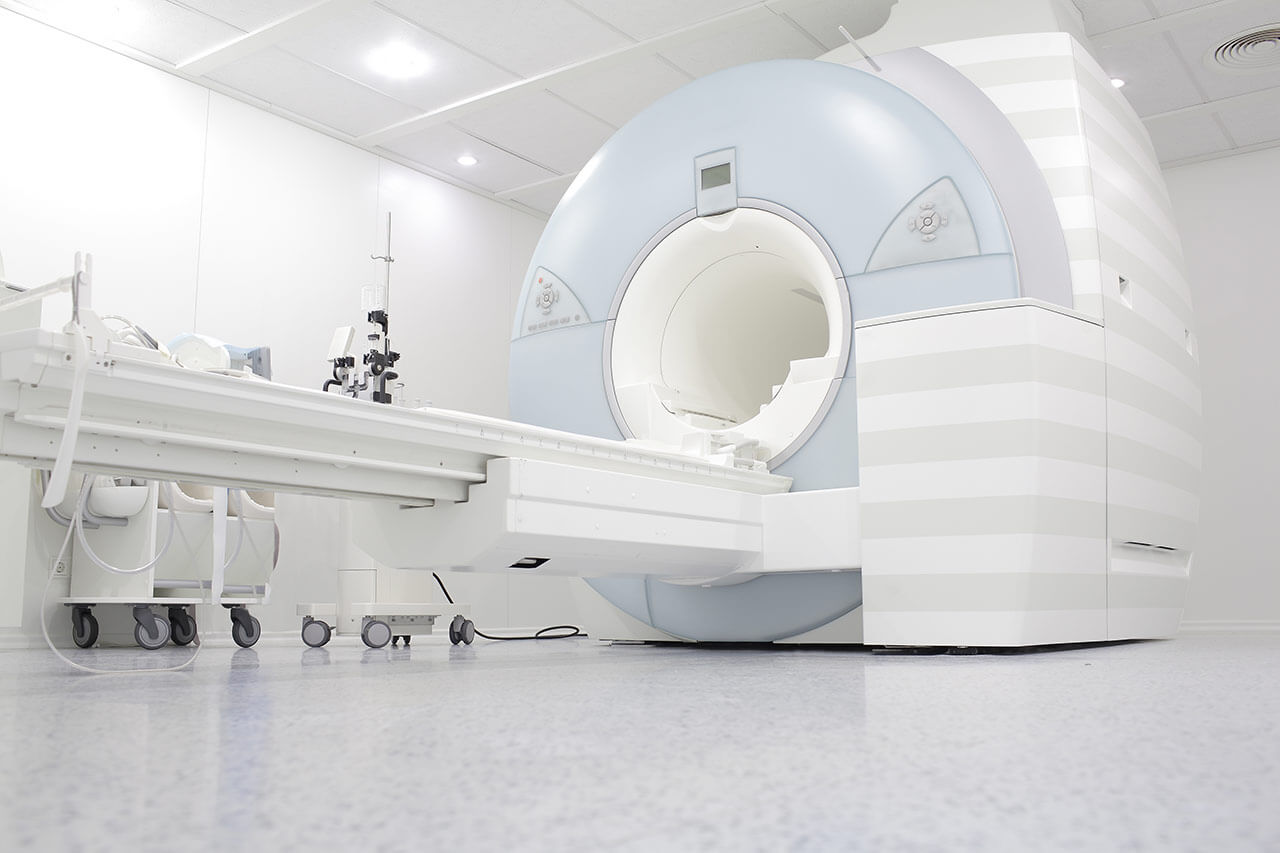
During the first visit, the doctor will conduct a clinical examination and go through the results of the available diagnostic tests. After that, you will undergo the necessary additional examination, such as the assessment of liver and kidney function, ultrasound scan, CT scan and MRI. This will allow the doctor to determine which vessels are feeding the tumor and its metastases, as well as determine how well you will tolerate the procedure.
Chemoembolization begins with local anesthesia and catheterization of the femoral artery. The thin catheter is inserted through a few centimeters long incision of the blood vessel. The doctor gradually moves the catheter to the vessel feeding the primary tumor or its metastases. The procedure is carried out under visual control, an angiographic device is used for this. The vascular bed and the position of the catheter in it are displayed on the screen of the angiograph.
When the catheter reaches a suspected artery, a contrast agent is injected through it. Due to the introduction of the contrast agent, the doctor clearly sees the smallest vessels of the tumor and the surrounding healthy tissues on the screen of the angiograph. After that, he injects emboli into the tumor vessels through the same catheter.
Emboli are the spirals or the liquid microspheres. The type of embolus is selected individually, taking into account the diameter of the target vessel. When carrying out chemoembolization, a solution of a chemotherapy drug is additionally injected into the tumor vessel. Due to the subsequent closure of the vessel lumen with an embolus, the chemotherapy drug influences the tumor for a long time. In addition, the drug does not enter the systemic circulation, which allows doctors to use high doses of chemotherapeutic agents without the development of serious side effects. Chemoembolization leads to the destruction of the tumor or slowing down its progression.
After that, the catheter is removed from the artery. The doctor puts a vascular suture on the femoral artery and closes it with a sterile dressing. During chemoembolization, you will be awake. General anesthesia is not used, which significantly reduces the risks of the procedure and allows performing it on an outpatient basis, avoiding long hospital stay.
After the first procedure, you will stay under the supervision of an interventional oncologist and general practitioner. If necessary, you will receive symptomatic treatment. As a rule, a second chemoembolization procedure is performed in 3-5 days after the first one in order to consolidate the therapeutic effect. After that, you will receive recommendations for further follow-up and treatment.
Required documents
Medical records
MRI/CT scan (not older than 3 months)
Biopsy results (if available)
Service
Price from:
Type of program :
Price for 1 day:
Expected duration of the program:
The minimum duration of the program:
Select
You may also book:
Hospital directly:
Saving:
BookingHealth Price from:
About the department
The Department of Interventional Radiology at the University Hospital Rechts der Isar Munich offers the full range of imaging examinations, as well as innovative image-guided minimally invasive techniques for the treatment of tumors, vascular diseases and internal pathologies (for example, CT, MRI, PET-CT, SPECT). The department's doctors have deep knowledge and colossal experience in the field of interventional radiological methods of treatment, which represent an excellent alternative to open surgical interventions. Despite the high level of technical equipment and the presence of advanced computerized systems, the focus is always on the person with his individual needs. Compliance with current clinical protocols and high professionalism of the department's specialists contribute to the successful clinical practice, as well as the reputability of the department among the best medical facilities of this kind in Germany. The department is headed by Prof. Dr. med. Marcus Makowski.
All modern imaging tests are available in the department to assess the state and function of the internal organs, various anatomical structures. Patients can undergo X-ray examinations, fluoroscopy, sonography, angiography, computed tomography and magnetic resonance imaging. With the appropriate clinical indications, doctors conduct contrast-enhanced imaging tests. The innovative technical equipment meets all the requirements of radiation protection, therefore, during the examination, the patient receives a minimum dose of radiation that does not harm health.
The department's service range includes not only diagnostic tests. The specialists of the medical facility also carry out a number of highly effective image-guided therapeutic interventional procedures. Particular attention is paid to the interventional treatment of benign and malignant tumors. Doctors perform ablation procedures to treat benign bone neoplasms (osteoid osteomas), malignant tumors of the liver and kidneys, metastases in these organs, radiofrequency ablation and irreversible electroporation for malignant tumors (most often, if there are contraindications to surgery), and tumor embolization. Percutaneous transluminal angioplasty is also successfully performed here to eliminate stenoses and obstructions in the blood vessels. In addition, patients with traumatic, degenerative and inflammatory musculoskeletal lesions can receive pain management in the department (local injections of anesthetic and anti-inflammatory drugs). Pain management is carried out under the guidance of ultrasound or CT, which ensures high safety and effectiveness of treatment.
The department's range of diagnostic and therapeutic options includes:
Diagnostic examinations
Ultrasound scans
Classical X-ray scans
Fluoroscopy
Angiography
Computed tomography
Magnetic resonance imaging
Image-guided biopsy of the internal organs and anatomical structures
Image-guided therapeutic interventional procedures on the genitourinary system
Uterine artery embolization for the treatment of uterine fibroids
Prostatic artery embolization for the treatment of prostate adenoma
Sclerotherapy for varicocele
Image-guided therapeutic interventional procedures on the intestine
Selective prostaglandin infusion
Embolization to stop bleeding
Percutaneous transluminal angioplasty
Stenting
Image-guided therapeutic interventional procedures on the bones
Radiofrequency ablation for osteoid osteoma
Sacroiliac joint osteosynthesis
Vertebroplasty
Image-guided therapeutic interventional procedures on the vessels
Arteriovenous malformation embolization
Lysis therapy
Port system implantation
Percutaneous transluminal angioplasty
Image-guided therapeutic interventional procedures on the nerve endings
Facet joint infiltration
Celiac plexus block
Sympatholysis
Nerve root infiltration
Other medical services
Curriculum vitae
Since 2020, Prof. Dr. med. Marcus Makowski has been heading the Department of Interventional Radiology at the University Hospital Rechts der Isar Munich. The professor's research activities are focused on quantitative imaging and the application of artificial intelligence methods in radiology. Of particular clinical interest is the assessment and development of new quantitative biomarkers in radiology.
After studying medicine, in 2007 Dr. Makowski defended his thesis at the Ludwig Maximilian University of Munich. In 2010, he successfully completed his PhD at King's College London. In 2013, the professor's habilitation work was distinguished with the Wilhelm Conrad Roentgen Award (Charite University Hospital Berlin). In 2016, Dr. Marcus Makowski received an offer to take the position of Professor W2 at the Charite. Prior to becoming the Head Physician of the Department of Interventional Radiology at the University Hospital Rechts der Isar Munich, the specialist held the position of Deputy Head of the Department of Radiology at the University Hospital Charite Berlin.
Prof. Marcus Makowski has won numerous prizes and awards, the most significant of which are the Marie Curie Prize (2018), the Heisenberg Award (2014) and the Wilhelm Conrad Roentgen Award (2013).
Photo of the doctor: (c) Klinikum rechts der Isar der Technischen Universität München
About hospital
The University Hospital Rechts der Isar Munich was founded in 1834. It combines long traditions with the very latest advances in modern medicine. The medical facility includes 33 specialized departments and 20 interdisciplinary centers, where patients can receive top-class medical care in all medical fields.
The hospital annually admits more than 65,000 inpatients for diagnostics and treatment, and about 250,000 outpatients receive effective medical care. The hospital also performs more than 40,000 surgical procedures every year, and about 2,100 babies are born here annually. One of the most significant achievements of the medical facility can be called the first transplantation of both arms above the elbow performed in 2008. The surgery that lasted 15 hours, and in which 40 doctors of various medical specialties took part, became a real sensation in the scientific world. Thanks to a unique surgical procedure, the doctors managed to give the patient new hands.
In addition, the employees of the hospital are actively involved in research activities, in which they study various diseases, as well as develop new therapeutic options for their treatment. It should be noted that the research institutes of the hospital are among the most reputable research organizations in the world. A striking example can be considered the Roman Herzog Comprehensive Cancer Center, whose specialists cooperate closely with the Comprehensive Cancer Center Munich in order to find new treatment methods for cancers.
The university hospital has a strict quality management system to maintain a high level of patient care. Since 2011, the hospital has been certified in accordance with DIN EN ISO 9001:2015 at the national and international level. The medical facility was also recertified by TÜV Rheinland in 2020.
The hospital annually provides medical services not only to German citizens, but also to thousands of patients from different countries of the world. This indicates that the hospital has an excellent reputation in the international medical arena and takes on the most complex clinical cases where other medical centers are unable to help the patient.
Photo: (с) depositphotos
Accommodation in hospital
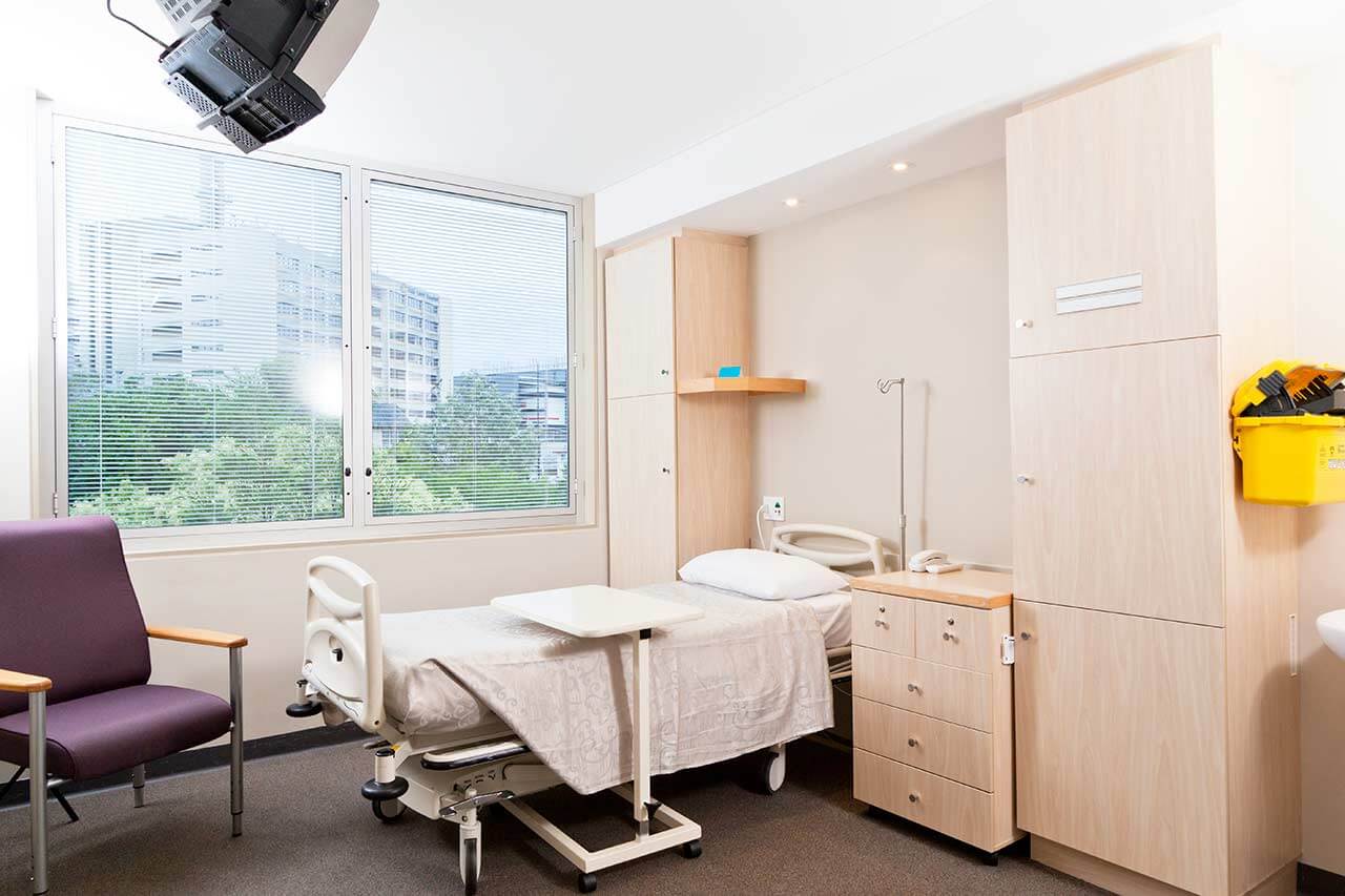
Patients rooms
The patients of the University Hospital Rechts der Isar Munich live in comfortable single and double rooms with modern design. An accompanying person may stay with the patient in the single room. All patient rooms have an ensuite bathroom with shower and toilet. The furnishings of a standard patient room include an automatically adjustable bed, a bedside table for personal belongings, a table and chairs for receiving visitors, a wardrobe, a telephone and a TV. The patient rooms also have Wi-Fi.
The hospital also has enhanced-comfort rooms, corresponding to the level of a high-end hotel. Such patient rooms have additional amenities: a safe, a mini fridge and upholstered furniture.
Meals and Menus
The patients of the hospital are offered a balanced and healthy three meals a day: breakfast, lunch and dinner. The patients have a choice of three different menus for lunch – a classic full menu, as well as a dietary and vegetarian one. When cooking meals, the chefs comply with the current recommendations of the German Society for Nutritional Medicine (DGEM) and the German Nutrition Society (DGE).
If for some reason you do not eat all the foods, you will be offered an individual menu. The hospital also houses a cafeteria with a large assortment of hot and cold drinks, snacks and desserts.
Further details
Standard rooms include:
Toilet
Shower
Wi-Fi
TV
Religion
Religious services are available upon request.
Accompanying person
Your accompanying person may stay with you in your patient room or at the hotel of your choice during the inpatient program.
Hotel
You may stay at the hotel of your choice during the outpatient program. Our managers will support you for selecting the best option.
The hospital offers a full range of laboratory tests (general, hormonal, tests for infections, antibodies, tumor markers, etc.), genetic tests, various modifications of ultrasound scans, CT scans, MRI and PET / CT, angiography, myelography, biopsy and other examinations. Treatment with medications, endoscopic and robotic operations, stereotaxic interventions is carried out here, modern types of radiation therapy are also used. The hospital offers patients all the necessary therapeutic techniques.
These are acute and chronic leukemias, solid malignant tumors, heart failure, cardiac arrhythmias, heart valves stenosis and insufficiency, vasculitis, benign prostatic hyperplasia, pathologies of retina and vitreous body, Guillain-Barré syndrome, myasthenia gravis and other pathologies.

 BookingHealth Price from: