{"translation_price":"50","translation_doc_price":"40","child_coefficient":"1.1","transfer_price":"2.00","transfer_price_vip":"5.00","constant_transfer_price_vip":350,"constant_transfer_price":150,"constant_transfer_distanse":60,"type":"treatment","program_full_story":"<ul>\n\t<li>Initial presentation in the clinic<\/li>\n\t<li>clinical history taking<\/li>\n\t<li>review of medical records<\/li>\n\t<li>physical examination<\/li>\n\t<li>laboratory tests:\n\t<ul>\n\t\t<li>complete blood count<\/li>\n\t\t<li>general urine analysis<\/li>\n\t\t<li>biochemical analysis of blood<\/li>\n\t\t<li>TSH-basal, fT3, fT4<\/li>\n\t\t<li>tumor markers (AFP, CEA, \u0421\u0410-19-9)<\/li>\n\t\t<li>inflammation indicators (CRP, ESR)<\/li>\n\t\t<li>indicators of blood coagulation<\/li>\n\t<\/ul>\n\t<\/li>\n\t<li>abdominal ultrasound\u200b scan<\/li>\n\t<li>CT\/MRI of abdomen<\/li>\n\t<li>preoperative care<\/li>\n\t<li>percutaneous embolization (coiling) or chemoembolization<\/li>\n\t<li>symptomatic treatment<\/li>\n\t<li>cost of essential medicines<\/li>\n\t<li>nursing services<\/li>\n\t<li>elaboration of further recommendations<\/li>\n<\/ul>\n<div class=\"program_indications_for_surgery\"><h4>Indications<\/h4><ul>\n\t<li>Inoperable liver metastases<\/li>\n\t<li>Poor response to systemic chemotherapy<\/li>\n<\/ul>\n\n<p><strong>Treatment is not indicated<\/strong> in:<\/p>\n\n<ul>\n\t<li>Presence of extrahepatic metastases<\/li>\n\t<li>Affection of more than 70% of the liver<\/div><div class=\"program_how_program_going mt-4\"><h4>How program is carried out<\/h4><p style=\"text-align:justify\"><strong>During the first visit<\/strong>, the doctor will conduct a clinical examination and go through the results of the available diagnostic tests. After that, you will undergo the necessary additional examination, such as the assessment of liver and kidney function, ultrasound scan, CT scan and MRI. This will allow the doctor to determine which vessels are feeding the tumor and its metastases, as well as determine how well you will tolerate the procedure.<\/p>\n\n<p style=\"text-align:justify\"><strong>Chemoembolization <\/strong>begins with local anesthesia and catheterization of the femoral artery. The thin catheter is inserted through a few centimeters long incision of the blood vessel. The doctor gradually moves the catheter to the vessel feeding the primary tumor or its metastases. The procedure is carried out under visual control, an angiographic device is used for this. The vascular bed and the position of the catheter in it are displayed on the screen of the angiograph.<\/p>\n\n<p style=\"text-align:justify\">When the catheter reaches a suspected artery, a contrast agent is injected through it. Due to the introduction of the contrast agent, the doctor clearly sees the smallest vessels of the tumor and the surrounding healthy tissues on the screen of the angiograph. After that, he injects emboli into the tumor vessels through the same catheter.<\/p>\n\n<p style=\"text-align:justify\">Emboli are the spirals or the liquid microspheres. The type of embolus is selected individually, taking into account the diameter of the target vessel. When carrying out chemoembolization, a solution of a chemotherapy drug is additionally injected into the tumor vessel. Due to the subsequent closure of the vessel lumen with an embolus, the chemotherapy drug influences the tumor for a long time. In addition, the drug does not enter the systemic circulation, which allows doctors to use high doses of chemotherapeutic agents without the development of serious side effects. Chemoembolization leads to the destruction of the tumor or slowing down its progression.<\/p>\n\n<p style=\"text-align:justify\">After that, the catheter is removed from the artery. The doctor puts a vascular suture on the femoral artery and closes it with a sterile dressing. During chemoembolization, you will be awake. General anesthesia is not used, which significantly reduces the risks of the procedure and allows performing it on an outpatient basis, avoiding long hospital stay.<\/p>\n\n<p style=\"text-align:justify\"><strong>After the first procedure<\/strong>, you will stay under the supervision of an interventional oncologist and general practitioner. If necessary, you will receive symptomatic treatment. As a rule, a second chemoembolization procedure is performed in 3-5 days after the first one in order to consolidate the therapeutic effect. After that, you will receive recommendations for further follow-up and treatment.<\/p>\n<\/div><div class=\"program_required_documents mt-4\"><h4>Required documents<\/h4><ul>\n\t<li style=\"text-align: justify;\">Medical records<\/li>\n\t<li style=\"text-align: justify;\">Abdominal ultrasound (if available)<\/li>\n\t<li style=\"text-align: justify;\">MRI\/CT scan of the abdomen (if available)<\/li>\n\t<li style=\"text-align: justify;\">Biopsy results (if available)<\/li>\n<\/ul>\n<\/div>","program_full_story_crm":"<ul>\n\t<li>Initial presentation in the clinic<\/li>\n\t<li>clinical history taking<\/li>\n\t<li>review of medical records<\/li>\n\t<li>physical examination<\/li>\n\t<li>laboratory tests:\n\t<ul>\n\t\t<li>complete blood count<\/li>\n\t\t<li>general urine analysis<\/li>\n\t\t<li>biochemical analysis of blood<\/li>\n\t\t<li>TSH-basal, fT3, fT4<\/li>\n\t\t<li>tumor markers (AFP, CEA, \u0421\u0410-19-9)<\/li>\n\t\t<li>inflammation indicators (CRP, ESR)<\/li>\n\t\t<li>indicators of blood coagulation<\/li>\n\t<\/ul>\n\t<\/li>\n\t<li>abdominal ultrasound\u200b scan<\/li>\n\t<li>CT\/MRI of abdomen<\/li>\n\t<li>preoperative care<\/li>\n\t<li>percutaneous embolization (coiling) or chemoembolization<\/li>\n\t<li>symptomatic treatment<\/li>\n\t<li>cost of essential medicines<\/li>\n\t<li>nursing services<\/li>\n\t<li>elaboration of further recommendations<\/li>\n<\/ul>\n<div class=\"program_indications_for_surgery\"><h4>Indications<\/h4><ul>\n\t<li>Inoperable liver metastases<\/li>\n\t<li>Poor response to systemic chemotherapy<\/li>\n<\/ul>\n\n<p><strong>Treatment is not indicated<\/strong> in:<\/p>\n\n<ul>\n\t<li>Presence of extrahepatic metastases<\/li>\n\t<li>Affection of more than 70% of the liver<\/div><div class=\"program_how_program_going mt-4\"><h4>How program is carried out<\/h4><p style=\"text-align:justify\"><strong>During the first visit<\/strong>, the doctor will conduct a clinical examination and go through the results of the available diagnostic tests. After that, you will undergo the necessary additional examination, such as the assessment of liver and kidney function, ultrasound scan, CT scan and MRI. This will allow the doctor to determine which vessels are feeding the tumor and its metastases, as well as determine how well you will tolerate the procedure.<\/p>\n\n<p style=\"text-align:justify\"><strong>Chemoembolization <\/strong>begins with local anesthesia and catheterization of the femoral artery. The thin catheter is inserted through a few centimeters long incision of the blood vessel. The doctor gradually moves the catheter to the vessel feeding the primary tumor or its metastases. The procedure is carried out under visual control, an angiographic device is used for this. The vascular bed and the position of the catheter in it are displayed on the screen of the angiograph.<\/p>\n\n<p style=\"text-align:justify\">When the catheter reaches a suspected artery, a contrast agent is injected through it. Due to the introduction of the contrast agent, the doctor clearly sees the smallest vessels of the tumor and the surrounding healthy tissues on the screen of the angiograph. After that, he injects emboli into the tumor vessels through the same catheter.<\/p>\n\n<p style=\"text-align:justify\">Emboli are the spirals or the liquid microspheres. The type of embolus is selected individually, taking into account the diameter of the target vessel. When carrying out chemoembolization, a solution of a chemotherapy drug is additionally injected into the tumor vessel. Due to the subsequent closure of the vessel lumen with an embolus, the chemotherapy drug influences the tumor for a long time. In addition, the drug does not enter the systemic circulation, which allows doctors to use high doses of chemotherapeutic agents without the development of serious side effects. Chemoembolization leads to the destruction of the tumor or slowing down its progression.<\/p>\n\n<p style=\"text-align:justify\">After that, the catheter is removed from the artery. The doctor puts a vascular suture on the femoral artery and closes it with a sterile dressing. During chemoembolization, you will be awake. General anesthesia is not used, which significantly reduces the risks of the procedure and allows performing it on an outpatient basis, avoiding long hospital stay.<\/p>\n\n<p style=\"text-align:justify\"><strong>After the first procedure<\/strong>, you will stay under the supervision of an interventional oncologist and general practitioner. If necessary, you will receive symptomatic treatment. As a rule, a second chemoembolization procedure is performed in 3-5 days after the first one in order to consolidate the therapeutic effect. After that, you will receive recommendations for further follow-up and treatment.<\/p>\n<\/div>","is_ambulant":"1","bh_fee":0,"only_for_children":"0","no_service":"0","with_prepayment":"1","show_calculator":"1","paket_type":"1","btn_type":"2","clinic_icon":"60055135c7899.jpg","city":"St. Gallen","clinic_site":"https:\/\/www.hirslanden.ch\/global\/de\/startseite\/kliniken_zentren\/klinik_stephanshorn.html","department_recommend":"0","country":"Switzerland","country_id":"2","clinic_name":"Hirslanden Clinic Stephanshorn St. Gallen","cinic_name":"Hirslanden Clinic Stephanshorn St. Gallen","department_id":"254","duration":"5","direction":"Interventional radiology","min_duration":0,"clinic_id":"69","paketPrice":"0.00","paket":"<ul>\n <li> Interpreter up to 8 hours<\/li>\n <li>Translation of up to 5 pages<\/li>\n <li>Visa support<\/li>\n\n<\/ul>","title":"Treatment of liver metastases with percutaneous embolization (coiling) or chemoembolization","price":{"val":0,"type":"val"},"price_surcharge":0,"price_surcharge_clear":0,"extra_service_clinic":[],"extra_service":[],"translation_hours":"0","translation_doc_count":null,"roads":[{"id":"18","distance":"175","airport_title":"Basel"}],"pakets":[],"lang":{"day":"Day","days":"days","ambulatory":"Outpatient","stationaryProgram":"Inpatient"}}
Treatment of liver metastases with percutaneous embolization (coiling) or chemoembolization in Hirslanden Clinic Stephanshorn St. Gallen
percutaneous embolization (coiling) or chemoembolization
symptomatic treatment
cost of essential medicines
nursing services
elaboration of further recommendations
Indications
Inoperable liver metastases
Poor response to systemic chemotherapy
Treatment is not indicated in:
Presence of extrahepatic metastases
Affection of more than 70% of the liver
How program is carried out
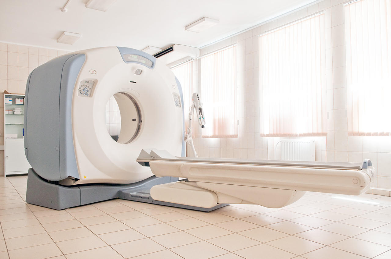
During the first visit, the doctor will conduct a clinical examination and go through the results of the available diagnostic tests. After that, you will undergo the necessary additional examination, such as the assessment of liver and kidney function, ultrasound scan, CT scan and MRI. This will allow the doctor to determine which vessels are feeding the tumor and its metastases, as well as determine how well you will tolerate the procedure.
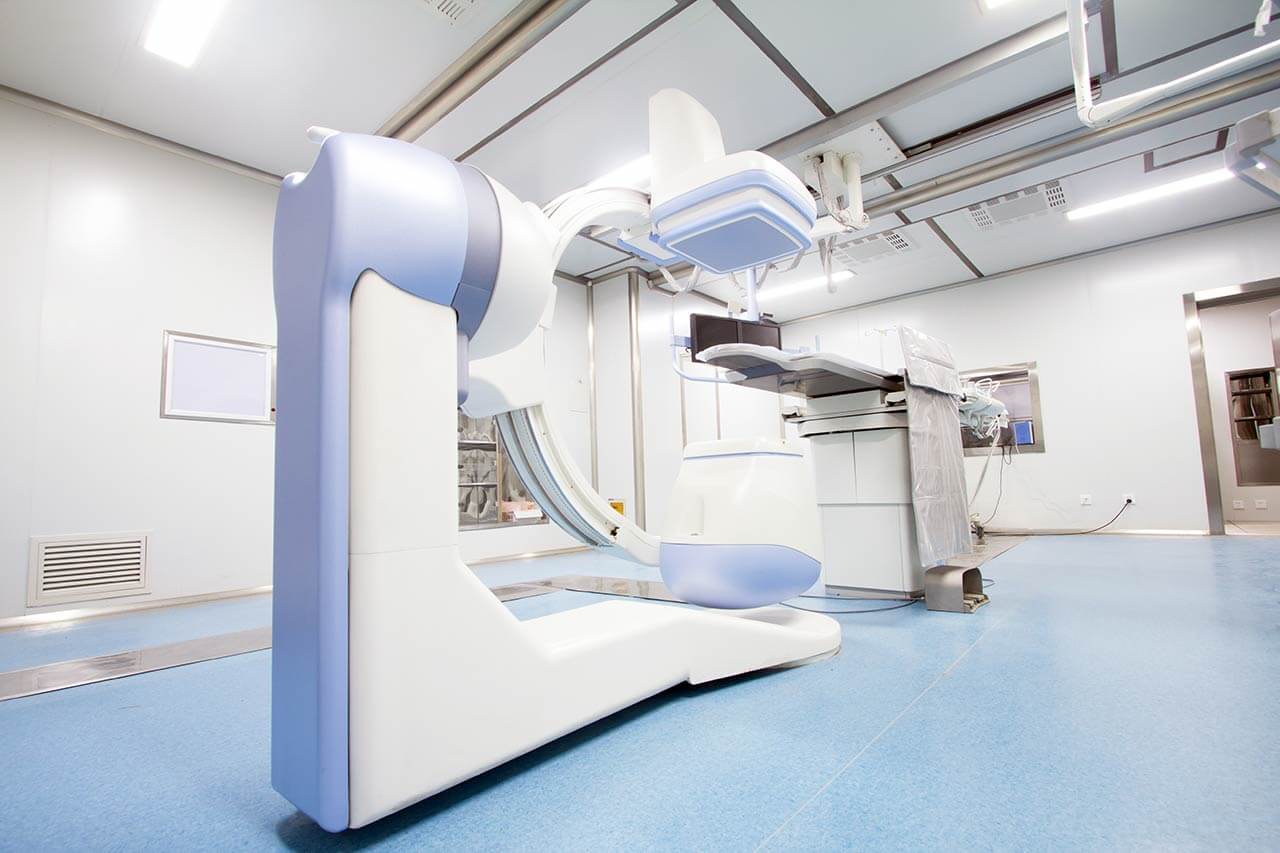
Chemoembolization begins with local anesthesia and catheterization of the femoral artery. The thin catheter is inserted through a few centimeters long incision of the blood vessel. The doctor gradually moves the catheter to the vessel feeding the primary tumor or its metastases. The procedure is carried out under visual control, an angiographic device is used for this. The vascular bed and the position of the catheter in it are displayed on the screen of the angiograph.
When the catheter reaches a suspected artery, a contrast agent is injected through it. Due to the introduction of the contrast agent, the doctor clearly sees the smallest vessels of the tumor and the surrounding healthy tissues on the screen of the angiograph. After that, he injects emboli into the tumor vessels through the same catheter.
Emboli are the spirals or the liquid microspheres. The type of embolus is selected individually, taking into account the diameter of the target vessel. When carrying out chemoembolization, a solution of a chemotherapy drug is additionally injected into the tumor vessel. Due to the subsequent closure of the vessel lumen with an embolus, the chemotherapy drug influences the tumor for a long time. In addition, the drug does not enter the systemic circulation, which allows doctors to use high doses of chemotherapeutic agents without the development of serious side effects. Chemoembolization leads to the destruction of the tumor or slowing down its progression.
After that, the catheter is removed from the artery. The doctor puts a vascular suture on the femoral artery and closes it with a sterile dressing. During chemoembolization, you will be awake. General anesthesia is not used, which significantly reduces the risks of the procedure and allows performing it on an outpatient basis, avoiding long hospital stay.
After the first procedure, you will stay under the supervision of an interventional oncologist and general practitioner. If necessary, you will receive symptomatic treatment. As a rule, a second chemoembolization procedure is performed in 3-5 days after the first one in order to consolidate the therapeutic effect. After that, you will receive recommendations for further follow-up and treatment.
Required documents
Medical records
Abdominal ultrasound (if available)
MRI/CT scan of the abdomen (if available)
Biopsy results (if available)
Service
Price from:
Type of program :
Price for 1 day:
Expected duration of the program:
The minimum duration of the program:
Select
You may also book:
Hospital directly:
Saving:
BookingHealth Price from:
About the department
Radiology is a branch of medicine which studies the effect of ionizing radiation on human health. Radiology Institute is a center equipped with up-to-date devices for imaging diagnosis. Our Radiology Institute examines the patients using all the techniques and methods that currently exist in diagnostic radiology. We provide services to both inpatients of Hirslanden Klinik Stephanshorn and outpatients. The outpatients with basic medical insurance can easily be diagnosed without additional payment.
In addition to conventional radiology, mammography, sonography, computed tomography and magnetic resonance imaging, the radiology in Klinik Stephanshorn also offers interventional radiology. Radiology Institute aims at providing patients with competent medical services. Thanks to a highly qualified and motivated team and modern technical infrastructure, we guarantee a high level of service and quality standards.
Curriculum vitae
2013 Klinik Stephanshorn, St.Gallen
2012 – 2013 Leitender Arzt der Radiologie im Spital Bülach
2010 Universitätslehrgang für die Krankenhausleitung, Donau-Universität Krems
2010 Prüfungskommission der Österreichischen Röntgengesellschaft für das Sonderfach Radiologie
2009 – 2012 Leitender Oberarzt der Abteilung für Radiologie im LKH Klosterneuburg/Niederösterreich mit Subspezialisierung für Neuroradiologie, im Exzellenzzentrum für Neurologie und Neuroradiologie, Stroke Unit LKH Tulln
2008 – 2010 Bereichsleitender Oberarzt für Uroradiologie und Stv. Berreichsleiter für Frauenbildgebung an der Abteilung Allgemeine und Kinderradiologie, Universitätsklinik für Radiodiagnostik, Medizinische Universität Wien
2008 Habilitation für Medizinische Radiologie-Diagnostik, Medizinische Universität Wien Thema: Stones and Bones: Multidetector CT Applications in Emergency Medicine
2005 Facharztprüfung für Medizinische Radiologie-Diagnostik
2000 – 2005 Ausbildung zum Facharzt für das Fach Medizinische Radiologie-Diagnostik an der Universitätsklinik für Radiodiagnostik, Medizinische Universität Wien
1998 – 2000 Ausbildung zum Facharzt für Chirurgie und klinische Physiologie an der Universitätsklinik für Chirurgie, Universität Wien
1998 – 1998 Assistenzarzt an der Medizinischen und Unfallchirurgischen Abteilungen des Aö. Krankenhauses Neunkirchen
1997 – 1998 Assistenzarzt an der Chirurgischen Abteilung des Heeresspitals, Wien
1997 Promotion zum Doktor der gesamten Heilkunde
1989 – 1996 Medizinstudium an der Medizinischen Fakultät der Universität Wien
Photo of the doctor: (c) Hirslanden AG
About hospital
Today, many people prefer healthcare in Europe, for example, in Switzerland. Medical infrastructure and a wide range of medical services in Swiss clinics, including Klinik Stephanshorn, provide optimal medical services. Our clinic offers exactly the treatment that meets all the parameters and the individual needs of the patient. Healthcare in Europe is known for its high quality, therefore, in order to ensure the best medical care for our patients, the clinic cooperates exclusively with experienced professionals and practitioners who rent beds in the hospital. The advantage of free medical practitioners system is that the patient may choose his/her personal doctor.
Free medical practitioners perform surgeries in the clinic, as well as all the necessary procedures for the treatment of inpatients and outpatients. In addition, these doctors work in their own consultation rooms. Close cooperation with our accredited free medical practitioners in the competent centers gives an opportunity to provide even more comprehensive medical care for the benefits of our patients. Thus, you can take advantage of a whole range of high-quality health services that can meet a wide variety of needs. At your request, we will be happy to advise you on matters of choice of a qualified physician or other specified specialist.
Photo: (с) depositphotos
Accommodation in hospital
Patients rooms
Each room in the clinic has modern, comfortable bathrooms (with a toilet, a wash stand and a shower). A hair dryer can also be found in every room. There is soap and terry towelling that can be replaced daily upon request. A direct dial phone and radio and TV power buttons are next to the bed. The Internet connection can be set up upon request. The clinic offers single and double rooms. If you choose a single room, an accompanying person will be able to stay with you. A double room is shared with a patient of the same gender. The enhanced comfort rooms are equipped with a safety box, a fridge and upholstered furniture.
Meals and Menus
The patient and the accompanying person are offered a daily choice of three menus. If you are on a specific diet for any reason, you will be offered an individual menu. Please inform the medical staff about your dietary preferences prior to the treatment.
Further details
Standard rooms include:
Toilet
Shower
Wi-Fi
TV
Religion
Christian priests are available for the patients at any time. Representatives of other religions may be requested at any time.
Accompanying person
Your companion may stay with you in your room or at a hotel of your choice during the fixed program.
Hotel
You may stay at the hotel during the outpatient program. Our employees will support you for selecting the best option.
Select your currency:
Your guarantee
One year of support after the treatment
Insurance to cover unforeseen expenses arising from complications during and 48 months after treatment (coverage up to 200,000 €)
Reduced costs by 40-70% (contracts with Hospitals)
In addition, we are the only TÜV-certified company with an ISO 9001:2015 certificate
thank you for reaching out! One of our medical advisors is currently reviewing your details. You can expect a call from us within one business day to discuss your request.
Note: The incoming call will appear as a German number or your local area code. This call is entirely free of charge.
Booking Health guarantees:
Expert Placement
We use rigorous statistical analysis to ensure you are treated at the best-rated facility for your specific diagnosis.
Total Cost Protection
No hidden fees or unexpected bills. We provide a fixed price guarantee, backed by insurance that covers any additional complications.
12-Month Extended Care
Your recovery is our priority. Benefit from a full year of direct medical support following your procedure.
 BookingHealth Price from:
