google_counter
Video-assisted thoracoscopic (VATS) lobectomy (687608) | Specialized Pulmonary Clinic Asklepios Munich-Gauting - BookingHealth
{"translation_price":"50","translation_doc_price":"40","child_coefficient":"1.1","transfer_price":"2.00","transfer_price_vip":"5.00","constant_transfer_price_vip":350,"constant_transfer_price":150,"constant_transfer_distanse":60,"type":"treatment","program_full_story":"<ul>\n\t<li>Initial presentation in the clinic<\/li>\n\t<li>Clinical history taking<\/li>\n\t<li>Physical examination<\/li>\n\t<li>Review of medical records<\/li>\n\t<li>Laboratory tests:\n\t<ul>\n\t\t<li>Complete blood count<\/li>\n\t\t<li>General urine analysis<\/li>\n\t\t<li>Biochemical analysis of blood<\/li>\n\t\t<li>Tumor markers<\/li>\n\t\t<li>Indicators of inflammation<\/li>\n\t\t<li>Indicators blood coagulation<\/li>\n\t<\/ul>\n\t<\/li>\n\t<li>Spirogram<\/li>\n\t<li>CT scan\/MRI of the chest&nbsp;<\/li>\n\t<li>Bronchoscopy with biopsy<\/li>\n\t<li>Histological and immunohistochemical examination of the harvested tissue<\/li>\n\t<li>Preoperative care<\/li>\n\t<li>Video-assisted thoracoscopic lobectomy (VATS)<\/li>\n\t<li>Histological and immunohistochemical examination of the removed tissues<\/li>\n\t<li>Symptomatic treatment<\/li>\n\t<li>Control examinations<\/li>\n\t<li>Full hospital accommodation<\/li>\n\t<li>Cost of essential medicines and materials<\/li>\n\t<li>Nursing services<\/li>\n\t<li>Explanation of future recommendations<\/li>\n<\/ul>\n<div class=\"program_indications_for_surgery\"><h4>Indications<\/h4><ul>\n\t<li>Lung cancer with peripheral tumor localization and tumor size less than 4 cm, the possibility of performing radical surgery.<\/li>\n<\/ul>\n<\/div><div class=\"program_how_program_going mt-4\"><h4>How program is carried out<\/h4><p style=\"text-align:justify\"><strong>Day 1-2<\/strong>. Initial presentation at the clinic. After consultation with the attending physician and physical examination, the patient undergoes the necessary laboratory and instrumental examination.<\/p>\n\n<p style=\"text-align:justify\"><strong>Day 3<\/strong>. On the 2-3 day of hospital stay, after receiving the examination results and their assessment by the attending physician, the operation is performed under general anesthesia. A two-channel breathing tube must be installed, since during the intervention, the patient breathes only with a healthy lung (the operation is performed on a non-working lung). Surgical instruments and a video camera are inserted into the chest cavity through 3 small incisions on the sides of the chest. The affected part of the lung is removed and sent for histological examination. The operation takes no more than 2-3 hours. After its completion, the patient is transferred to the intensive care unit.<\/p>\n\n<p style=\"text-align:justify\"><strong>Day 4<\/strong>. In the absence of complications and in good general condition, the patient is transferred from the intensive care unit to the regular unit. Due to the minimal trauma of the VATS procedure, early mobilization is possible &ndash; the patient can start exercises in bed and move within the ward in a day after the operation.<\/p>\n\n<p style=\"text-align:justify\"><strong>From day 5<\/strong>. Follow-up and rehabilitation are continued. With positive dynamics of the condition, the patient is discharged from the hospital on the 7-10th day.<\/p>\n<\/div><div class=\"program_required_documents mt-4\"><h4>Required documents<\/h4><ul>\n\t<li>Medical records<\/li>\n\t<li>MRI \/ CT scan not older than 3 months<\/li>\n\t<li>Biopsy (if available)<\/li>\n<\/ul>\n<\/div>","program_full_story_crm":"<ul>\n\t<li>Initial presentation in the clinic<\/li>\n\t<li>Clinical history taking<\/li>\n\t<li>Physical examination<\/li>\n\t<li>Review of medical records<\/li>\n\t<li>Laboratory tests:\n\t<ul>\n\t\t<li>Complete blood count<\/li>\n\t\t<li>General urine analysis<\/li>\n\t\t<li>Biochemical analysis of blood<\/li>\n\t\t<li>Tumor markers<\/li>\n\t\t<li>Indicators of inflammation<\/li>\n\t\t<li>Indicators blood coagulation<\/li>\n\t<\/ul>\n\t<\/li>\n\t<li>Spirogram<\/li>\n\t<li>CT scan\/MRI of the chest&nbsp;<\/li>\n\t<li>Bronchoscopy with biopsy<\/li>\n\t<li>Histological and immunohistochemical examination of the harvested tissue<\/li>\n\t<li>Preoperative care<\/li>\n\t<li>Video-assisted thoracoscopic lobectomy (VATS)<\/li>\n\t<li>Histological and immunohistochemical examination of the removed tissues<\/li>\n\t<li>Symptomatic treatment<\/li>\n\t<li>Control examinations<\/li>\n\t<li>Full hospital accommodation<\/li>\n\t<li>Cost of essential medicines and materials<\/li>\n\t<li>Nursing services<\/li>\n\t<li>Explanation of future recommendations<\/li>\n<\/ul>\n<div class=\"program_indications_for_surgery\"><h4>Indications<\/h4><ul>\n\t<li>Lung cancer with peripheral tumor localization and tumor size less than 4 cm, the possibility of performing radical surgery.<\/li>\n<\/ul>\n<\/div><div class=\"program_how_program_going mt-4\"><h4>How program is carried out<\/h4><p style=\"text-align:justify\"><strong>Day 1-2<\/strong>. Initial presentation at the clinic. After consultation with the attending physician and physical examination, the patient undergoes the necessary laboratory and instrumental examination.<\/p>\n\n<p style=\"text-align:justify\"><strong>Day 3<\/strong>. On the 2-3 day of hospital stay, after receiving the examination results and their assessment by the attending physician, the operation is performed under general anesthesia. A two-channel breathing tube must be installed, since during the intervention, the patient breathes only with a healthy lung (the operation is performed on a non-working lung). Surgical instruments and a video camera are inserted into the chest cavity through 3 small incisions on the sides of the chest. The affected part of the lung is removed and sent for histological examination. The operation takes no more than 2-3 hours. After its completion, the patient is transferred to the intensive care unit.<\/p>\n\n<p style=\"text-align:justify\"><strong>Day 4<\/strong>. In the absence of complications and in good general condition, the patient is transferred from the intensive care unit to the regular unit. Due to the minimal trauma of the VATS procedure, early mobilization is possible &ndash; the patient can start exercises in bed and move within the ward in a day after the operation.<\/p>\n\n<p style=\"text-align:justify\"><strong>From day 5<\/strong>. Follow-up and rehabilitation are continued. With positive dynamics of the condition, the patient is discharged from the hospital on the 7-10th day.<\/p>\n<\/div>","is_ambulant":"0","bh_fee":"0","only_for_children":"0","no_service":"0","with_prepayment":"1","show_calculator":"1","paket_type":"1","btn_type":"0","clinic_icon":"5ff85d317665e.jpg","city":"Gauting","clinic_site":"https:\/\/www.asklepios.com\/gauting\/","department_recommend":"0","country":"Germany","country_id":"1","clinic_name":"Specialized Pulmonary Clinic Asklepios Munich-Gauting","cinic_name":"Specialized Pulmonary Clinic Asklepios Munich-Gauting","department_id":"3660","duration":"10","direction":"Thoracic surgery","min_duration":0,"clinic_id":"2520","paketPrice":7800,"paket":"<ul>\n <li>Interpreter up to 17 hours<\/li>\n <li>Translation up to 10 pages<\/li>\n <li>Visa support<\/li>\n\n \n<\/ul>","title":"Video-assisted thoracoscopic (VATS) lobectomy","price":{"val":29862.85,"type":"val"},"price_surcharge":0,"price_surcharge_clear":0,"extra_service_clinic":[{"id":"need_to_use_head_doctor","title":"Treatment by leading experts","type":"val","val":"5790.53","req":0}],"extra_service":[],"translation_hours":"0","translation_doc_count":null,"roads":[{"id":"2","distance":"58","airport_title":"Munich"},{"id":"27","distance":"201","airport_title":"N\u00fcrnberg"},{"id":"19","distance":"215","airport_title":"Stuttgart"}],"pakets":[],"lang":{"day":"Day","days":"days","ambulatory":"Outpatient","stationaryProgram":"Inpatient"}}

Video-assisted thoracoscopic (VATS) lobectomy

Specialized Pulmonary Clinic Asklepios Munich-Gauting

Gauting, Germany
Program id # 687608
Doctor photo
Prof. Dr. med. Rudolf Hatz
Department of Thoracic Surgery
Specialized in: thoracic surgery

The program includes:

  • Initial presentation in the clinic
  • Clinical history taking
  • Physical examination
  • Review of medical records
  • Laboratory tests:
    • Complete blood count
    • General urine analysis
    • Biochemical analysis of blood
    • Tumor markers
    • Indicators of inflammation
    • Indicators blood coagulation
  • Spirogram
  • CT scan/MRI of the chest 
  • Bronchoscopy with biopsy
  • Histological and immunohistochemical examination of the harvested tissue
  • Preoperative care
  • Video-assisted thoracoscopic lobectomy (VATS)
  • Histological and immunohistochemical examination of the removed tissues
  • Symptomatic treatment
  • Control examinations
  • Full hospital accommodation
  • Cost of essential medicines and materials
  • Nursing services
  • Explanation of future recommendations

Indications

  • Lung cancer with peripheral tumor localization and tumor size less than 4 cm, the possibility of performing radical surgery.

How program is carried out

Day 1-2. Initial presentation at the clinic. After consultation with the attending physician and physical examination, the patient undergoes the necessary laboratory and instrumental examination.

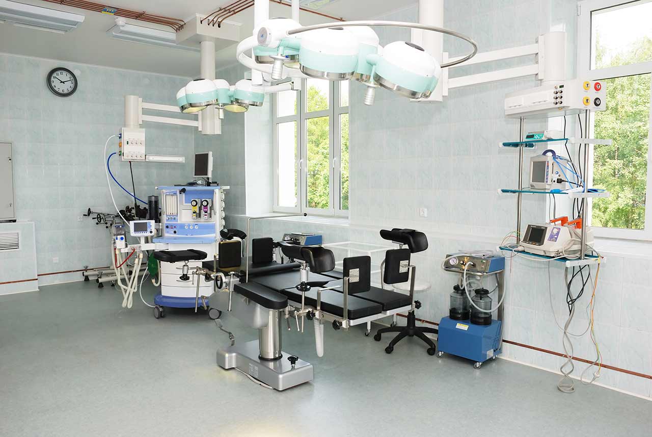
Day 3. On the 2-3 day of hospital stay, after receiving the examination results and their assessment by the attending physician, the operation is performed under general anesthesia. A two-channel breathing tube must be installed, since during the intervention, the patient breathes only with a healthy lung (the operation is performed on a non-working lung). Surgical instruments and a video camera are inserted into the chest cavity through 3 small incisions on the sides of the chest. The affected part of the lung is removed and sent for histological examination. The operation takes no more than 2-3 hours. After its completion, the patient is transferred to the intensive care unit.

Day 4. In the absence of complications and in good general condition, the patient is transferred from the intensive care unit to the regular unit. Due to the minimal trauma of the VATS procedure, early mobilization is possible – the patient can start exercises in bed and move within the ward in a day after the operation.

From day 5. Follow-up and rehabilitation are continued. With positive dynamics of the condition, the patient is discharged from the hospital on the 7-10th day.

Required documents

  • Medical records
  • MRI / CT scan not older than 3 months
  • Biopsy (if available)

Service

Price:
Type of program :
Price for 1 day:
Expected duration of the program:
The minimum duration of the program:
Select

You may also book:

Guarantee Price:

About the department

The Department of Thoracic Surgery at the Specialized Pulmonary Clinic Asklepios Munich-Gauting offers top-class surgical treatment for the full range of chest diseases. The department has the status of one of the largest medical facilities of this kind in Germany. In addition, the department is the country's leading center for surgery to treat lung cancer, which is one of the most common and life-threatening types of oncological diseases. Patients are treated in two high-tech operating rooms, where all hygiene and safety standards are strictly met. Video-assisted thoracoscopic surgical techniques are widely used in clinical practice, which contributes to the rapid recovery of patients in the postoperative period. At the same time, the effectiveness of such operations is in no way inferior to classical open surgical techniques. The department annually performs more than 1,000 surgical interventions of varying complexity on the chest organs. The department's special therapeutic offer is hyperthermic intrathoracic chemotherapy (HITHOC), which is used for treating pleural mesothelioma.

The department is headed by Prof. Dr. med. Rudolf Hatz. The specialist has more than 35 years of successful clinical practice. He is a Fellow of the American College of Surgeons (FACS) and many German professional societies. Prof. Hatz has also repeatedly won prestigious prizes and awards, confirming his high competence in the field of thoracic surgery.

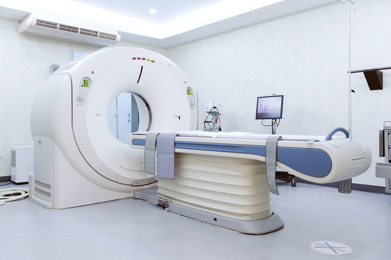
Today, lung cancer is one of the most common oncological diseases worldwide. Malignant lung tumors often do not cause any symptoms, so a person may not even be aware of the presence of the disease until it reaches an advanced stage. Chronic cough, hemoptysis, shortness of breath, and chest pain are reasons for a chest X-ray to be performed. If the results of this examination are unsatisfactory, chest computed tomography (CT), bronchoscopy, biopsy, and other tests will be performed additionally. During the diagnostics, it is important for the department's surgeons to determine the exact type of malignant tumor (small or non-small cell lung cancer), since the treatment tactics for these cancers are fundamentally different. Most small-cell lung cancers are treated with chemotherapy or chemoradiation therapy, while for non-small cell lung cancers, surgery is mostly preferred. At the preparation stage for surgery, the department's specialists perform CT scans of the head and upper abdomen, skeletal scintigraphy, and PET-CT scans (if clinically indicated) to detect metastases. In addition, a patient undergoes pulmonary and cardiac function tests, the results of which also play an important role in deciding whether to perform the operation.

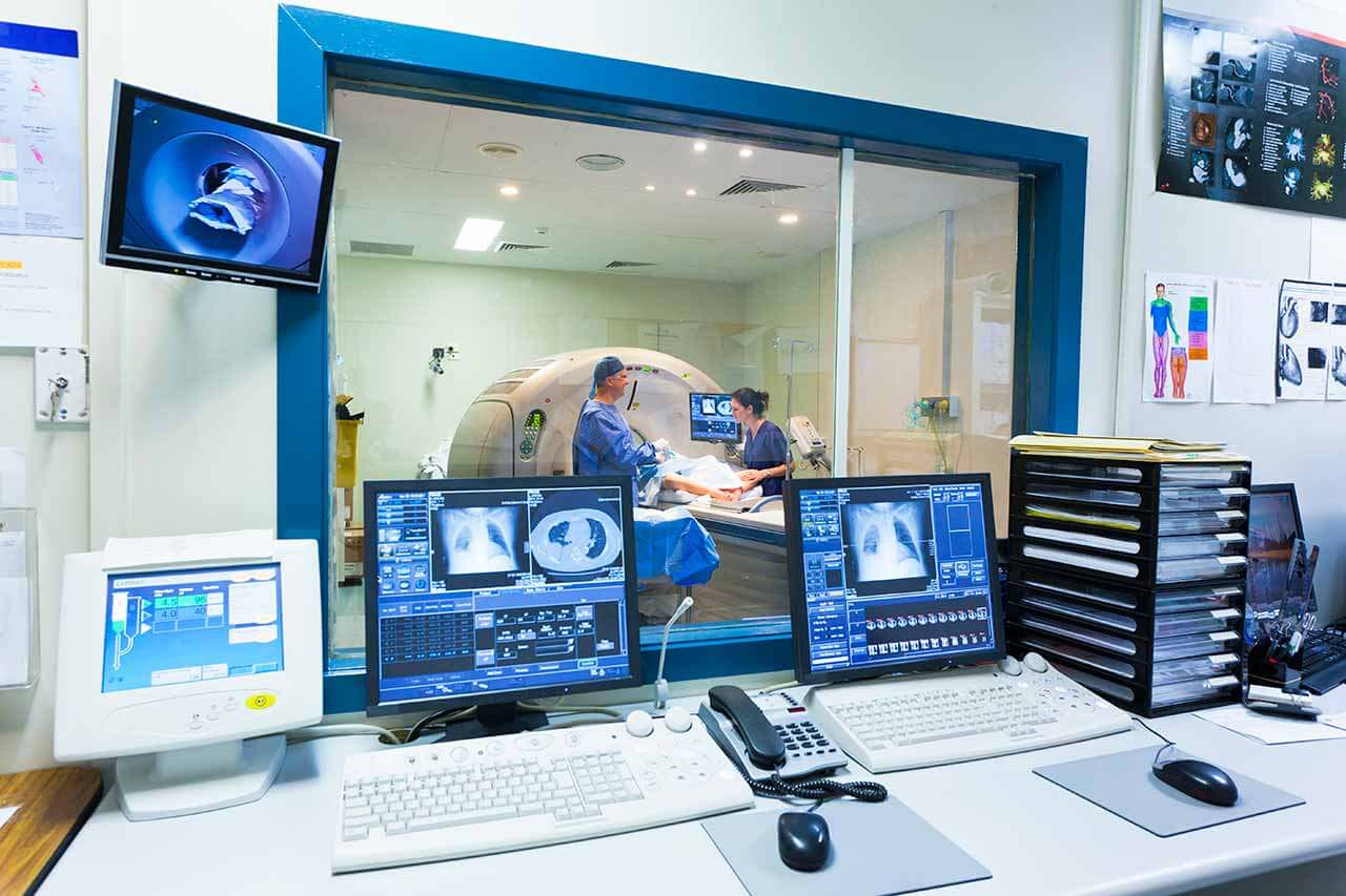
In many cases, the department's thoracic surgeons manage to perform minimally invasive video-assisted thoracoscopic surgery without a thoracotomy. When performing such interventions, it is enough for surgeons to make one or several small incisions through which a thoracoscope with a camera and miniature instruments are delivered to the tumor. The images of the surgical field are continuously transmitted on a large screen with multiple zooming, which ensures the highest accuracy of surgical procedures. Following video-assisted thoracoscopic surgery, patients experience almost no pain and recover in the shortest possible time. Such interventions provide a good aesthetic result, as they leave minor scars.

The department often admits patients with pleural mesothelioma (pleural cancer). In almost all cases, a malignant neoplasm develops in the pleura due to asbestos exposure. Typical symptoms of pleural mesothelioma are difficulty breathing and chest pain. Whenever possible, the department's thoracic surgeons prefer a pleurectomy (a surgical procedure to partially or totally remove the pleura) followed by hyperthermic intrathoracic chemotherapy (HITHOC). The essence of HITHOC is as follows: a solution of chemotherapy agents heated to a high temperature is injected into the chest cavity, and the drugs easily penetrate into the remaining tumor foci due to the heating and destroy them. It should be noted that hyperthermic intrathoracic chemotherapy is a rather complicated procedure, and therefore it is carried out only in advanced German medical centers. A pleurectomy combined with HITHOC ensures relief of symptoms and significantly prolongs the life expectancy of patients, but complete cure of pleural mesothelioma by these methods rarely occurs. A complete cure for cancer usually requires radical surgery, namely an extrapleural pleuropneumonectomy. During this operation, the department's surgeons totally remove the lung, pericardium, diaphragm, and pleura, which is followed by pericardial and diaphragmatic plastic repair. Extrapleural pleuropneumonectomy is one of the most complex operations in thoracic surgery. Irradiation is usually indicated after pleurectomy and extrapleural pleuropneumonectomy.

The department's doctors have vast experience in helping patients with spontaneous pneumothorax. The pathology is characterized by the penetration of air into the pleural cavity due to an impairment of the pleural integrity. If left untreated, spontaneous pneumothorax may lead to severe complications such as pleurisy, pleural empyema, emphysema, and acute respiratory failure. The main diagnostic method for suspected pneumothorax is an X-ray scan. In some cases, computed tomography may also be performed. The treatment tactics are determined individually. The first-line therapy for spontaneous pneumothorax is most often drainage, when a tube is inserted into the pleural cavity through a puncture in the chest wall. After placing the drainage tube, it is fixed with a small suture and connected to the drainage system, after which the air begins to leave the pleural cavity. The treatment process takes about 5 days. However, in 30% of cases, patients have recurrent spontaneous pneumothorax. In some cases, even in the case of primary spontaneous pneumothorax, the department's doctors prefer the surgical treatment option since the risk of recurrence after surgery does not exceed 5%. Surgery for pneumothorax is performed using low-traumatic video-assisted thoracoscopic techniques.

The department's main clinical activities include:

  • Video-assisted thoracoscopic surgery (VATS)
    • Surgery for pleurisy
    • Surgery for lung, mediastinal, and pleural tumors
    • Lung lobe resection for lung cancer
    • Surgery for spontaneous pneumothorax
    • Surgery for pleural empyema
    • Lung volume reduction surgery
    • Surgery for hyperhidrosis
    • Nuss procedure to repair pectus excavatum
  • Pleurectomy combined with hyperthermic intrathoracic chemotherapy (HITHOC) and extrapleural pleuropneumonectomy for malignant pleural mesothelioma
  • Surgical treatment of tracheal stenosis and tracheal cancer
  • Surgical removal of lung metastases, including laser surgery
  • Thoracotomy (open surgery with a chest opening)
  • Other surgical services

Curriculum vitae

Higher Education

  • 1985 Medical Studies at Ludwig Maximilian University of Munich.
  • 1983 Summer Semester at the Faculty of Medicine at Stanford University, USA.

Professional Сareer

  • 1986 Assistant Physician, Department of Surgery, University Hospital of Ludwig Maximilian University of Munich, Campus Grosshadern.
  • 1987 Research Fellow at the Massachusetts General Hospital, Harvard Medical School.
  • 1988 Assistant Physician, Department of Surgery at the University Hospital of Ludwig Maximilian University of Munich, Campus Grosshadern.
  • 1998 Venia Legendi, PD, and Senior Physician.
  • 1998 Habilitation.
  • 2001 Specialization in Thoracic and Abdominal Surgery.
  • 2002 Qualification Certificate: Quality Management, Bavarian State Medical Association.
  • 2003 Head of the Section of Thoracic Surgery with responsibility for advanced training, Bavarian State Medical Association.
  • 2004 Managing Senior Physician, Department of Surgery, University Hospital of Ludwig Maximilian University of Munich, Campus Grosshadern.
  • 2005 Professorship at Ludwig Maximilian University of Munich.
  • 2006 Head of the Department of Thoracic Surgery at the Specialized Pulmonary Clinic Asklepios Munich-Gauting.
  • 2007 Responsible for advanced training courses and Medical Consultant in Thoracic Surgery, Bavarian State Medical Association.
  • 2008 Full University Professorship (W2) for Thoracic Surgery.

Memberships in Professional Medical Societies

  • 2015 Member of the American Surgical Association as Fellow of the American College of Surgeons (FACS).
  • Association of Bavarian Surgeons.
  • Professional Association of German Surgeons.
  • Surgical Working Group on Endoscopy of the German Society of Surgery.
  • Surgical Working Group on Wound Treatment of the German Society of Surgery.
  • German Society of Surgery.
  • German Society of Thoracic Surgery.
  • German Society for Wound Healing, Secretary, and since 1996 till 1998, Member of the Advisory Council.
  • International Society for Heart and Lung Transplantation.
  • Head of the Munich Lung Transplant Group.
  • Project Group "Gastrointestinal infections" of the German Society for Hygiene and Microbiology.
  • Project Member of the Cancer Center Munich.

Scientific Awards, Prizes and Honors

  • 1993 Prize from the European Research Group on Gastroduodenal Pathologies, Brussels, Belgium.
  • 1994 Prize from the European Research Group on Gastroduodenal Pathologies, Barcelona, Spain.
  • 1998 and 2000 Prize from the European Research Group on Immunology and Pathogenesis in Mucosal Infectious Lesions, Elsinore, Denmark.
  • 2001 Award for Video on Minimally Invasive Surgery from the German Society of Surgery, Munich.
  • 2002 Rudolf Schindler Prize, Society for Endoscopy and Imaging, Munich.
  • 2007 Georg Heberer Prize.
  • 2007 Walter Schulz Prize.

Photo of the doctor: (c) Asklepios Fachkliniken München-Gauting


About hospital

According to the reputable Focus magazine, the Specialized Pulmonary Clinic Asklepios Munich-Gauting ranks among the top German medical facilities specializing in lung cancer treatment! 

The clinic has earned the status of one of the leading medical centers in Europe in the area of its specialization. It is part of the Ludwig Maximilian University of Munich, which makes it possible to gain access to all the innovations in diagnostics and treatment. The medical facility has more than 70 years of history, so the clinic can be proud of its rich and successful clinical experience. The patients of the clinic receive medical care that meets the highest European standards. At the same time, the doctors at the clinic always provide patients with moral support and treat their life situation with understanding.

The clinic provides top-class conservative and surgical treatment for diseases of the lungs and other chest organs. In addition, innovative laser techniques are available at the clinic, and bronchoscopic procedures are successfully performed. Particular attention is paid to the treatment of lung cancer, chronic obstructive pulmonary disease, infectious lung diseases, pulmonary hypertension, emphysema, pulmonary fibrosis, mediastinal tumors, etc. A big advantage is the presence of the Department of Radiology at the clinic, where lung imaging is performed. In collaboration with the University Hospital of Ludwig Maximilian University of Munich, the specialists of the clinic founded the Lung Cancer Center. This was certified by the German Cancer Society.

A specific feature will be the fact that the clinic is located at a distance of 21 km from Munich, which is one of the largest and most beautiful cities in Germany. And what is more, Munich is home to many eminent academic and medical facilities, as well as outstanding historical monuments. In addition, the patients of the clinic will live in enhanced comfort rooms, corresponding to the level of a five-star hotel.

The Specialized Pulmonary Clinic Asklepios Munich-Gauting is an etalon Pulmonary Center that treats both simple and complex, extremely rare pulmonary diseases using innovative conservative and minimally invasive techniques. The clinic is known throughout Germany and far beyond its borders, and therefore it regularly admits patients from abroad.

Photo: (с) depositphotos

Accommodation in hospital

Patients rooms

The patients of the Specialized Pulmonary Clinic Asklepios Munich-Gauting live in comfortable single and double rooms. The patient rooms in the clinic are cozy and bright and also have small balconies, which offer a beautiful view of the park. Each patient room has an ensuite bathroom with a shower and a toilet. Standard patient room furnishings include an automatically adjustable bed, a bedside table, a TV, and a telephone. Wi-Fi is also available in the patient rooms.

The clinic also offers enhanced-comfort patient rooms, corresponding to the level of a five-star hotel. The patient room furnishing includes a comfortable bed, beautiful upholstered furniture, a desk with a reading lamp, a spacious wardrobe with a built-in safe, a free mini-bar, a TV, a telephone, a radio, and a CD/DVD player. The patient room is decorated with paintings and has modern lighting. The bathroom includes the necessary toiletries, a bathrobe, and changeable towels.

Meals and Menus

The clinic offers tasty and healthy meals three times a day. Breakfast and dinner are served as buffets, and for lunch, patients are provided with first and second courses to choose from. Selected, high-quality products are used for cooking, which guarantees excellent taste.

Patients who live in enhanced-comfort rooms are additionally offered fresh fruit, coffee and tea with desserts, snacks, and a rich selection of main dishes for lunch and dinner.

Further details

Standard rooms include:

Toilet
Shower
Wi-Fi
TV

Accompanying person

Your accompanying person may stay with you in your patient room or at the hotel of your choice during the inpatient program.

Hotel

You may stay at the hotel of your choice during the outpatient program. Our managers will support you in selecting the best option.