The program includes:
- Initial presentation in the hospital
- Clinical history taking
- Review of available medical records
- Physical examination
- Laboratory tests:
- Complete blood count
- General urine analysis
- Biochemical analysis of blood
- Tumor markers
- Inflammation indicators (CRP, ESR)
- Coagulogram
- Ultrasound scan
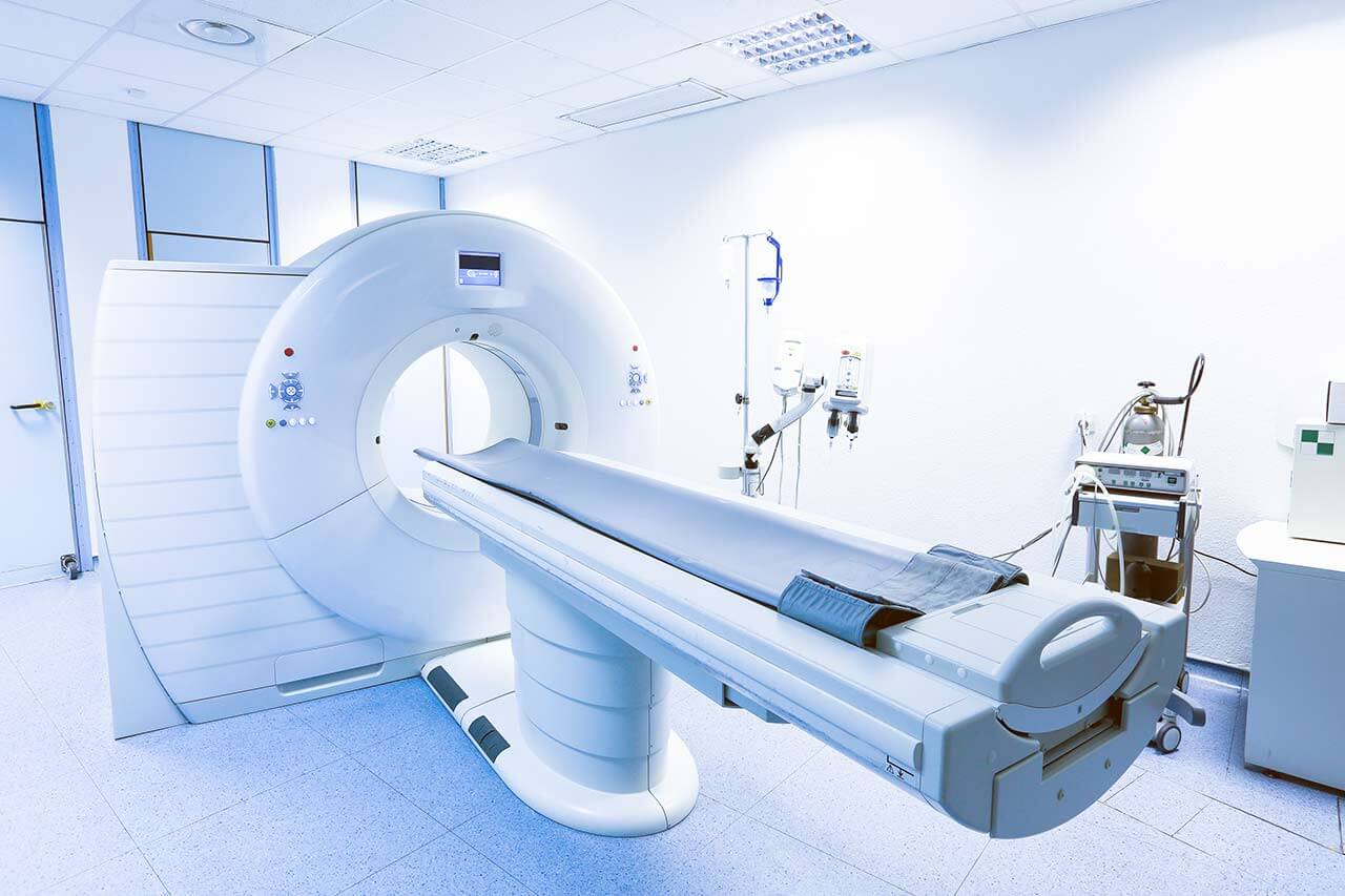
- CT scan / MRI
- Preoperative care
- Embolization or chemoembolization, 2 procedures
- Symptomatic treatment
- Cost of essential medicines
- Nursing services
- Elaboration of further recommendations
How program is carried out
During the first visit, the doctor will conduct a clinical examination and go through the results of the available diagnostic tests. After that, you will undergo the necessary additional examination, such as the assessment of liver and kidney function, ultrasound scan, CT scan and MRI. This will allow the doctor to determine which vessels are feeding the tumor and its metastases, as well as determine how well you will tolerate the procedure.
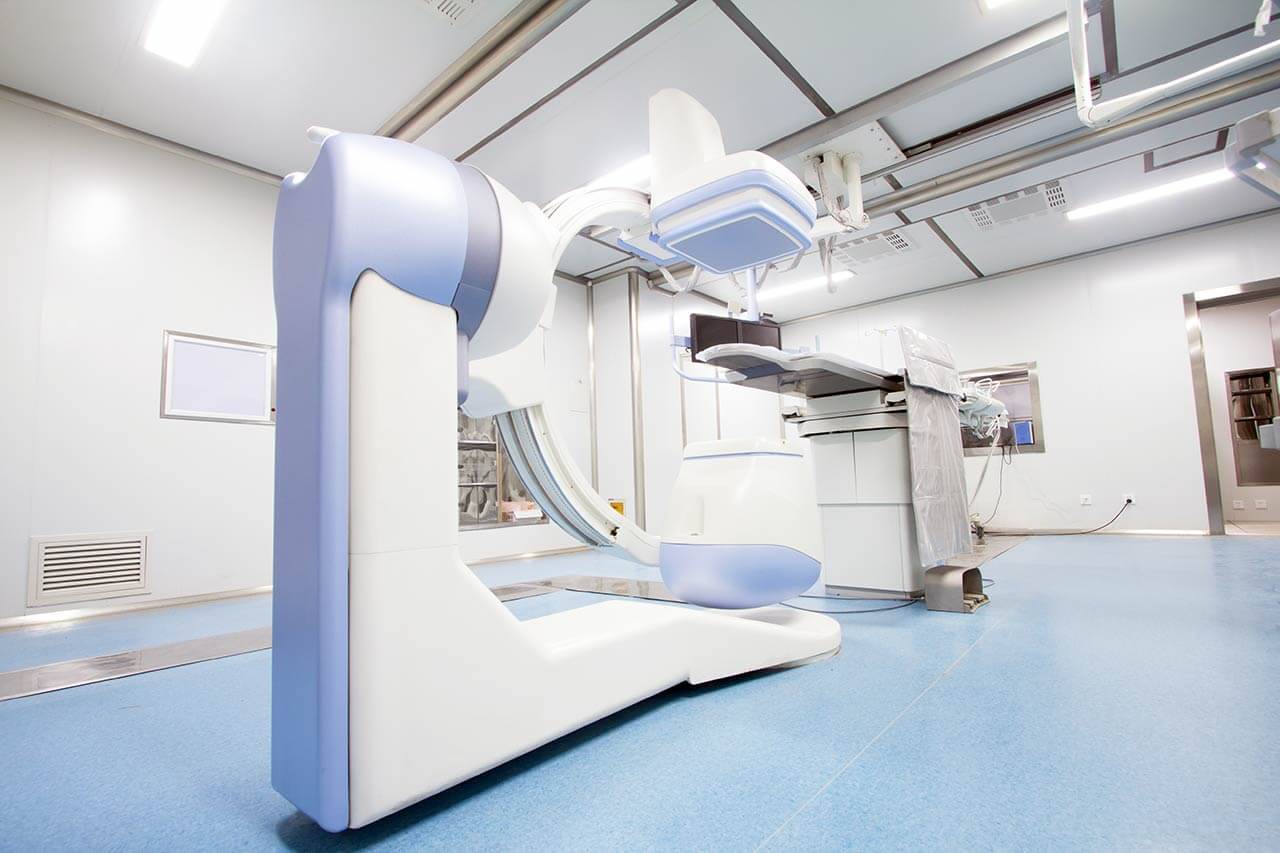
Chemoembolization begins with local anesthesia and catheterization of the femoral artery. The thin catheter is inserted through a few centimeters long incision of the blood vessel. The doctor gradually moves the catheter to the vessel feeding the primary tumor or its metastases. The procedure is carried out under visual control, an angiographic device is used for this. The vascular bed and the position of the catheter in it are displayed on the screen of the angiograph.
When the catheter reaches a suspected artery, a contrast agent is injected through it. Due to the introduction of the contrast agent, the doctor clearly sees the smallest vessels of the tumor and the surrounding healthy tissues on the screen of the angiograph. After that, he injects emboli into the tumor vessels through the same catheter.
Emboli are the spirals or the liquid microspheres. The type of embolus is selected individually, taking into account the diameter of the target vessel. When carrying out chemoembolization, a solution of a chemotherapy drug is additionally injected into the tumor vessel. Due to the subsequent closure of the vessel lumen with an embolus, the chemotherapy drug influences the tumor for a long time. In addition, the drug does not enter the systemic circulation, which allows doctors to use high doses of chemotherapeutic agents without the development of serious side effects. Chemoembolization leads to the destruction of the tumor or slowing down its progression.
After that, the catheter is removed from the artery. The doctor puts a vascular suture on the femoral artery and closes it with a sterile dressing. During chemoembolization, you will be awake. General anesthesia is not used, which significantly reduces the risks of the procedure and allows performing it on an outpatient basis, avoiding long hospital stay.
After the first procedure, you will stay under the supervision of an interventional oncologist and general practitioner. If necessary, you will receive symptomatic treatment. As a rule, a second chemoembolization procedure is performed in 3-5 days after the first one in order to consolidate the therapeutic effect. After that, you will receive recommendations for further follow-up and treatment.
Service
You may also book:
 BookingHealth Price from:
About the department
The Department of Urology and Andrology at the University Hospital Greifswald offers the full range of medical services for the treatment of diseases of the organs of the reproductive system in men as well as urinary system pathologies in men and women. In addition, men with erectile dysfunction, infertility, and hormonal imbalances are treated here. The key focus of the work of the department's doctors is the treatment of prostate, testicular, penile, kidney, and bladder cancers. The medical facility is certified as a Urologic Oncology Center in accordance with the standards of the German Cancer Society (DKG), which indicates the excellent quality of treatment and its high success rates. The department's specialists have a perfect command of modern laparoscopic techniques and laser technologies, which allow them to perform treatment with minimal tissue trauma. The team of doctors at the medical facility devotes enough time to personal communication with the patient and always strives to carry out treatment in the most comfortable conditions for the patient. Many minor interventions and therapeutic procedures are performed on an outpatient basis without the need for hospitalization, which is an additional advantage.
The Head Physician of the department is Prof. Dr. med. Martin Burchardt, who has proven himself as a top-class expert at the national and international levels. He is the co-author of the current S3 guideline for prostate cancer treatment of the German Society of Urology. The specialist actively participates in the work of professional urology societies.
The department's particular specialty is prostate cancer treatment. This type of cancer is one of the most common in men. A complete cure for prostate cancer can be achieved in the early stages, when the tumor is localized and does not spread beyond the organ capsule. When metastases spread, the therapeutic process becomes complex, and in most cases, doctors manage to achieve a long-term remission but not a complete cure. Since the oncological disease is asymptomatic in its early stages, the department's doctors strongly recommend patients over 45 undergo an annual PSA test, which is the first-line screening for prostate cancer. If the test results are abnormal and cause concern for the doctor, the man may be advised to undergo a prostate biopsy. The procedure is performed on an outpatient basis under local anesthesia. Since 2019, the department has also been performing modern MRI fusion biopsies. For localized prostate cancer, the main treatment is surgery or external beam radiation therapy. It is possible to undergo a surgical resection of a prostate tumor using robot-assisted techniques in the department. In the advanced stages of the oncological process, drug therapy methods come to the fore, including hormone therapy, chemotherapy, immunotherapy, radionuclide therapy, etc.
Since 2012, the department has had the status of a certified Prostate Cancer Center with a certificate from the German Cancer Society. The certified medical facilities of this kind in Germany are considered the best choice for patients with cancer, as highly qualified specialists and advanced treatment methods are available here and excellent therapeutic results are regularly achieved.
The department also admits patients with kidney cancer. Like most cancers, kidney cancer does not cause any symptoms in the early stages, so it is often detected during an ultrasound scan for another problem. After that, CT or MRI scans and scintigraphy scans may be required. A complete blood count and a urinalysis are also performed. The first-line treatment for localized kidney tumors is a partial or total resection of the organ, depending on the size of the neoplasm. The department's doctors perform both interventions using minimally invasive techniques, thanks to which patients recover in the shortest possible time and have a low risk of surgical complications. Patients with metastatic kidney cancer are usually recommended for immunotherapy or targeted therapy with angiogenesis inhibitors.
Another focus of the department's clinical practice is medical care for patients with bladder cancer. If this pathology is suspected, the diagnostics include ultrasound scans, cystoscopy, contrast-enhanced X-ray scans, and laboratory tests. Transurethral resection is the main treatment for superficial bladder tumors. Nevertheless, patients with advanced tumors that have invaded the muscular layer of the bladder require total removal of the organ. If the bladder is completely removed, an orthotopic neobladder will be formed after surgery, or urine will be diverted through the ileal conduit. Surgery may be supplemented with chemotherapy, immunotherapy, radiation therapy, and other methods.
The department's medical team successfully treats benign urologic diseases. Benign prostatic hyperplasia is one of the most common among them. The pathology is manifested by the overgrowth of prostate tissue and its significant increase in size. The main manifestation of the disease is a urinary disorder. In some cases, acute urinary retention may develop, requiring emergency treatment. In the early stages of prostate adenoma, the department's doctors use only drug therapy, and in the advanced stages, it is often impossible to do without surgery. The department's specialists perform transurethral resection of the prostate or thulium laser enucleation of the prostate (ThuLEP). The optimal type of intervention is selected depending on the patient's clinical case. The department's urologists perform operations for benign prostatic hyperplasia using minimally invasive techniques.
The department's specialists in andrology provide consultations for men with erectile dysfunction, infertility, hormonal imbalances caused by age-related changes, and other pathologies in this spectrum. As a rule, patients with such disorders receive individually selected drug therapy.
The department's main clinical activities include:
- Diagnostics and treatment of malignant urologic diseases
- Prostate cancer
- Testicular cancer
- Penile cancer
- Kidney cancer
- Bladder cancer
- Diagnostics and treatment of benign urologic diseases
- Benign prostatic hyperplasia
- Kidney stone disease
- Urethral stricture
- Phimosis
- Hydrocele
- Urinary incontinence
- Diagnostics and treatment of andrologic pathologies
- Erectile dysfunction
- Male infertility
- Hormonal imbalances due to age-related changes in men
- Sexual disorders due to diseases of the reproductive system and their treatment
- Diagnostics and treatment of other diseases
The department's range of surgical services includes:
- Endourological interventions
- Transurethral resection, including laser therapies
- Transurethral resection of the prostate (TURP), including laser one
- Bladder neck resection using Turner Warwick procedure
- Transvesical enucleation of prostate adenoma
- Transurethral resection of the bladder
- Endoscopic procedures for urinary system diseases
- Extracorporeal shock wave lithotripsy
- Ureterorenoscopy (semi-rigid and flexible)
- Percutaneous nephrolitholapaxy
- Lithotripsy for bladder stones
- Ureteral splinting (for example, a double J stent)
- Percutaneous nephrostomy
- Transurethral resection, including laser therapies
- Laparoscopic interventions
- Laparoscopic removal of the prostate gland
- Laparoscopic removal of the kidney (nephrectomy): total and partial
- Laparoscopic plastic repair of the kidney pelvis
- Laparoscopic nephropexy
- Laparoscopic removal of a kidney cyst
- Ureteral intraperitonealization (for Ormond's disease)
- Laparoscopic removal of the kidneys and ureters (nephroureterectomy)
- Laparoscopic removal of the adrenal glands (adrenalectomy)
- Classic open interventions
- Abdominal operations
- Open nerve-sparing radical prostatectomy
- Cystectomy using modern types of urinary diversion
- Open nephrectomy
- Open surgery to remove retroperitoneal lymph nodes
- Operations on the external male genitalia
- Open orchiectomy
- Open penectomy: total and partial
- Open testicular prosthetic repair
- Open surgery for hydrocele
- Open surgery for spermatocele
- Open surgery for varicocele
- Open plastic repair of the frenulum of the foreskin
- Vasectomy
- Vasovasostomy
- Circumcision
- Abdominal operations
- Reconstructive interventions
- Urethroplasty: open and endoscopic techniques
- Implantation of an artificial urethral sphincter (Scott-Sphincter®, Flow Secure®)
- Ligament implantation for urinary incontinence (TOT, Advance®)
- Ureter formation (Boari flap procedure, Psoas Hitch procedure)
- Penile curvature repair in patients with Peyronie's disease
- Other surgical options
Curriculum vitae
Higher Education and Professional Experience
- Till 1995 Studied Human Medicine at the University of Hamburg.
- Till 1997 Intern, Department of General Surgery, Alton General Hospital, Hamburg.
- Till 1999 Postdoctoral Fellow, Urology, Columbia University, New York.
- Till 2004 Training for board certification in Urology, Department of Urology, University Hospital Duesseldorf.
- Since 01.2005 Senior Physician in the Department of Adult and Pediatric Urology, Hannover Medical School.
- Since 04.2006 Head of the Department of Adult and Pediatric Urology, Hannover Medical School.
- Since 12.2009 Professor in the Chair of Urology and Head of the Department of Urology and Andrology at the University Hospital Greifswald.
Scientific Awards
- 1999 First Poster Award from the Society for the Study of Impotence (SSI Meeting, Boston, 10.99): "Study of the microvascular bed of the rat's vagina using the technique of vascular corrosion casting." Int J Impot Res 1999; Suppl1:23-30.
- 1999 Travel Award from the Society for Basic Urologic Research (SBUR Meeting, Paris, 10.99): "Effects of modulated p53 expression in LNCaP cells in vitro and in vivo using the induced antisense approach". Prostate 2001; 48(4):225-230.
- 2001 Travel Award from the Society for Basic Urologic Research (SBUR Meeting, Phoenix 11.01): "Phenotypic effects of modulated expression of p53 in LNCaP cells using the induced antisense approach".
- 2003 Third Poster Award: "The inhibition of p53 function reduces the androgen receptor signaling in prostate cancer cell lines". Urologe 2003(A);42(Suppl.1):16.
Main Clinical Focuses
- Tumor surgery and oncology.
- Laparoscopy
- Radical prostatectomy.
- Radical / partial nephrectomy.
- Pelvic plastic surgery.
- Counseling in uro-oncology.
- Counseling in laparoscopy and pediatric urology.
Memberships in Professional Societies
- German Society of Urology.
- European Association of Urology.
- Endourological Society.
- Association of North German Urologists.
- European Society of Urological Technology.
- Urological Research Society.
- Working Group on Urological Oncology.
- Working Group on Laparoscopy and Endourology.
- European Urological Scholarship Programme (EUSP) of the European Association of Urology (EAU).
Photo of the doctor: (c) Universitätsmedizin Greifswald
About hospital
According to the reputable Focus magazine, the University Hospital Greifswald is included in the ranking of the best medical complexes throughout Germany!
The hospital is one of the oldest healthcare facilities in Germany, with long traditions and an excellent reputation. The history of the hospital begins in 1456, when the Faculty of Medicine at the University of Greifswald was founded. During this time, the hospital has managed to earn recognition in the national medical arena and gain prestige abroad. The hospital has 19 institutes and 21 specialized departments. The key to successful clinical practice is the combination of state-of-the-art equipment and highly qualified medical personnel who are actively engaged in the development of effective medical techniques and implement them in everyday practice.
The medical team of the university hospital has more than 4,400 employees, including world-famous professors who regularly undergo advanced training in the leading European and American hospitals, where they share their experience with foreign specialists. The doctors at the medical facility annually treat about 180,000 patients. At the same time, the specialists often provide medical care to patients with complex clinical cases, even in those cases that doctors at other medical centers consider hopeless. The hospital has more than 1,000 beds for inpatients, and many highly-specialized outpatient clinics are available in the medical facility for counseling and outpatient medical care.
The hospital presents all the fields of modern medicine. According to Focus magazine, the medical complex is recognized as one of the best in Germany for treating bowel cancer, bladder cancer, malignant brain tumors, skin cancer, multiple sclerosis, Parkinson's disease, dementia, cardiovascular pathologies, knee and hip pathologies, as well as ophthalmic and proctologic diseases. Diagnostic and therapeutic procedures are carried out in strict accordance with national and international standards, which ensures top-class medical care.
The medical team at the hospital makes sure that each patient feels as comfortable as possible during the therapeutic process. Doctors and nursing staff show humanity and understanding, striving to support each patient in every possible way on the path to recovery.
Photo: (с) depositphotos
Accommodation in hospital
Patients rooms
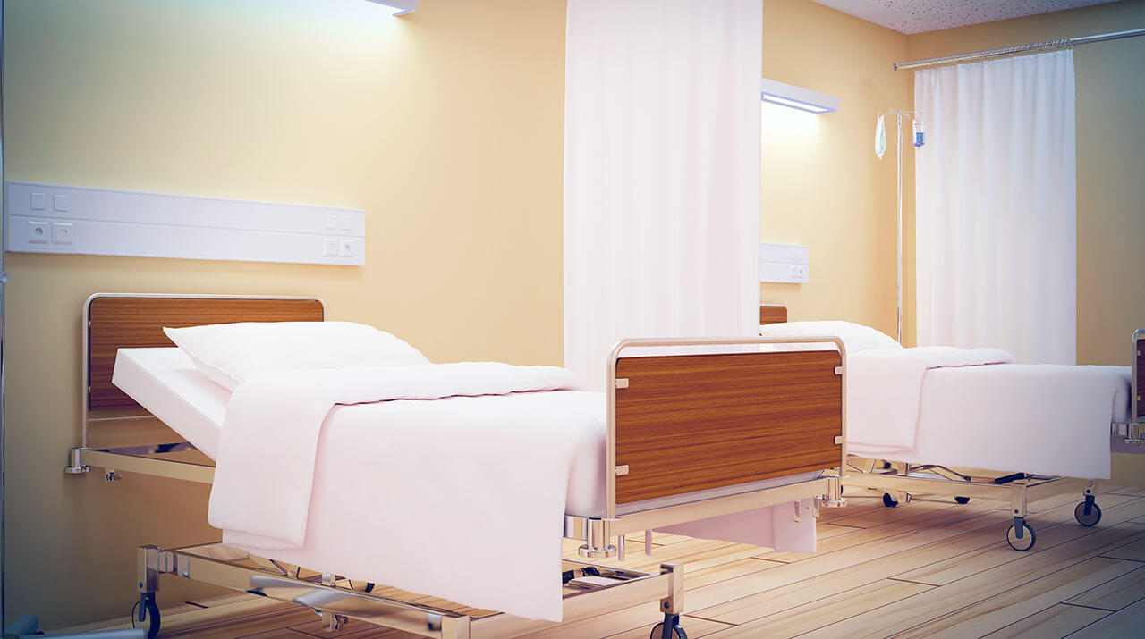
The patients of the University Hospital Greifswald live in comfortable single and double rooms. The patient rooms are made in bright colors and are quite cozy. The furnishings of a standard patient room include an automatically adjustable bed, a bedside table, a TV, and a telephone. The patient room has a table and chairs for receiving visitors. There is also free Wi-Fi in the patient rooms.
If desired, patients can live in enhanced-comfort rooms. In such rooms, patients are additionally offered toiletries, a bathrobe, and a change of towels.
Meals and Menus
The patients of the hospital are offered three healthy and tasty meals a day: a buffet breakfast, a hearty lunch, and dinner. The hospital also houses a cafeteria where one can have a tasty snack, a cup of aromatic coffee, tea, or soft drinks.
Patients staying in enhanced-comfort rooms are offered a special menu with a wide range of main courses, appetizers, desserts, and drinks.
Further details
Standard rooms include:
Religion
The services of representatives of religions are available upon request.
Accompanying person
Your accompanying person may stay with you in your patient room or at the hotel of your choice during the inpatient program.
Hotel
You may stay at the hotel of your choice during the outpatient program. Our managers will support you for selecting the best option.


