The program includes:
- Initial presentation in the clinic
- clinical history taking
- review of medical records
- physical examination
- laboratory tests:
- complete blood count
- general urine analysis
- biochemical analysis of blood
- TSH-basal, fT3, fT4
- tumor markers (AFP, CEA, СА-19-9)
- inflammation indicators (CRP, ESR)
- indicators of blood coagulation
- abdominal ultrasound scan
- CT/MRI of abdomen
- preoperative care
- percutaneous embolization (coiling) or chemoembolization
- symptomatic treatment
- cost of essential medicines
- nursing services
- elaboration of further recommendations
Indications
- Inoperable liver metastases
- Poor response to systemic chemotherapy
Treatment is not indicated in:
- Presence of extrahepatic metastases
- Affection of more than 70% of the liver
How program is carried out
During the first visit, the doctor will conduct a clinical examination and go through the results of the available diagnostic tests. After that, you will undergo the necessary additional examination, such as the assessment of liver and kidney function, ultrasound scan, CT scan and MRI. This will allow the doctor to determine which vessels are feeding the tumor and its metastases, as well as determine how well you will tolerate the procedure.
Chemoembolization begins with local anesthesia and catheterization of the femoral artery. The thin catheter is inserted through a few centimeters long incision of the blood vessel. The doctor gradually moves the catheter to the vessel feeding the primary tumor or its metastases. The procedure is carried out under visual control, an angiographic device is used for this. The vascular bed and the position of the catheter in it are displayed on the screen of the angiograph.
When the catheter reaches a suspected artery, a contrast agent is injected through it. Due to the introduction of the contrast agent, the doctor clearly sees the smallest vessels of the tumor and the surrounding healthy tissues on the screen of the angiograph. After that, he injects emboli into the tumor vessels through the same catheter.
Emboli are the spirals or the liquid microspheres. The type of embolus is selected individually, taking into account the diameter of the target vessel. When carrying out chemoembolization, a solution of a chemotherapy drug is additionally injected into the tumor vessel. Due to the subsequent closure of the vessel lumen with an embolus, the chemotherapy drug influences the tumor for a long time. In addition, the drug does not enter the systemic circulation, which allows doctors to use high doses of chemotherapeutic agents without the development of serious side effects. Chemoembolization leads to the destruction of the tumor or slowing down its progression.
After that, the catheter is removed from the artery. The doctor puts a vascular suture on the femoral artery and closes it with a sterile dressing. During chemoembolization, you will be awake. General anesthesia is not used, which significantly reduces the risks of the procedure and allows performing it on an outpatient basis, avoiding long hospital stay.
After the first procedure, you will stay under the supervision of an interventional oncologist and general practitioner. If necessary, you will receive symptomatic treatment. As a rule, a second chemoembolization procedure is performed in 3-5 days after the first one in order to consolidate the therapeutic effect. After that, you will receive recommendations for further follow-up and treatment.
Required documents
- Medical records
- Abdominal ultrasound (if available)
- MRI/CT scan of the abdomen (if available)
- Biopsy results (if available)
Service
You may also book:
 BookingHealth Price from:
About the department
The Department of Interventional Radiology at the Brothers of Mercy Hospital Munich offers the full range of diagnostic and treatment methods with ionizing radiation. In addition to conventional X-ray diagnostics, computed tomography (CT), and magnetic resonance imaging (MRI), the tasks of the department's doctors include minimally invasive image-guided interventions. Therapeutic procedures are performed using computed tomography and angiography. The department has state-of-the-art medical equipment, which makes it possible to provide patients with not only high-precision diagnostics and the highest treatment standards, but also maximum protection of the body from negative radiation exposure. In most cases, patients receive medical care on an outpatient basis. The department's specialists always devote time to personal communication with their patients and answering all their questions. The department is headed by PD Dr. med. Tobias Jakobs.
The department's main therapeutic options include the treatment of vascular diseases (for example, occlusive peripheral arterial disease), dialysis shunt revascularization, minimally invasive methods for treating liver pathologies (for example, chemoembolization, thermal ablation), minimally invasive treatment of kidney tumors (in cooperation with the Department of Urology), and image-guided punctures and drainages.
Interventional therapy is carried out under CT or angiography guidance. Angiographic guidance is commonly used during vascular interventions. The image-guided treatment gives excellent results and, unlike classical surgery, does not require any traumatic skin and soft tissue incisions. In addition, interventional procedures are performed under local anesthesia, which reduces the load on the heart and, accordingly, the risks to the patient's health.
The department has an innovative picture archiving and communication system, which provides access to diagnostic data for all hospital departments, specialists from partner hospitals and doctors in private practices. This system allows the specialists to store all images for 10 years, which also helps them to lead proper follow-up monitoring of patients after treatment completion (comparing old and current images in dynamics).
The department's main clinical activities include:
- Minimally invasive CT-guided interventions
- Diagnostic procedures
- High-speed punch biopsy of suspicious tissues in all organs and anatomical structures of the body (liver, lungs, bones, kidneys, adrenal glands, pancreas, lymph nodes, and muscles)
- Malignant tissue marking before further treatment (surgery, radiation therapy, and CyberKnife therapy)
- Therapeutic procedures
- Drainage and fluid removal (for example, in abscesses, seromas, biliomas, and lymphoceles)
- Radiofrequency and microwave ablation for liver, kidney, lung and bone tumors
- Cryoablation for kidney, lung, bone and soft tissue tumors
- Vertebroplasty and kyphoplasty for osteoporotic vertebral fractures and osteolysis, which can lead to impaired spinal stability
- Percutaneous pelvic bone osteosynthesis for fractures and treatment of traumatic sacral fractures
- Pain management for spinal diseases: periradicular therapy and facet joint block
- Nerve plexus block for advanced tumors accompanied by severe pain
- Diagnostic procedures
- Minimally invasive angiography-guided catheter-based interventions
- Therapeutic procedures
- Treatment of arterial circulation disorders with balloon dilatation (percutaneous transluminal angioplasty) and stent implantation
- Interventions for vascular occlusions in aneurysms, internal bleeding, uterine fibroids, vascular malformations, varicocele, and benign prostatic hyperplasia
- Catheter-based interventions for cancer: embolization of tumors of various localization, transarterial chemoembolization of primary and secondary liver tumors
- Endovascular aneurysm repair (EVAR)
- Implantation of port systems
- Percutaneous transhepatic cholangiodrainage
- Transjugular intrahepatic portosystemic shunt
- Therapeutic procedures
- Other diagnostic and therapeutic options
Curriculum vitae
Higher Education and Professional Career
- 1994 - 1996 Preclinical medical training at the Georg August University of Goettingen and the University of Vienna.
- 1996 - 2001 Medical studies at Ludwig Maximilian University of Munich and the University of California, San Francisco.
- 2001 - 2002 Internship, Institute of Clinical Radiology, University Hospital of Ludwig Maximilian University of Munich.
- 2002 - 2008 Research Fellow, Institute of Clinical Radiology, University Hospital of Ludwig Maximilian University of Munich.
- 2004 Thesis defense (magna cum laude), Ludwig Maximilian University of Munich. Subject: "Quantitative determination of calcium levels in the coronary arteries using electron beam tomography and conventional computed tomography".
- 2007 Research Fellowship in Interventional Radiology, Anderson Cancer Center, Houston, Texas, USA.
- 2008 - 2010 Senior Physician and Head of the Section of Angiography and Interventional Radiology at the Institute of Clinical Radiology, University Hospital of Ludwig Maximilian University of Munich.
- 2009 Board certification in Radiology.
- 2010 Habilitation, Ludwig Maximilian University of Munich. Subject: "Local image-guided minimally invasive therapy for liver tumors".
- 2011 Head of the Department of Interventional Radiology at the Brothers of Mercy Hospital Munich.
Clinical and Research Focuses
- Musculoskeletal injuries and diseases.
- Liver and abdominal MRI.
- Lung and abdominal CT.
- CT-guided minimally invasive surgical treatment of cancer.
- Minimally invasive treatment of oncological diseases with the help of catheter-based techniques.
Memberships in Professional Societies
- German Society of Radiology (DRG).
- German Society of Interventional Radiology (DeGIR).
- Cardiovascular and Interventional Radiological Society of Europe (CIRSE).
- European Society of Radiology (ESR).
- Radiological Society of North America (RSNA).
- European Society of Gastrointestinal and Abdominal Radiology (ESGAR).
Review Activities
- Member of the Editorial Board of European Radiology (Eur Radiol).
- Journal of Vascular and Interventional Radiology (JVIR).
- Journal of CardioVascular and Interventional Radiology (CVIR).
- Fortschritte auf dem Gebiet der Röntgenstrahlen und bildgebenden Verfahren (RöFo).
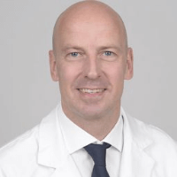
Photo of the doctor: (c) Barmherzige Brüder Krankenhaus München
About hospital
According to the Focus magazine, the Brothers of Mercy Hospital Munich ranks among the top German hospitals in the Federal State of Bavaria!
The hospital is a modern medical facility with the highest level of medical care and long traditions. The hospital operates on the basis of the Technical University of Munich and the German Academy of Nutrition, which contributes to the introduction of progressive diagnostic techniques and therapeutic innovations into clinical practice. The history of the medical facility has more than 100 years, and throughout this time, the specialists of the hospital manage not only to keep abreast of the very latest advances in medicine, but also remain true to the principles of charity and human compassion.
With more than 1,100 employees, the department's competent medical team admits about 18,300 inpatients and more than 31,500 outpatients every year. The hospital employs about 200 specialized physicians who work for the benefit of their patients and provide them with the best quality of treatment, but, at the same time, pay due attention to the patient's emotional state, try to support him in every possible way, and tune him to a positive treatment outcome.
The hospital's main focuses include general and abdominal surgery, gastroenterology, hepatology, oncology, urology, cardiology and pulmonology, orthopedics and traumatology, palliative care, diagnostic and interventional radiology, and others. The hospital regularly demonstrates high treatment success rates in all areas of its specialization, thanks to which it has gained an excellent reputation not only in Germany, but also in the European medical arena.
Many hospital's departments are awarded with prestigious quality certificates, including the endoCert certificate in joint replacement surgery from the German Society for Orthopedics and Orthopedic Surgery (DGOOC), the certificate of the German Hernia Society (DHG), the certificate of the German Cardiac Society (DGK), the certificates of the German Cancer Society (DKG) in bowel and liver cancer treatment. Patients can be sure that they trust their health to the best professionals who successfully cope with clinical tasks of any complexity.
Photo: (с) depositphotos
Accommodation in hospital
Patients rooms
The patients of the Brothers of Mercy Hospital Munich live in comfortable, spacious single and double rooms with bright colors. The standard room furnishing includes an automatically adjustable bed, a bedside table, a wardrobe, a telephone, a TV and a radio. Single rooms also have a mini-fridge and a safe. There is also a table and chairs for receiving visitors. The hospital offers access to Wi-Fi. Each patient room has an ensuite bathroom with a shower and a toilet. The bathroom has a cosmetic mirror, a hair dryer and toiletries, towels, etc.
Meals and Menus
The patients of the hospital are offered healthy and tasty three meals a day: breakfast, lunch and dinner. The menu also features vegetarian dishes. If you do not eat all the food for any reason, you will be provided with an individual menu.
Further details
Standard rooms include:
Religion
There is a small chapel on the territory of the hospital, where worship is regularly held. The chapel is also open for independent visits and prayers.
Accompanying person
Your accompanying person may stay with you in your patient room or at the hotel of your choice during the inpatient program.
Hotel
You may stay at the hotel of your choice during the outpatient program. Our managers will support you for selecting the best option.


