{"translation_price":"50","translation_doc_price":"40","child_coefficient":"1.1","transfer_price":"2.00","transfer_price_vip":"5.00","constant_transfer_price_vip":350,"constant_transfer_price":150,"constant_transfer_distanse":60,"type":"treatment","program_full_story":"<ul>\n\t<li>Initial presentation in the clinic<\/li>\n\t<li>clinical history taking<\/li>\n\t<li>review of medical records<\/li>\n\t<li>physical examination<\/li>\n\t<li>laboratory tests:\n\t<ul>\n\t\t<li>complete blood count<\/li>\n\t\t<li>general urine analysis<\/li>\n\t\t<li>biochemical analysis of blood<\/li>\n\t\t<li>TSH-basal, fT3, fT4<\/li>\n\t\t<li>tumor markers (AFP, CEA, \u0421\u0410-19-9)<\/li>\n\t\t<li>inflammation indicators (CRP, ESR)<\/li>\n\t\t<li>indicators of blood coagulation<\/li>\n\t<\/ul>\n\t<\/li>\n\t<li>abdominal ultrasound\u200b scan<\/li>\n\t<li>CT\/MRI of abdomen<\/li>\n\t<li>preoperative care<\/li>\n\t<li>percutaneous embolization (coiling) or chemoembolization<\/li>\n\t<li>symptomatic treatment<\/li>\n\t<li>cost of essential medicines<\/li>\n\t<li>nursing services<\/li>\n\t<li>elaboration of further recommendations<\/li>\n<\/ul>\n<div class=\"program_how_program_going mt-4\"><h4>How program is carried out<\/h4><p style=\"text-align: justify;\"><strong>During the first visit<\/strong>, the doctor will conduct a clinical examination and go through the results of the available diagnostic tests. After that, you will undergo the necessary additional examination, such as the assessment of liver and kidney function, ultrasound scan, CT scan and MRI. This will allow the doctor to determine which vessels are feeding the tumor and its metastases, as well as determine how well you will tolerate the procedure.<\/p>\n\n<p style=\"text-align: justify;\"><strong>Chemoembolization <\/strong>begins with local anesthesia and catheterization of the femoral artery. The thin catheter is inserted through a few centimeters long incision of the blood vessel. The doctor gradually moves the catheter to the vessel feeding the primary tumor or its metastases. The procedure is carried out under visual control, an angiographic device is used for this. The vascular bed and the position of the catheter in it are displayed on the screen of the angiograph.<\/p>\n\n<p style=\"text-align: justify;\">When the catheter reaches a suspected artery, a contrast agent is injected through it. Due to the introduction of the contrast agent, the doctor clearly sees the smallest vessels of the tumor and the surrounding healthy tissues on the screen of the angiograph. After that, he injects emboli into the tumor vessels through the same catheter.<\/p>\n\n<p style=\"text-align: justify;\">Emboli are the spirals or the liquid microspheres. The type of embolus is selected individually, taking into account the diameter of the target vessel. When carrying out chemoembolization, a solution of a chemotherapy drug is additionally injected into the tumor vessel. Due to the subsequent closure of the vessel lumen with an embolus, the chemotherapy drug influences the tumor for a long time. In addition, the drug does not enter the systemic circulation, which allows doctors to use high doses of chemotherapeutic agents without the development of serious side effects. Chemoembolization leads to the destruction of the tumor or slowing down its progression.<\/p>\n\n<p style=\"text-align: justify;\">After that, the catheter is removed from the artery. The doctor puts a vascular suture on the femoral artery and closes it with a sterile dressing. During chemoembolization, you will be awake. General anesthesia is not used, which significantly reduces the risks of the procedure and allows performing it on an outpatient basis, avoiding long hospital stay.<\/p>\n\n<p style=\"text-align: justify;\"><strong>After the first procedure<\/strong>, you will stay under the supervision of an interventional oncologist and general practitioner. If necessary, you will receive symptomatic treatment. As a rule, a second chemoembolization procedure is performed in 3-5 days after the first one in order to consolidate the therapeutic effect. After that, you will receive recommendations for further follow-up and treatment.<\/p>\n<\/div><div class=\"program_required_documents mt-4\"><h4>Required documents<\/h4><ul>\n\t<li style=\"text-align: justify;\">Medical records<\/li>\n\t<li style=\"text-align: justify;\">MRI\/CT scan (not older than 3 months)<\/li>\n\t<li style=\"text-align: justify;\">Biopsy results (if available)<\/li>\n<\/ul>\n<\/div>","program_full_story_crm":"<ul>\n\t<li>Initial presentation in the clinic<\/li>\n\t<li>clinical history taking<\/li>\n\t<li>review of medical records<\/li>\n\t<li>physical examination<\/li>\n\t<li>laboratory tests:\n\t<ul>\n\t\t<li>complete blood count<\/li>\n\t\t<li>general urine analysis<\/li>\n\t\t<li>biochemical analysis of blood<\/li>\n\t\t<li>TSH-basal, fT3, fT4<\/li>\n\t\t<li>tumor markers (AFP, CEA, \u0421\u0410-19-9)<\/li>\n\t\t<li>inflammation indicators (CRP, ESR)<\/li>\n\t\t<li>indicators of blood coagulation<\/li>\n\t<\/ul>\n\t<\/li>\n\t<li>abdominal ultrasound\u200b scan<\/li>\n\t<li>CT\/MRI of abdomen<\/li>\n\t<li>preoperative care<\/li>\n\t<li>percutaneous embolization (coiling) or chemoembolization<\/li>\n\t<li>symptomatic treatment<\/li>\n\t<li>cost of essential medicines<\/li>\n\t<li>nursing services<\/li>\n\t<li>elaboration of further recommendations<\/li>\n<\/ul>\n<div class=\"program_how_program_going mt-4\"><h4>How program is carried out<\/h4><p style=\"text-align: justify;\"><strong>During the first visit<\/strong>, the doctor will conduct a clinical examination and go through the results of the available diagnostic tests. After that, you will undergo the necessary additional examination, such as the assessment of liver and kidney function, ultrasound scan, CT scan and MRI. This will allow the doctor to determine which vessels are feeding the tumor and its metastases, as well as determine how well you will tolerate the procedure.<\/p>\n\n<p style=\"text-align: justify;\"><strong>Chemoembolization <\/strong>begins with local anesthesia and catheterization of the femoral artery. The thin catheter is inserted through a few centimeters long incision of the blood vessel. The doctor gradually moves the catheter to the vessel feeding the primary tumor or its metastases. The procedure is carried out under visual control, an angiographic device is used for this. The vascular bed and the position of the catheter in it are displayed on the screen of the angiograph.<\/p>\n\n<p style=\"text-align: justify;\">When the catheter reaches a suspected artery, a contrast agent is injected through it. Due to the introduction of the contrast agent, the doctor clearly sees the smallest vessels of the tumor and the surrounding healthy tissues on the screen of the angiograph. After that, he injects emboli into the tumor vessels through the same catheter.<\/p>\n\n<p style=\"text-align: justify;\">Emboli are the spirals or the liquid microspheres. The type of embolus is selected individually, taking into account the diameter of the target vessel. When carrying out chemoembolization, a solution of a chemotherapy drug is additionally injected into the tumor vessel. Due to the subsequent closure of the vessel lumen with an embolus, the chemotherapy drug influences the tumor for a long time. In addition, the drug does not enter the systemic circulation, which allows doctors to use high doses of chemotherapeutic agents without the development of serious side effects. Chemoembolization leads to the destruction of the tumor or slowing down its progression.<\/p>\n\n<p style=\"text-align: justify;\">After that, the catheter is removed from the artery. The doctor puts a vascular suture on the femoral artery and closes it with a sterile dressing. During chemoembolization, you will be awake. General anesthesia is not used, which significantly reduces the risks of the procedure and allows performing it on an outpatient basis, avoiding long hospital stay.<\/p>\n\n<p style=\"text-align: justify;\"><strong>After the first procedure<\/strong>, you will stay under the supervision of an interventional oncologist and general practitioner. If necessary, you will receive symptomatic treatment. As a rule, a second chemoembolization procedure is performed in 3-5 days after the first one in order to consolidate the therapeutic effect. After that, you will receive recommendations for further follow-up and treatment.<\/p>\n<\/div>","is_ambulant":"1","bh_fee":"0","only_for_children":"0","no_service":"0","with_prepayment":"1","show_calculator":"1","paket_type":"1","btn_type":"2","clinic_icon":"6005433e1e502.jpg","city":"Munich","clinic_site":"http:\/\/www.klinikum-muenchen.de\/","department_recommend":"0","country":"Germany","country_id":"1","clinic_name":"Hospital Neuperlach Munich","cinic_name":"Hospital Neuperlach Munich","department_id":"4515","duration":"6","direction":"Interventional radiology","min_duration":0,"clinic_id":"81","paketPrice":3200,"paket":"<ul>\n <li> Interpreter up to 8 hours<\/li>\n <li>Translation of up to 5 pages<\/li>\n <li>Visa support<\/li>\n\n<\/ul>","title":"Treatment of liver cancer with percutaneous embolization (coiling) or chemoembolization","price":{"val":8497.26,"type":"val"},"price_surcharge":0,"price_surcharge_clear":0,"extra_service_clinic":[],"extra_service":[],"translation_hours":"0","translation_doc_count":null,"roads":[{"id":"2","distance":"36","airport_title":"Munich"},{"id":"19","distance":"219","airport_title":"Stuttgart"}],"pakets":[],"lang":{"day":"Day","days":"days","ambulatory":"Outpatient","stationaryProgram":"Inpatient"}}
Treatment of liver cancer with percutaneous embolization (coiling) or chemoembolization in Hospital Neuperlach Munich
percutaneous embolization (coiling) or chemoembolization
symptomatic treatment
cost of essential medicines
nursing services
elaboration of further recommendations
How program is carried out
During the first visit, the doctor will conduct a clinical examination and go through the results of the available diagnostic tests. After that, you will undergo the necessary additional examination, such as the assessment of liver and kidney function, ultrasound scan, CT scan and MRI. This will allow the doctor to determine which vessels are feeding the tumor and its metastases, as well as determine how well you will tolerate the procedure.
Chemoembolization begins with local anesthesia and catheterization of the femoral artery. The thin catheter is inserted through a few centimeters long incision of the blood vessel. The doctor gradually moves the catheter to the vessel feeding the primary tumor or its metastases. The procedure is carried out under visual control, an angiographic device is used for this. The vascular bed and the position of the catheter in it are displayed on the screen of the angiograph.
When the catheter reaches a suspected artery, a contrast agent is injected through it. Due to the introduction of the contrast agent, the doctor clearly sees the smallest vessels of the tumor and the surrounding healthy tissues on the screen of the angiograph. After that, he injects emboli into the tumor vessels through the same catheter.
Emboli are the spirals or the liquid microspheres. The type of embolus is selected individually, taking into account the diameter of the target vessel. When carrying out chemoembolization, a solution of a chemotherapy drug is additionally injected into the tumor vessel. Due to the subsequent closure of the vessel lumen with an embolus, the chemotherapy drug influences the tumor for a long time. In addition, the drug does not enter the systemic circulation, which allows doctors to use high doses of chemotherapeutic agents without the development of serious side effects. Chemoembolization leads to the destruction of the tumor or slowing down its progression.
After that, the catheter is removed from the artery. The doctor puts a vascular suture on the femoral artery and closes it with a sterile dressing. During chemoembolization, you will be awake. General anesthesia is not used, which significantly reduces the risks of the procedure and allows performing it on an outpatient basis, avoiding long hospital stay.
After the first procedure, you will stay under the supervision of an interventional oncologist and general practitioner. If necessary, you will receive symptomatic treatment. As a rule, a second chemoembolization procedure is performed in 3-5 days after the first one in order to consolidate the therapeutic effect. After that, you will receive recommendations for further follow-up and treatment.
Required documents
Medical records
MRI/CT scan (not older than 3 months)
Biopsy results (if available)
Service
Price from:
Type of program :
Price for 1 day:
Expected duration of the program:
The minimum duration of the program:
Select
You may also book:
Hospital directly:
Saving:
BookingHealth Price from:
About the department
The Department of Diagnostic and Interventional Radiology at the Hospital Neuperlach Munich offers the full range of image-guided diagnostic and interventional therapeutic procedures at the highest level of modern medicine. Medical care can be provided both on an inpatient and outpatient basis. The department's specialists annually perform more than 70,000 diagnostic tests, and therefore they have tremendous experience and guarantee an accurate diagnosis, which determines the success of follow-up treatment. The doctors use in their work a modern picture archiving and communication system, thanks to which the specialists from various departments of the hospital have access to the results of patient's diagnostic examination at any time. The Chief Physician of the department is Prof. Dr. med. Andreas Saleh.
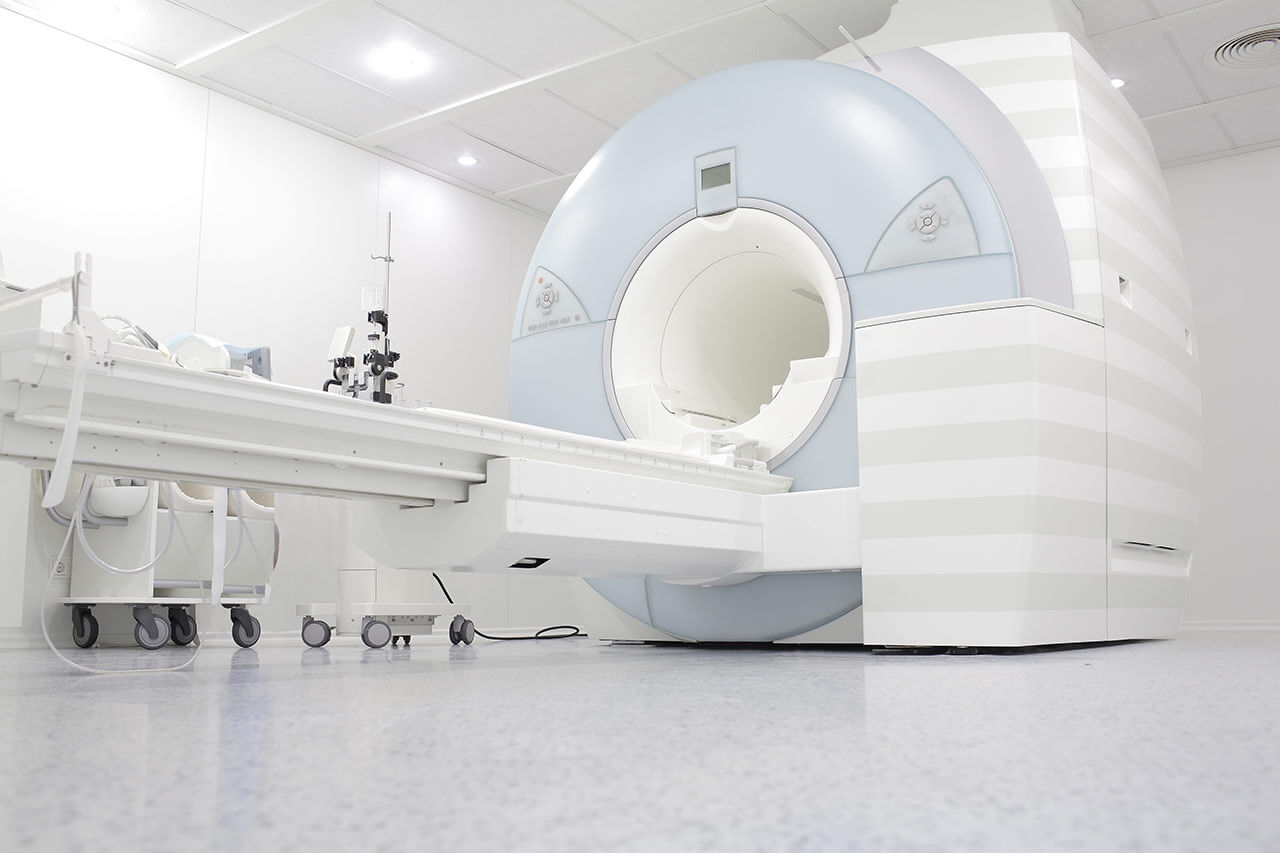
Radiologists have state-of-the-art systems for digital radiography, digital fluoroscopy, sonography, mammography, computed tomography, magnetic resonance imaging, angiography and other examinations. The radiation protection standards are strictly observed in the department's clinical practice. In addition to non-invasive diagnostic tests, the department's specialists also perform invasive diagnostic ultrasound- and CT-guided procedures, including puncture, drainage and biopsy.
In the field of interventional radiology, the department's medical team conducts therapeutic procedures, which allow the patient to avoid open surgery, while guaranteeing high efficiency of treatment. One of the primary focuses of the specialists' work is balloon dilatation and stent implantation for occlusions and stenoses of the vessels in the abdominal cavity, pelvis and lower limbs, stenoses of the carotid and renal arteries. Of particular interest is also the treatment of tumors using radiofrequency or microwave ablation and chemoembolization. In addition, the department's doctors have a special competence in the placement of port systems for chemotherapy and the formation of vascular access for dialysis. The range of services in interventional radiology is also complemented by uterine artery embolization in women.
The department specializes in such diagnostic tests and interventional therapeutic procedures:
Diagnostic tests
Digital radiography
Digital fluoroscopy
Sonography
Mammography
Computed tomography
Magnetic resonance imaging, including cardiac MRI
Angiography
Ultrasound- and CT-guided biopsy, puncture and drainage
Bone mineral density measurement (densitometry)
Brain scintigraphy
Interventional therapeutic procedures
Embolization to stop gastrointestinal bleeding
Pain therapy
Sympatholysis
Facet joint block
Nerve root block
Ganglionic block
Vertebroplasty
Radiofrequency and microwave ablation for tumor treatment
Photo of the doctor: (c) München Klinik Neuperlach
About hospital
The Hospital Neuperlach Munich provides modern medical services of the highest quality. The medical center is an Academic Hospital of the Ludwig Maximilian University of Munich, and therefore it can offer its patients innovative diagnostic and therapeutic methods available only in the best German hospitals. The medical complex opened its doors to patients on September 12, 1972 and to this day holds a leading position in the country's medical arena. The medical facility has 500 beds. The highly qualified doctors of the hospital annually admit over 59,000 patients for both diagnostics and treatment, including patients from foreign countries.
The hospital is distinguished by its rich experience and high success rates in conservative and surgical treatment of diseases of the gastrointestinal tract, liver, metabolic disorders, cancers and blood diseases, heart diseases, as well as vascular pathologies. The hospital operates a highly specialized Colon Cancer Center certified by the German Cancer Society. The center is one of the largest medical facilities of this kind in Germany and has advanced therapeutic options for the treatment of life-threatening colon disease. In addition, the hospital provides high-quality obstetric services – more than 1,000 babies are born here annually.
For outstanding achievements in various medical specialties, the hospital was awarded with many prestigious certificates, including the IQM certificate, the DIN EN ISO 9001:2008 certificate, the DGAV certificate as a Competence Center for Liver and Pancreatic Surgery, the DDG certificate of the German Diabetes Society and others. The above mentioned certificates testify to the excellent quality of medical service.
Special attention should be paid to the medical staff of the hospital who have extensive clinical experience and do everything possible to achieve a complete recovery of the patient. During the treatment, both doctors and nursing staff surround the patient with maximum care, support him in every possible way and show a humane attitude. The work of physicians is based on an individual approach to each patient and his clinical case.
Photo: (с) depositphotos
Accommodation in hospital
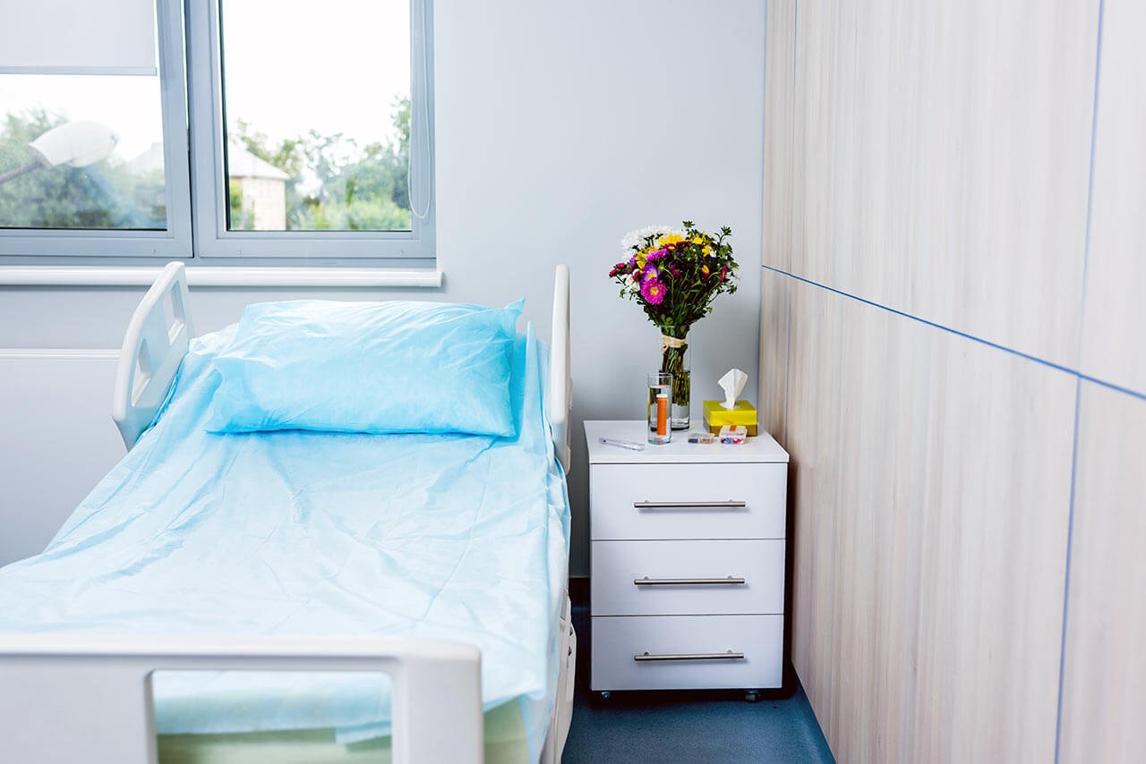
Patients rooms
The patients of the Hospital Neuperlach Munich live in single, double and triple rooms with all the amenities for a comfortable stay. The patient rooms have a modern and cozy design. Each patient room has an ensuite bathroom with shower and toilet. The furnishings of a standard patient room include an automatically adjustable bed, a bedside table, a wardrobe, a modern multimedia system with a TV and telephone, a table and chairs for receiving visitors. Wi-Fi is also available in the patient rooms.
The patients can also stay in the enhanced comfort patient rooms, which additionally include a safe, a minifridge and upholstered furniture.
The hospital also has a library with a large assortment of books, magazines, CDs and DVDs. In addition, there is a cozy cafe on the territory of the hospital, which offers its guests a variety of delicious snacks, pastries, cakes, ice cream and various drinks. The cafe also has a takeout service.
Meals and Menus
The patient and his accompanying person are offered tasty and healthy three meals a day. All dishes are cooked only from fresh and high quality ingredients. Breakfast and dinner are served buffet style, and the patient can choose the dishes that he likes on his own. For lunch the patient has a choice of three menus, one of which is vegetarian.
If you are on a specific diet for some reason, you will be offered an individual menu. Please inform the medical staff about your dietary preferences prior to the treatment.
Further details
Standard rooms include:
Toilet
Shower
Wi-Fi
TV
Religion
The religious services are available upon request.
Accompanying person
During the inpatient program, the accompanying person can live with the patient in a patient room or a hotel of his choice. Our managers will help you choose the most suitable option.
Hotel
During the outpatient program, the patient can stay at the hotel of his choice. Our managers will help you choose the most suitable option.
Select your currency:
Your guarantee
One year of support after the treatment
Insurance to cover unforeseen expenses arising from complications during and 48 months after treatment (coverage up to 200,000 €)
Reduced costs by 40-70% (contracts with Hospitals)
In addition, we are the only TÜV-certified company with an ISO 9001:2015 certificate
thank you for reaching out! One of our medical advisors is currently reviewing your details. You can expect a call from us within one business day to discuss your request.
Note: The incoming call will appear as a German number or your local area code. This call is entirely free of charge.
Booking Health guarantees:
Expert Placement
We use rigorous statistical analysis to ensure you are treated at the best-rated facility for your specific diagnosis.
Total Cost Protection
No hidden fees or unexpected bills. We provide a fixed price guarantee, backed by insurance that covers any additional complications.
12-Month Extended Care
Your recovery is our priority. Benefit from a full year of direct medical support following your procedure.

 BookingHealth Price from:
