{"translation_price":"50","translation_doc_price":"40","child_coefficient":"1.1","transfer_price":"2.00","transfer_price_vip":"5.00","constant_transfer_price_vip":350,"constant_transfer_price":150,"constant_transfer_distanse":60,"type":"treatment","program_full_story":"<ul>\n\t<li>Initial presentation in the clinic<\/li>\n\t<li>clinical history taking<\/li>\n\t<li>review of medical records<\/li>\n\t<li>physical examination<\/li>\n\t<li>laboratory tests:\n\t<ul>\n\t\t<li>complete blood count<\/li>\n\t\t<li>general urine analysis<\/li>\n\t\t<li>biochemical analysis of blood<\/li>\n\t\t<li>TSH-basal, fT3, fT4<\/li>\n\t\t<li>tumor markers (AFP, CEA, \u0421\u0410-19-9)<\/li>\n\t\t<li>inflammation indicators (CRP, ESR)<\/li>\n\t\t<li>indicators of blood coagulation<\/li>\n\t<\/ul>\n\t<\/li>\n\t<li>abdominal ultrasound\u200b scan<\/li>\n\t<li>CT\/MRI of abdomen<\/li>\n\t<li>preoperative care<\/li>\n\t<li>percutaneous embolization (coiling) or chemoembolization<\/li>\n\t<li>symptomatic treatment<\/li>\n\t<li>cost of essential medicines<\/li>\n\t<li>nursing services<\/li>\n\t<li>elaboration of further recommendations<\/li>\n<\/ul>\n<div class=\"program_how_program_going mt-4\"><h4>How program is carried out<\/h4><p style=\"text-align: justify;\"><strong>During the first visit<\/strong>, the doctor will conduct a clinical examination and go through the results of the available diagnostic tests. After that, you will undergo the necessary additional examination, such as the assessment of liver and kidney function, ultrasound scan, CT scan and MRI. This will allow the doctor to determine which vessels are feeding the tumor and its metastases, as well as determine how well you will tolerate the procedure.<\/p>\n\n<p style=\"text-align: justify;\"><strong>Chemoembolization <\/strong>begins with local anesthesia and catheterization of the femoral artery. The thin catheter is inserted through a few centimeters long incision of the blood vessel. The doctor gradually moves the catheter to the vessel feeding the primary tumor or its metastases. The procedure is carried out under visual control, an angiographic device is used for this. The vascular bed and the position of the catheter in it are displayed on the screen of the angiograph.<\/p>\n\n<p style=\"text-align: justify;\">When the catheter reaches a suspected artery, a contrast agent is injected through it. Due to the introduction of the contrast agent, the doctor clearly sees the smallest vessels of the tumor and the surrounding healthy tissues on the screen of the angiograph. After that, he injects emboli into the tumor vessels through the same catheter.<\/p>\n\n<p style=\"text-align: justify;\">Emboli are the spirals or the liquid microspheres. The type of embolus is selected individually, taking into account the diameter of the target vessel. When carrying out chemoembolization, a solution of a chemotherapy drug is additionally injected into the tumor vessel. Due to the subsequent closure of the vessel lumen with an embolus, the chemotherapy drug influences the tumor for a long time. In addition, the drug does not enter the systemic circulation, which allows doctors to use high doses of chemotherapeutic agents without the development of serious side effects. Chemoembolization leads to the destruction of the tumor or slowing down its progression.<\/p>\n\n<p style=\"text-align: justify;\">After that, the catheter is removed from the artery. The doctor puts a vascular suture on the femoral artery and closes it with a sterile dressing. During chemoembolization, you will be awake. General anesthesia is not used, which significantly reduces the risks of the procedure and allows performing it on an outpatient basis, avoiding long hospital stay.<\/p>\n\n<p style=\"text-align: justify;\"><strong>After the first procedure<\/strong>, you will stay under the supervision of an interventional oncologist and general practitioner. If necessary, you will receive symptomatic treatment. As a rule, a second chemoembolization procedure is performed in 3-5 days after the first one in order to consolidate the therapeutic effect. After that, you will receive recommendations for further follow-up and treatment.<\/p>\n<\/div><div class=\"program_required_documents mt-4\"><h4>Required documents<\/h4><ul>\n\t<li style=\"text-align: justify;\">Medical records<\/li>\n\t<li style=\"text-align: justify;\">MRI\/CT scan (not older than 3 months)<\/li>\n\t<li style=\"text-align: justify;\">Biopsy results (if available)<\/li>\n<\/ul>\n<\/div>","program_full_story_crm":"<ul>\n\t<li>Initial presentation in the clinic<\/li>\n\t<li>clinical history taking<\/li>\n\t<li>review of medical records<\/li>\n\t<li>physical examination<\/li>\n\t<li>laboratory tests:\n\t<ul>\n\t\t<li>complete blood count<\/li>\n\t\t<li>general urine analysis<\/li>\n\t\t<li>biochemical analysis of blood<\/li>\n\t\t<li>TSH-basal, fT3, fT4<\/li>\n\t\t<li>tumor markers (AFP, CEA, \u0421\u0410-19-9)<\/li>\n\t\t<li>inflammation indicators (CRP, ESR)<\/li>\n\t\t<li>indicators of blood coagulation<\/li>\n\t<\/ul>\n\t<\/li>\n\t<li>abdominal ultrasound\u200b scan<\/li>\n\t<li>CT\/MRI of abdomen<\/li>\n\t<li>preoperative care<\/li>\n\t<li>percutaneous embolization (coiling) or chemoembolization<\/li>\n\t<li>symptomatic treatment<\/li>\n\t<li>cost of essential medicines<\/li>\n\t<li>nursing services<\/li>\n\t<li>elaboration of further recommendations<\/li>\n<\/ul>\n<div class=\"program_how_program_going mt-4\"><h4>How program is carried out<\/h4><p style=\"text-align: justify;\"><strong>During the first visit<\/strong>, the doctor will conduct a clinical examination and go through the results of the available diagnostic tests. After that, you will undergo the necessary additional examination, such as the assessment of liver and kidney function, ultrasound scan, CT scan and MRI. This will allow the doctor to determine which vessels are feeding the tumor and its metastases, as well as determine how well you will tolerate the procedure.<\/p>\n\n<p style=\"text-align: justify;\"><strong>Chemoembolization <\/strong>begins with local anesthesia and catheterization of the femoral artery. The thin catheter is inserted through a few centimeters long incision of the blood vessel. The doctor gradually moves the catheter to the vessel feeding the primary tumor or its metastases. The procedure is carried out under visual control, an angiographic device is used for this. The vascular bed and the position of the catheter in it are displayed on the screen of the angiograph.<\/p>\n\n<p style=\"text-align: justify;\">When the catheter reaches a suspected artery, a contrast agent is injected through it. Due to the introduction of the contrast agent, the doctor clearly sees the smallest vessels of the tumor and the surrounding healthy tissues on the screen of the angiograph. After that, he injects emboli into the tumor vessels through the same catheter.<\/p>\n\n<p style=\"text-align: justify;\">Emboli are the spirals or the liquid microspheres. The type of embolus is selected individually, taking into account the diameter of the target vessel. When carrying out chemoembolization, a solution of a chemotherapy drug is additionally injected into the tumor vessel. Due to the subsequent closure of the vessel lumen with an embolus, the chemotherapy drug influences the tumor for a long time. In addition, the drug does not enter the systemic circulation, which allows doctors to use high doses of chemotherapeutic agents without the development of serious side effects. Chemoembolization leads to the destruction of the tumor or slowing down its progression.<\/p>\n\n<p style=\"text-align: justify;\">After that, the catheter is removed from the artery. The doctor puts a vascular suture on the femoral artery and closes it with a sterile dressing. During chemoembolization, you will be awake. General anesthesia is not used, which significantly reduces the risks of the procedure and allows performing it on an outpatient basis, avoiding long hospital stay.<\/p>\n\n<p style=\"text-align: justify;\"><strong>After the first procedure<\/strong>, you will stay under the supervision of an interventional oncologist and general practitioner. If necessary, you will receive symptomatic treatment. As a rule, a second chemoembolization procedure is performed in 3-5 days after the first one in order to consolidate the therapeutic effect. After that, you will receive recommendations for further follow-up and treatment.<\/p>\n<\/div>","is_ambulant":"1","bh_fee":"0","only_for_children":"0","no_service":"0","with_prepayment":"1","show_calculator":"1","paket_type":"1","btn_type":"2","clinic_icon":"5ff834a10bcb3.jpg","city":"Berlin","clinic_site":"https:\/\/www.helios-gesundheit.de\/kliniken\/berlin-buch\/","department_recommend":"0","country":"Germany","country_id":"1","clinic_name":"Helios Hospital Berlin-Buch","cinic_name":"Helios Hospital Berlin-Buch","department_id":"3834","duration":"6","direction":"Interventional radiology","min_duration":0,"clinic_id":"2574","paketPrice":3200,"paket":"<ul>\n <li> Interpreter up to 8 hours<\/li>\n <li>Translation of up to 5 pages<\/li>\n <li>Visa support<\/li>\n\n<\/ul>","title":"Treatment of liver cancer with percutaneous embolization (coiling) or chemoembolization","price":{"val":9090.25,"type":"val"},"price_surcharge":0,"price_surcharge_clear":0,"extra_service_clinic":[],"extra_service":[],"translation_hours":"0","translation_doc_count":null,"roads":[{"id":"8","distance":"70","airport_title":"Berlin"},{"id":"20","distance":"191","airport_title":"Leipzig"},{"id":"9","distance":"295","airport_title":"Hamburg"}],"pakets":[],"lang":{"day":"Day","days":"days","ambulatory":"Outpatient","stationaryProgram":"Inpatient"}}
Treatment of liver cancer with percutaneous embolization (coiling) or chemoembolization in Helios Hospital Berlin-Buch
percutaneous embolization (coiling) or chemoembolization
symptomatic treatment
cost of essential medicines
nursing services
elaboration of further recommendations
How program is carried out
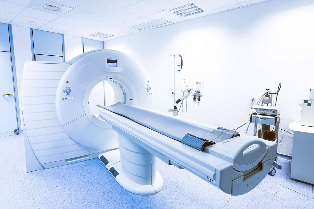
During the first visit, the doctor will conduct a clinical examination and go through the results of the available diagnostic tests. After that, you will undergo the necessary additional examination, such as the assessment of liver and kidney function, ultrasound scan, CT scan and MRI. This will allow the doctor to determine which vessels are feeding the tumor and its metastases, as well as determine how well you will tolerate the procedure.
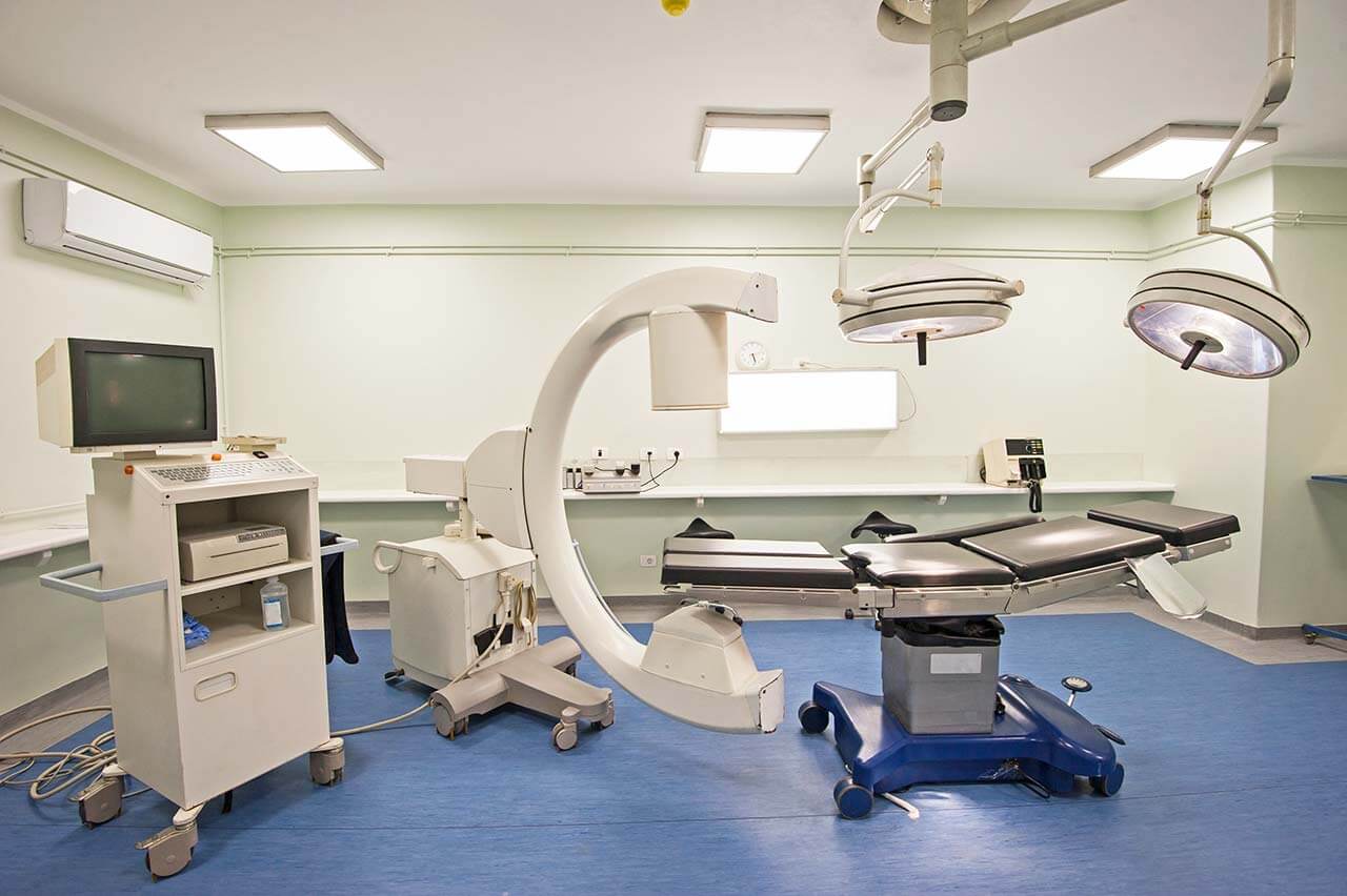
Chemoembolization begins with local anesthesia and catheterization of the femoral artery. The thin catheter is inserted through a few centimeters long incision of the blood vessel. The doctor gradually moves the catheter to the vessel feeding the primary tumor or its metastases. The procedure is carried out under visual control, an angiographic device is used for this. The vascular bed and the position of the catheter in it are displayed on the screen of the angiograph.
When the catheter reaches a suspected artery, a contrast agent is injected through it. Due to the introduction of the contrast agent, the doctor clearly sees the smallest vessels of the tumor and the surrounding healthy tissues on the screen of the angiograph. After that, he injects emboli into the tumor vessels through the same catheter.
Emboli are the spirals or the liquid microspheres. The type of embolus is selected individually, taking into account the diameter of the target vessel. When carrying out chemoembolization, a solution of a chemotherapy drug is additionally injected into the tumor vessel. Due to the subsequent closure of the vessel lumen with an embolus, the chemotherapy drug influences the tumor for a long time. In addition, the drug does not enter the systemic circulation, which allows doctors to use high doses of chemotherapeutic agents without the development of serious side effects. Chemoembolization leads to the destruction of the tumor or slowing down its progression.
After that, the catheter is removed from the artery. The doctor puts a vascular suture on the femoral artery and closes it with a sterile dressing. During chemoembolization, you will be awake. General anesthesia is not used, which significantly reduces the risks of the procedure and allows performing it on an outpatient basis, avoiding long hospital stay.
After the first procedure, you will stay under the supervision of an interventional oncologist and general practitioner. If necessary, you will receive symptomatic treatment. As a rule, a second chemoembolization procedure is performed in 3-5 days after the first one in order to consolidate the therapeutic effect. After that, you will receive recommendations for further follow-up and treatment.
Required documents
Medical records
MRI/CT scan (not older than 3 months)
Biopsy results (if available)
Service
Price from:
Type of program :
Price for 1 day:
Expected duration of the program:
The minimum duration of the program:
Select
You may also book:
Hospital directly:
Saving:
BookingHealth Price from:
About the department
The Department of Adult and Pediatric Diagnostic, Interventional Radiology at the Helios Hospital Berlin-Buch offers all modern methods of high-precision imaging diagnostics and minimally invasive imaging-guided therapeutic procedures. The department meets all radiation protection standards, which are especially important in the diagnostics of children and adolescents. The basis of successful clinical practice of the medical facility is a progressive technological base and the best highly qualified specialists. The department is headed by Prof. Dr. med. Thomas Herold.
One of the department's priority focuses is imaging diagnostics and interventional therapy of bone tumors and soft tissue sarcomas. To detect these types of neoplasms, the doctors use such examinations as sonography, classical radiography, computed tomography and magnetic resonance imaging. The service range is complemented by endovascular diagnostic procedures for the detection of benign soft tissue tumors (such examinations are carried out within the modern hybrid operating rooms), as well as PET-CT skeletal imaging (in collaboration with the Department of Nuclear Medicine). The therapeutic options in this field cover many minimally invasive CT-guided interventions, particularly, tumor biopsy, radiofrequency ablation for the treatment of osteoid osteomas, endovascular interventions (for example, embolization of hemangiomas).
It is worth noting that the department's diagnostic rooms have special medical equipment adapted for conducting examinations in children. Young patients can undergo such examinations as ultrasound scanning, classic radiography, fluoroscopy, CT and MRI. Depending on the specific clinical indications, the diagnostic methods are prescribed to each child individually. The diagnostic process also often involves the participation of necessary specialists from the Department of Pediatric and Adolescent Medicine.
The department's range of medical services includes:
Diagnostic radiology
Classic radiography
Ultrasound examinations (also with contrast enhancement)
Computed tomography (also with contrast enhancement)
Magnetic resonance imaging (also with contrast enhancement)
Fluoroscopy
Myelography
Arthrography
Angiography (also with contrast enhancement)
Pediatric radiology
Classic radiography
Ultrasound diagnostics
Fluoroscopy
Computed tomography
Magnetic resonance imaging
Interventional radiology
CT-guided biopsy (for example, for the detection of causes of lymphadenopathy, metastasis of cancer of unknown primary, metastasis in multiple tumors for treatment planning)
Diagnostic nerve ending and joint blocks for the detection of the cause of pain and therapeutic block for its elimination
Drainage of abscesses and cysts (for example, in Crohn's disease in order to avoid open surgery)
Radiofrequency ablation for tumor destruction
Interventional procedures for CT-guided treatment of lymphocele
Percutaneous CT-guided insertion of feeding tubes into the stomach
Interventional procedures for the treatment of vascular obstruction
Occlusive peripheral arterial disease
Renal artery stenosis in uncontrolled arterial hypertension or impaired kidney function
Aortic intestinal stenosis
Dialysis graft occlusion or stenosis
Thromboses
Embolisms
Superior vena cava syndrome with venous blood stagnation (for example, in cancers or after irradiation)
Catheter therapeutic procedures
Classical balloon angioplasty
Implantation of drug-coated stents
Implantation of classic stents (including flexible and self-expanding ones)
Atherectomy
Mechanical thrombectomy (removal of blood clots)
Classical thrombolysis (blood clot dissolution)
Ultrasound-guided thrombolysis
Catheter treatment of chronic vascular occlusion
Minimally invasive treatment of aneurysms (helix placement)
Therapeutic procedures for blood vessel closure
Benign or malignant tumors (for example, angiomyolipomas)
Varicocele
Impaired venous outflow
Vascular malformations
Arteriovenous malformations
Elimination of endoleaks after vascular prosthetics
Installation of port systems for chemotherapy, dialysis procedures, parenteral nutrition, etc.
Highly specialized procedures
Radiofrequency ablation of the renal artery in poorly controlled arterial hypertension
Implantation of cava filters into the inferior vena cava for the prevention of pulmonary embolism
Extraction of foreign bodies from the vascular system
Transarterial chemoembolization, radiofrequency ablation and selective internal radiation therapy for the treatment of liver tumors
Other diagnostic and therapeutic options
Photo of the doctor: (c) Helios Klinikum Berlin-Buch
About hospital
According to the reputable Focus magazine, the Helios Hospital Berlin-Buch ranks among the top medical facilities in Germany!
The hospital is proud of its rich history, which dates back over 100 years, as well as the status of a maximum care medical center with exceptionally high success treatment rates. The medical institution is an academic hospital of the Charite Medical Complex, which is one of the best in Europe and around the world. To provide its services to patients, the hospital has over 1000 beds, over 60 specialized departments, centers and institutes, including emergency service and a helipad, as well as 23 state-of-the-art operating rooms.
The medical institution presents almost all branches of modern medicine, many of which are certified by professional German societies (for example, certification of the German Cancer Society, German Diabetes Society).
The hospital diagnoses and treats about 52,000 inpatients and more than 144,000 outpatients every year. The medical services and patient care are provided by world-renowned highly competent doctors and qualified nursing staff. To achieve the best treatment results, the doctors of related medical disciplines work closely together and jointly develop optimal treatment regimens.
It is worth noting that the hospital is located in a beautiful green park area. In the immediate vicinity of the hospital one can find Buch Castle, Buch Forest and Barnim Nature Park. All this has a beneficial effect on patients, as they have the opportunity to stroll through beautiful places that inspire and help to gain strength for the successful overcoming of the therapeutic process.
Photo: (c) depositphotos
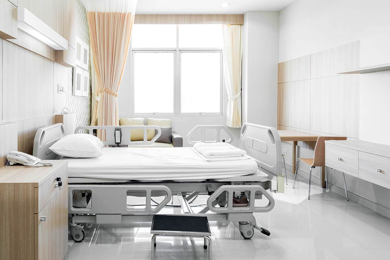
Accommodation in hospital
Patients rooms
The patients of the Helios Hospital Berlin-Buch live in comfortable rooms made in bright colors. Each patient room is equipped with an ensuite bathroom with shower and toilet. The standard room includes an automatically adjustable bed with a system for calling nursing staff (this system also serves to use TV, radio, lamps), a bedside table, a locker for storing personal belongings, a TV, a radio. The hospital offers WI-FI.
Meals and Menus
The patients of the hospital are offered tasty and balanced three meals a day. Breakfast is served as a buffet with a wide selection of pastries, cereal, sausages and cheese. For lunch, the patients usually have a choice of three menus, including a vegetarian menu. Also, the patient can independently combine a lunch menu from various meat, fish and vegetarian dishes and side dishes. Dinner includes a standard menu and dishes that the patient can choose on his own to his taste.
In addition, the hospital houses a cozy cafe with an excellent assortment of pastries, fresh salads, sandwiches, as well as traditional Berlin dishes. Here you can enjoy a cup of aromatic coffee, hot tea or refreshment drinks.
Further details
Standard rooms include:
Toilet
Shower
Wi-Fi
TV
Select your currency:
Your guarantee
We are the only TÜV-certified company with an ISO 9001:2015 certificate
thank you for reaching out! One of our medical advisors is currently reviewing your details. You can expect a call from us within one business day to discuss your request.
Note: The incoming call will appear as a German number or your local area code. This call is entirely free of charge.
Booking Health guarantees:
Expert Placement
We use rigorous statistical analysis to ensure you are treated at the best-rated facility for your specific diagnosis.
Total Cost Protection
No hidden fees or unexpected bills. We provide a fixed price guarantee, backed by insurance that covers any additional complications.
12-Month Extended Care
Your recovery is our priority. Benefit from a full year of direct medical support following your procedure.

 BookingHealth Price from:
