{"translation_price":"50","translation_doc_price":"40","child_coefficient":"1.1","transfer_price":"2.00","transfer_price_vip":"5.00","constant_transfer_price_vip":350,"constant_transfer_price":150,"constant_transfer_distanse":60,"type":"treatment","program_full_story":"<ul>\n\t<li>Initial presentation in the clinic<\/li>\n\t<li>clinical history taking<\/li>\n\t<li>review of medical records<\/li>\n\t<li>physical examination<\/li>\n\t<li>laboratory tests:\n\t<ul>\n\t\t<li>complete blood count<\/li>\n\t\t<li>general urine analysis<\/li>\n\t\t<li>biochemical analysis of blood<\/li>\n\t\t<li>TSH-basal, fT3, fT4<\/li>\n\t\t<li>tumor markers (AFP, CEA, \u0421\u0410-19-9)<\/li>\n\t\t<li>inflammation indicators (CRP, ESR)<\/li>\n\t\t<li>indicators of blood coagulation<\/li>\n\t<\/ul>\n\t<\/li>\n\t<li>abdominal ultrasound\u200b scan<\/li>\n\t<li>CT\/MRI of abdomen<\/li>\n\t<li>preoperative care<\/li>\n\t<li>percutaneous embolization (coiling) or chemoembolization<\/li>\n\t<li>symptomatic treatment<\/li>\n\t<li>cost of essential medicines<\/li>\n\t<li>nursing services<\/li>\n\t<li>elaboration of further recommendations<\/li>\n<\/ul>\n<div class=\"program_indications_for_surgery\"><h4>Indications<\/h4><ul>\n\t<li>Inoperable liver metastases<\/li>\n\t<li>Poor response to systemic chemotherapy<\/li>\n<\/ul>\n\n<p><strong>Treatment is not indicated<\/strong> in:<\/p>\n\n<ul>\n\t<li>Presence of extrahepatic metastases<\/li>\n\t<li>Affection of more than 70% of the liver<\/div><div class=\"program_how_program_going mt-4\"><h4>How program is carried out<\/h4><p style=\"text-align:justify\"><strong>During the first visit<\/strong>, the doctor will conduct a clinical examination and go through the results of the available diagnostic tests. After that, you will undergo the necessary additional examination, such as the assessment of liver and kidney function, ultrasound scan, CT scan and MRI. This will allow the doctor to determine which vessels are feeding the tumor and its metastases, as well as determine how well you will tolerate the procedure.<\/p>\n\n<p style=\"text-align:justify\"><strong>Chemoembolization <\/strong>begins with local anesthesia and catheterization of the femoral artery. The thin catheter is inserted through a few centimeters long incision of the blood vessel. The doctor gradually moves the catheter to the vessel feeding the primary tumor or its metastases. The procedure is carried out under visual control, an angiographic device is used for this. The vascular bed and the position of the catheter in it are displayed on the screen of the angiograph.<\/p>\n\n<p style=\"text-align:justify\">When the catheter reaches a suspected artery, a contrast agent is injected through it. Due to the introduction of the contrast agent, the doctor clearly sees the smallest vessels of the tumor and the surrounding healthy tissues on the screen of the angiograph. After that, he injects emboli into the tumor vessels through the same catheter.<\/p>\n\n<p style=\"text-align:justify\">Emboli are the spirals or the liquid microspheres. The type of embolus is selected individually, taking into account the diameter of the target vessel. When carrying out chemoembolization, a solution of a chemotherapy drug is additionally injected into the tumor vessel. Due to the subsequent closure of the vessel lumen with an embolus, the chemotherapy drug influences the tumor for a long time. In addition, the drug does not enter the systemic circulation, which allows doctors to use high doses of chemotherapeutic agents without the development of serious side effects. Chemoembolization leads to the destruction of the tumor or slowing down its progression.<\/p>\n\n<p style=\"text-align:justify\">After that, the catheter is removed from the artery. The doctor puts a vascular suture on the femoral artery and closes it with a sterile dressing. During chemoembolization, you will be awake. General anesthesia is not used, which significantly reduces the risks of the procedure and allows performing it on an outpatient basis, avoiding long hospital stay.<\/p>\n\n<p style=\"text-align:justify\"><strong>After the first procedure<\/strong>, you will stay under the supervision of an interventional oncologist and general practitioner. If necessary, you will receive symptomatic treatment. As a rule, a second chemoembolization procedure is performed in 3-5 days after the first one in order to consolidate the therapeutic effect. After that, you will receive recommendations for further follow-up and treatment.<\/p>\n<\/div><div class=\"program_required_documents mt-4\"><h4>Required documents<\/h4><ul>\n\t<li style=\"text-align: justify;\">Medical records<\/li>\n\t<li style=\"text-align: justify;\">Abdominal ultrasound (if available)<\/li>\n\t<li style=\"text-align: justify;\">MRI\/CT scan of the abdomen (if available)<\/li>\n\t<li style=\"text-align: justify;\">Biopsy results (if available)<\/li>\n<\/ul>\n<\/div>","program_full_story_crm":"<ul>\n\t<li>Initial presentation in the clinic<\/li>\n\t<li>clinical history taking<\/li>\n\t<li>review of medical records<\/li>\n\t<li>physical examination<\/li>\n\t<li>laboratory tests:\n\t<ul>\n\t\t<li>complete blood count<\/li>\n\t\t<li>general urine analysis<\/li>\n\t\t<li>biochemical analysis of blood<\/li>\n\t\t<li>TSH-basal, fT3, fT4<\/li>\n\t\t<li>tumor markers (AFP, CEA, \u0421\u0410-19-9)<\/li>\n\t\t<li>inflammation indicators (CRP, ESR)<\/li>\n\t\t<li>indicators of blood coagulation<\/li>\n\t<\/ul>\n\t<\/li>\n\t<li>abdominal ultrasound\u200b scan<\/li>\n\t<li>CT\/MRI of abdomen<\/li>\n\t<li>preoperative care<\/li>\n\t<li>percutaneous embolization (coiling) or chemoembolization<\/li>\n\t<li>symptomatic treatment<\/li>\n\t<li>cost of essential medicines<\/li>\n\t<li>nursing services<\/li>\n\t<li>elaboration of further recommendations<\/li>\n<\/ul>\n<div class=\"program_indications_for_surgery\"><h4>Indications<\/h4><ul>\n\t<li>Inoperable liver metastases<\/li>\n\t<li>Poor response to systemic chemotherapy<\/li>\n<\/ul>\n\n<p><strong>Treatment is not indicated<\/strong> in:<\/p>\n\n<ul>\n\t<li>Presence of extrahepatic metastases<\/li>\n\t<li>Affection of more than 70% of the liver<\/div><div class=\"program_how_program_going mt-4\"><h4>How program is carried out<\/h4><p style=\"text-align:justify\"><strong>During the first visit<\/strong>, the doctor will conduct a clinical examination and go through the results of the available diagnostic tests. After that, you will undergo the necessary additional examination, such as the assessment of liver and kidney function, ultrasound scan, CT scan and MRI. This will allow the doctor to determine which vessels are feeding the tumor and its metastases, as well as determine how well you will tolerate the procedure.<\/p>\n\n<p style=\"text-align:justify\"><strong>Chemoembolization <\/strong>begins with local anesthesia and catheterization of the femoral artery. The thin catheter is inserted through a few centimeters long incision of the blood vessel. The doctor gradually moves the catheter to the vessel feeding the primary tumor or its metastases. The procedure is carried out under visual control, an angiographic device is used for this. The vascular bed and the position of the catheter in it are displayed on the screen of the angiograph.<\/p>\n\n<p style=\"text-align:justify\">When the catheter reaches a suspected artery, a contrast agent is injected through it. Due to the introduction of the contrast agent, the doctor clearly sees the smallest vessels of the tumor and the surrounding healthy tissues on the screen of the angiograph. After that, he injects emboli into the tumor vessels through the same catheter.<\/p>\n\n<p style=\"text-align:justify\">Emboli are the spirals or the liquid microspheres. The type of embolus is selected individually, taking into account the diameter of the target vessel. When carrying out chemoembolization, a solution of a chemotherapy drug is additionally injected into the tumor vessel. Due to the subsequent closure of the vessel lumen with an embolus, the chemotherapy drug influences the tumor for a long time. In addition, the drug does not enter the systemic circulation, which allows doctors to use high doses of chemotherapeutic agents without the development of serious side effects. Chemoembolization leads to the destruction of the tumor or slowing down its progression.<\/p>\n\n<p style=\"text-align:justify\">After that, the catheter is removed from the artery. The doctor puts a vascular suture on the femoral artery and closes it with a sterile dressing. During chemoembolization, you will be awake. General anesthesia is not used, which significantly reduces the risks of the procedure and allows performing it on an outpatient basis, avoiding long hospital stay.<\/p>\n\n<p style=\"text-align:justify\"><strong>After the first procedure<\/strong>, you will stay under the supervision of an interventional oncologist and general practitioner. If necessary, you will receive symptomatic treatment. As a rule, a second chemoembolization procedure is performed in 3-5 days after the first one in order to consolidate the therapeutic effect. After that, you will receive recommendations for further follow-up and treatment.<\/p>\n<\/div>","is_ambulant":"1","bh_fee":"0","only_for_children":"0","no_service":"0","with_prepayment":"1","show_calculator":"1","paket_type":"1","btn_type":"2","clinic_icon":"5ffc4ddff1ac4.jpg","city":"Wuppertal","clinic_site":"https:\/\/www.helios-gesundheit.de\/kliniken\/wuppertal\/","department_recommend":"1","country":"Germany","country_id":"1","clinic_name":"HELIOS University Hospital Wuppertal","cinic_name":"HELIOS University Hospital Wuppertal","department_id":"2293","duration":"5","direction":"Interventional radiology","min_duration":0,"clinic_id":"1671","paketPrice":3200,"paket":"<ul>\n <li> Interpreter up to 8 hours<\/li>\n <li>Translation of up to 5 pages<\/li>\n <li>Visa support<\/li>\n\n<\/ul>","title":"Treatment of liver metastases with percutaneous embolization (coiling) or chemoembolization","price":{"val":9880,"type":"val"},"price_surcharge":0,"price_surcharge_clear":0,"extra_service_clinic":[],"extra_service":[],"translation_hours":"0","translation_doc_count":null,"roads":[{"id":"4","distance":"48","airport_title":"Duesseldorf"},{"id":"7","distance":"61","airport_title":"Cologne-Bonn"},{"id":"6","distance":"222","airport_title":"Frankfurt a.M."}],"pakets":[],"lang":{"day":"Day","days":"days","ambulatory":"Outpatient","stationaryProgram":"Inpatient"}}
Treatment of liver metastases with percutaneous embolization (coiling) or chemoembolization in HELIOS University Hospital Wuppertal
percutaneous embolization (coiling) or chemoembolization
symptomatic treatment
cost of essential medicines
nursing services
elaboration of further recommendations
Indications
Inoperable liver metastases
Poor response to systemic chemotherapy
Treatment is not indicated in:
Presence of extrahepatic metastases
Affection of more than 70% of the liver
How program is carried out
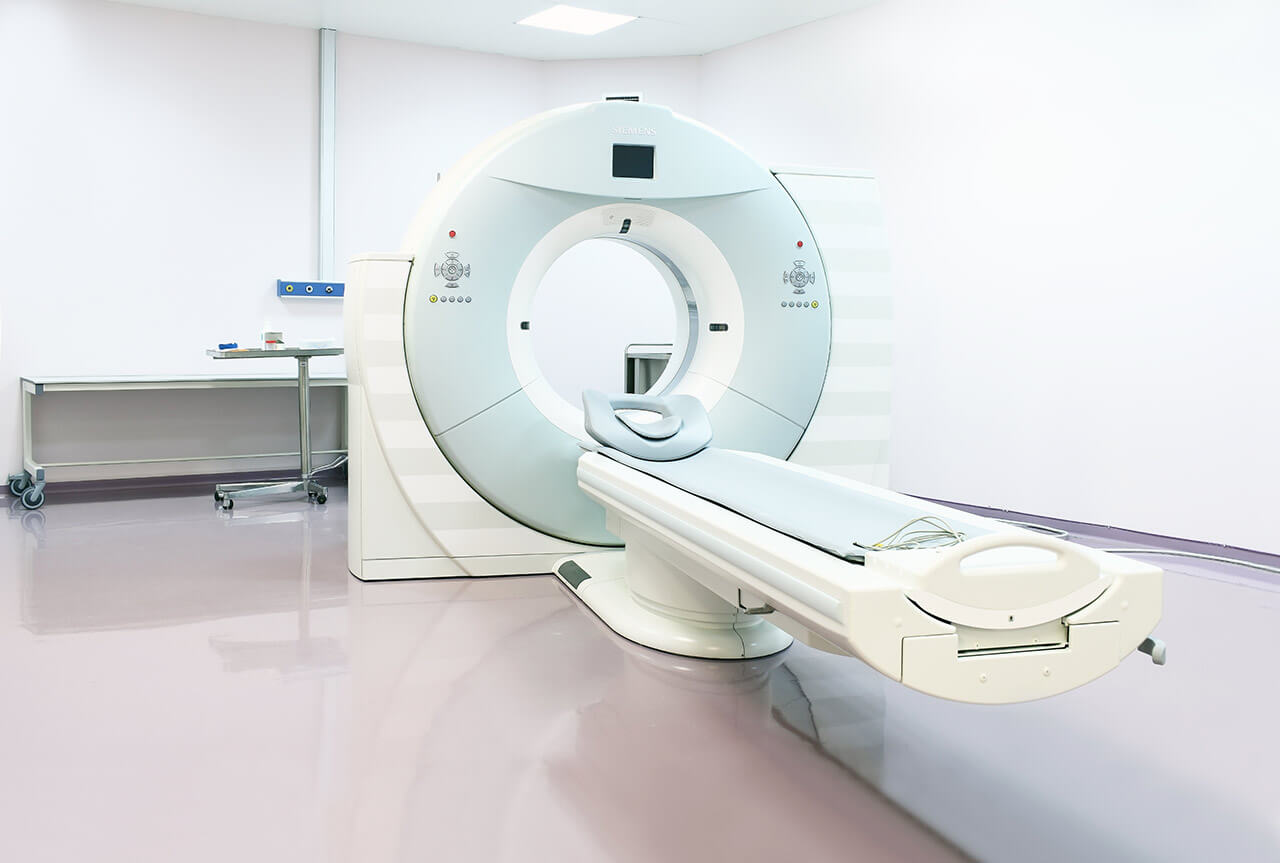
During the first visit, the doctor will conduct a clinical examination and go through the results of the available diagnostic tests. After that, you will undergo the necessary additional examination, such as the assessment of liver and kidney function, ultrasound scan, CT scan and MRI. This will allow the doctor to determine which vessels are feeding the tumor and its metastases, as well as determine how well you will tolerate the procedure.
Chemoembolization begins with local anesthesia and catheterization of the femoral artery. The thin catheter is inserted through a few centimeters long incision of the blood vessel. The doctor gradually moves the catheter to the vessel feeding the primary tumor or its metastases. The procedure is carried out under visual control, an angiographic device is used for this. The vascular bed and the position of the catheter in it are displayed on the screen of the angiograph.
When the catheter reaches a suspected artery, a contrast agent is injected through it. Due to the introduction of the contrast agent, the doctor clearly sees the smallest vessels of the tumor and the surrounding healthy tissues on the screen of the angiograph. After that, he injects emboli into the tumor vessels through the same catheter.
Emboli are the spirals or the liquid microspheres. The type of embolus is selected individually, taking into account the diameter of the target vessel. When carrying out chemoembolization, a solution of a chemotherapy drug is additionally injected into the tumor vessel. Due to the subsequent closure of the vessel lumen with an embolus, the chemotherapy drug influences the tumor for a long time. In addition, the drug does not enter the systemic circulation, which allows doctors to use high doses of chemotherapeutic agents without the development of serious side effects. Chemoembolization leads to the destruction of the tumor or slowing down its progression.
After that, the catheter is removed from the artery. The doctor puts a vascular suture on the femoral artery and closes it with a sterile dressing. During chemoembolization, you will be awake. General anesthesia is not used, which significantly reduces the risks of the procedure and allows performing it on an outpatient basis, avoiding long hospital stay.
After the first procedure, you will stay under the supervision of an interventional oncologist and general practitioner. If necessary, you will receive symptomatic treatment. As a rule, a second chemoembolization procedure is performed in 3-5 days after the first one in order to consolidate the therapeutic effect. After that, you will receive recommendations for further follow-up and treatment.
Required documents
Medical records
Abdominal ultrasound (if available)
MRI/CT scan of the abdomen (if available)
Biopsy results (if available)
Service
Price from:
Type of program :
Price for 1 day:
Expected duration of the program:
The minimum duration of the program:
Select
You may also book:
Hospital directly:
Saving:
BookingHealth Price from:
About the department
The Department of Interventional Radiology at the HELIOS University Hospital Wuppertal specializes in the full range of minimally invasive image-guided interventions for diagnostic and therapeutic purposes. Diagnostic interventional procedures allow obtaining tissue samples for further laboratory tests. Most minimally invasive interventional procedures are performed for the treatment of vascular obstruction or stenosis, as well as for tumor destruction. The surgeon inserts special catheters and/or probes through small skin punctures. All surgical procedures are performed under image guidance – X-ray, digital subtraction angiography, ultrasound, CT, MRI. The specialist can clearly see all the anatomical structures and a pathological focus, thanks to which he accurately controls the delivery of the catheter to the required site and all subsequent manipulations. It is worth noting that the department was awarded the quality certificate of the German Society for Interventional Radiology (DeGIR) in 2018, which guarantees effective treatment in accordance with modern medical standards. The department's doctors have the necessary skills and qualifications, which allow them to select the best treatment option for each patient and ensure its successful conduction. The department is headed by Prof. Dr. med. Patrick Haage.
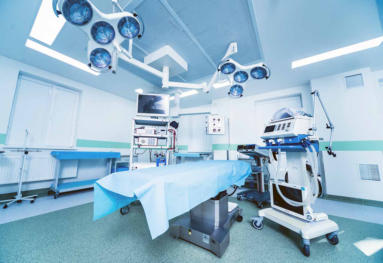
Upon admission to the department, the patient receives a medical consultation, during which the specialist studies his medical history and appoints the necessary imaging diagnostic tests (X-ray, ultrasound, CT, MRI, angiography) to assess the advisability of an interventional therapeutic procedure. Should indications for the intervention be confirmed, the next step will be treatment planning. The procedure is carried out in a special operating room, the patient is on the operating table with large screens to visualize the surgical field. The surgeon inserts the catheter and/or probe through several skin punctures without making extensive incisions. The advantage for the patient is the fact that the interventional procedures are usually performed under local anesthesia.
Most of the department's patients undergo treatment of vascular obstruction or stenosis. Prior to the intervention, angiography and other necessary imaging tests are performed to determine the exact location of the obstruction. After the injection of local anesthetic, the surgeon makes several skin punctures, through which a balloon catheter is inserted into the bloodstream. The specialist delivers the device to the pathological focus, while controlling its movement along a vein or artery using images on a large screen at multiple zoom. When the balloon catheter reaches the area of obstruction or narrowing of the vessel, it expands its lumen, thereby restoring normal blood flow. With severe types of stenosis and obstruction, during the same interventional procedure, stent implantation into the vessel is additionally performed – the placement of a special metal frame to strengthen the vessel walls and prevent recurrent obstruction.
The team of the department's doctors often admits patients suffering from cancers. Of particular interest is chemoembolization for primary liver tumors and liver metastases caused by kidney, pancreatic, breast, colorectal cancer and other types of oncology. Chemoembolization is a local chemotherapy. It involves inserting a catheter through the femoral artery puncture. Under the guidance of angiography, the catheter is directed to the artery supplying the tumor, and the surgeon inserts embolization microspheres into the pathological focus, which block the blood flow to the malignant tumor. The microspheres also contain a high concentration of chemotherapy agent, the gradual release of which destroys the tumor. The department also performs an innovative radioembolization procedure, the essence of which is similar to chemoembolization. The only difference is that the destruction of cancer cells occurs due to the administration of radioactive microspheres, but not microspheres with chemotherapy agents.
The department's specialists are deservedly proud of their successful experience in radiofrequency ablation for the treatment of lung metastases. The essence of the procedure is in the exposure of high-energy radio waves to the metastases. During the procedure, pathological tissues are heated to high temperatures (about 90°C), due to which they are destroyed. Radiofrequency ablation is performed under general anesthesia and with the use of CT guidance. The access to the lungs is provided by puncturing the skin and soft tissues in the thorax, through which a special needle is inserted. With appropriate clinical indications, the department's specialists perform cryoablation – a similar procedure, during which metastases are destroyed by cold. Both procedures are used to remove single metastases in the lungs and are an excellent modern alternative to classic open surgery, which requires thoracotomy.
The department specializes in such minimally invasive interventional procedures:
Diagnostics
Assessment of the patency of arterial and venous vessels in all parts of the body
Biopsy: needle biopsy and fine needle aspiration biopsy
Treatment
Minimally invasive treatment of vascular pathologies
Minimally invasive treatment of peripheral arterial occlusive disease: lysis therapy, percutaneous transluminal angioplasty, stent implantation and thrombectomy
Minimally invasive closure of bleeding foci and correction of vascular malformations
Implantation of cava filters at high risk of pulmonary embolism
Minimally invasive treatment of oncological diseases
Treatment of malignant tumors of the liver and other organs: chemoembolization, catheter embolization and Yttrium-90 radioembolization
Ablation procedures for the treatment of lung metastases: radiofrequency ablation, cryoablation and laser therapy
Other medical services
Curriculum vitae
Higher Education and Postgraduate Training
2001 - 2003 Master of Business Administration (MBA), School of Business and Economics, Maastricht University.
1988 - 1994 Study of Human Medicine at the University of Duesseldorf.
Professional Career
Since June 2019 Medical Director of the HELIOS University Hospital Wuppertal.
Since January 2006 Head Physician of the Department of Interventional Radiology at the HELIOS University Hospital Wuppertal.
January 2010 - December 2018 Member of the Medical Advisory Board of the Helios Clinics Group.
January 2008 - December 2014 Medical Director of the St. Josefs-Hospital Bochum-Linden.
Since May 2014 Head of the Department of Clinical Radiology at the Witten-Herdecke University.
Since September 2015 Coordinator of the Educational Program of the Cardiovascular and Interventional Radiological Society of Europe (CIRSE).
2002 - 2005 Assistant Professor at the RWTH Aachen University.
Memberships in Professional Societies
Radiology Imaging Community.
Vascular Access Society of the Americas.
Society of Interventional Radiology.
Photo of the doctor: (c) Helios Universitätsklinikum Wuppertal
About hospital
According to the prestigious Focus magazine, the HELIOS University Hospital Wuppertal ranks among the top medical facilities in Germany!
The hospital rightfully enjoys the status of the maximum care medical facility and provides its high-quality services in all modern fields of medicine. The hospital operates on the basis of the Witten/Herdecke University, which was opened in 1982 and today is considered one of the best in Germany. Thus, many head physicians of the medical complex are in charge of the corresponding department at the university, which contributes to the close intertwining of research activities and clinical practice. The hospital has long traditions and its own values – the main goal of doctors is to provide comprehensive medical care focused not only on curing the disease, but also on the patient's personal needs.
The hospital has 1,000 beds. The doctors of the medical facility admit more than 50,000 inpatients annually. In addition, more than 100,000 outpatients undergo diagnostic and therapeutic procedures. Such high attendance rates speak for themselves and are undeniable proof of the high-quality medical service of the European level. The medical staff of the hospital has more than 2,500 employees, whose main task is to restore the patient's health and provide him with a decent quality of life.
The hospital has more than 26 specialized departments, as well as many narrowly focused centers and institutes dealing with the treatment of patients suffering from a particular group of diseases: Breast Center, Cancer Center, Cardiology Center, Trauma Center, Spine Center and others. The primary clinical focus of the medical center is cancer treatment.
For more than 25 years, the hospital has been running a special quality management system for medical care, which regulates the aspects of work of the medical staff, compliance with hygiene and safety standards during diagnostics and treatment. Consequently, patients can be sure that their health is in the safe hands of true professionals who work in accordance with the latest medical standards.
Special attention should be paid to the honors of the hospital for excellent patient care. The medical complex has quality certificates from the German Cancer Society (DKG), the German Trauma Society (DGU), the German Cardiac Society (DGK), the German Stroke Society (DSG) and other professional German societies.
Photo: (с) depositphotos
Accommodation in hospital
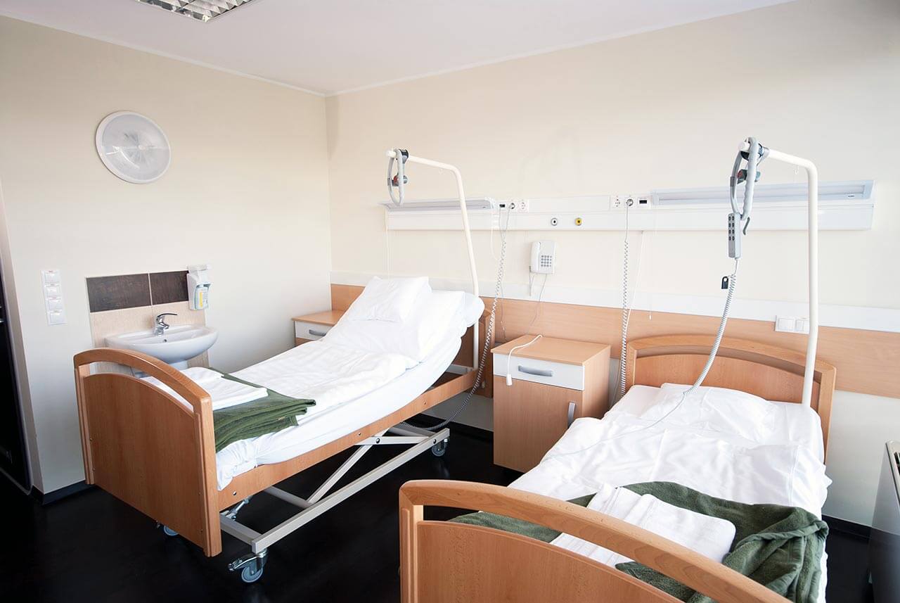
Patients rooms
The patients of the HELIOS University Hospital Wuppertal live in comfortable single, double, triple and quadruple rooms. Each patient room has an ensuite bathroom with shower and toilet. The standard room furnishings include a comfortable automatically adjustable bed, a bedside table, a wardrobe, a TV and a telephone. The hospital has Wi-Fi (free). For maximum patient comfort, there is a nurse call device on the bedside table. This device allows the patient to control the TV, radio, turn on or off the lights, and adjust the position of the bed.
The patients of the hospital are also offered accommodation in enhanced-comfort rooms. These rooms additionally provide a safe and a free minibar with soft drinks. The enhanced-comfort rooms also have a spacious bathroom with hairdryer, bathrobe, towels and toiletries.
Meals and Menus
The patients of the hospital are offered three meals a day: breakfast, lunch and dinner. The menu offers a variety of delicious dishes to suit all tastes, including dietary and vegetarian options.
The hospital also has a bistro where one can taste delicious hot dishes, cold snacks, desserts, as well as a cup of tea, coffee or refreshments.
The patients staying in enhanced-comfort rooms are offered a separate menu that includes a wider and more refined range of dishes. In addition, fresh fruit, tea, coffee and desserts are delivered to the patient room every day, if desired.
Further details
Standard rooms include:
Shower
Toilet
Wi-Fi
TV
Religion
The religious services are available upon request.
Accompanying person
During an inpatient program, your accompanying person can stay with you in the patient room or in the hotel of your choice.
Hotel
During an outpatient program, you can stay in the hotel of your choice. Our managers will help you choose the most suitable options.
Select your currency:
Your guarantee
One year of support after the treatment
Insurance to cover unforeseen expenses arising from complications during and 48 months after treatment (coverage up to 200,000 €)
Reduced costs by 40-70% (contracts with Hospitals)
In addition, we are the only TÜV-certified company with an ISO 9001:2015 certificate
thank you for reaching out! One of our medical advisors is currently reviewing your details. You can expect a call from us within one business day to discuss your request.
Note: The incoming call will appear as a German number or your local area code. This call is entirely free of charge.
Booking Health guarantees:
Expert Placement
We use rigorous statistical analysis to ensure you are treated at the best-rated facility for your specific diagnosis.
Total Cost Protection
No hidden fees or unexpected bills. We provide a fixed price guarantee, backed by insurance that covers any additional complications.
12-Month Extended Care
Your recovery is our priority. Benefit from a full year of direct medical support following your procedure.
 BookingHealth Price from:
