The program includes:
- Initial presentation in the clinic
- clinical history taking
- review of medical records
- physical examination
- laboratory tests:
- complete blood count
- general urine analysis
- biochemical analysis of blood
- TSH-basal, fT3, fT4
- tumor markers (AFP, CEA, СА-19-9)
- inflammation indicators (CRP, ESR)
- indicators of blood coagulation
- abdominal ultrasound scan
- CT/MRI of abdomen
- preoperative care
- percutaneous embolization (coiling) or chemoembolization
- symptomatic treatment
- cost of essential medicines
- nursing services
- elaboration of further recommendations
Indications
- Inoperable liver metastases
- Poor response to systemic chemotherapy
Treatment is not indicated in:
- Presence of extrahepatic metastases
- Affection of more than 70% of the liver
How program is carried out
During the first visit, the doctor will conduct a clinical examination and go through the results of the available diagnostic tests. After that, you will undergo the necessary additional examination, such as the assessment of liver and kidney function, ultrasound scan, CT scan and MRI. This will allow the doctor to determine which vessels are feeding the tumor and its metastases, as well as determine how well you will tolerate the procedure.
Chemoembolization begins with local anesthesia and catheterization of the femoral artery. The thin catheter is inserted through a few centimeters long incision of the blood vessel. The doctor gradually moves the catheter to the vessel feeding the primary tumor or its metastases. The procedure is carried out under visual control, an angiographic device is used for this. The vascular bed and the position of the catheter in it are displayed on the screen of the angiograph.
When the catheter reaches a suspected artery, a contrast agent is injected through it. Due to the introduction of the contrast agent, the doctor clearly sees the smallest vessels of the tumor and the surrounding healthy tissues on the screen of the angiograph. After that, he injects emboli into the tumor vessels through the same catheter.
Emboli are the spirals or the liquid microspheres. The type of embolus is selected individually, taking into account the diameter of the target vessel. When carrying out chemoembolization, a solution of a chemotherapy drug is additionally injected into the tumor vessel. Due to the subsequent closure of the vessel lumen with an embolus, the chemotherapy drug influences the tumor for a long time. In addition, the drug does not enter the systemic circulation, which allows doctors to use high doses of chemotherapeutic agents without the development of serious side effects. Chemoembolization leads to the destruction of the tumor or slowing down its progression.
After that, the catheter is removed from the artery. The doctor puts a vascular suture on the femoral artery and closes it with a sterile dressing. During chemoembolization, you will be awake. General anesthesia is not used, which significantly reduces the risks of the procedure and allows performing it on an outpatient basis, avoiding long hospital stay.
After the first procedure, you will stay under the supervision of an interventional oncologist and general practitioner. If necessary, you will receive symptomatic treatment. As a rule, a second chemoembolization procedure is performed in 3-5 days after the first one in order to consolidate the therapeutic effect. After that, you will receive recommendations for further follow-up and treatment.
Required documents
- Medical records
- Abdominal ultrasound (if available)
- MRI/CT scan of the abdomen (if available)
- Biopsy results (if available)
Service
You may also book:
 BookingHealth Price from:
About the department
The Department of Diagnostic and Interventional Radiology, Nuclear Medicine at the Hospital Kassel offers the full range of diagnostic and therapeutic procedures in the areas of its specialization. The department has high-tech equipment for all types of modern imaging tests, image-guided therapeutic procedures, as well as radioisotope examinations and treatment of thyroid diseases using radioiodine therapy. The patients are provided with both inpatient and outpatient medical care. However, hospitalization is usually required only in the case of complex therapeutic interventions. The patients' health is in the good hands of the highly qualified doctors. The specialists regularly undergo advanced training courses, attend medical congresses and symposia, where they find out about innovations in the area of their specialization and share their experience with colleagues. To provide maximum safety for patients, the department strictly adheres to radiation protection standards.
The department is headed by Prof. Dr. med. Walter Hundt, who is board certified in Diagnostic and Interventional Radiology, as well as Nuclear Medicine. The doctor is one of the best specialists in this medical field in Germany. Prof. Walter Hundt worked in such leading medical facilities as the University Hospital Heidelberg, the University Hospital Marburg UKGM and the University Hospital of Ludwig Maximilian University of Munich, where he gained invaluable clinical experience.
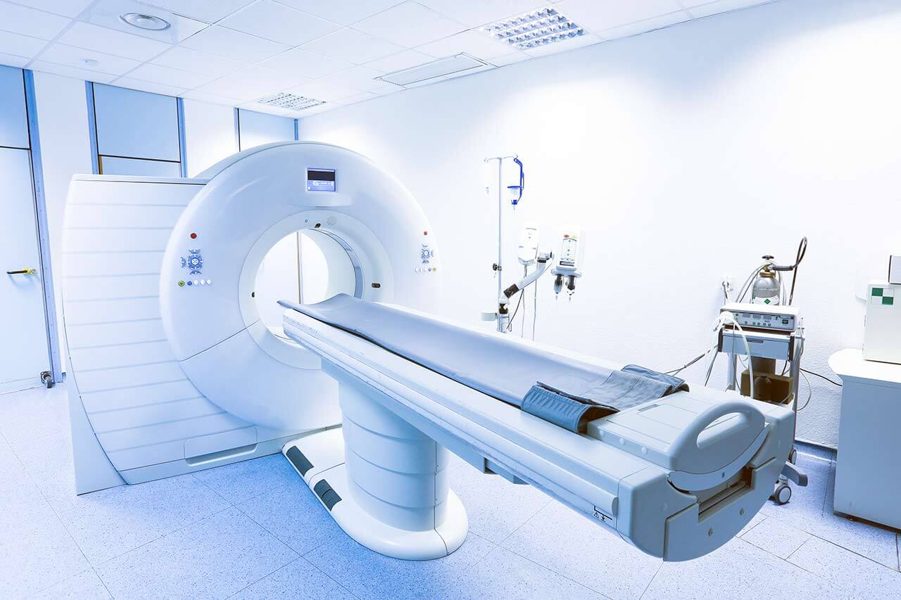
The department's medical team offers excellent diagnostic options to their patients. It carries out such examinations as X-ray, sonography, digital mammography, computed tomography, magnetic resonance imaging, angiography, positron emission tomography combined with computed tomography, scintigraphy, etc. All these diagnostic tests help to detect the slightest changes in the human body and accurately determine their origin, while positron emission tomography combined with computed tomography provides not only comprehensive information about changes in the anatomical structures of the human body, but also about metabolism. The radiological diagnostics and diagnostic examinations in nuclear medicine are of particular value for the accurate detection of malignant tumors and metastases.
In addition to high-quality diagnostic examinations, the department's patients are provided with a variety of image-guided therapeutic interventional procedures. The department's radiologists most often perform various catheter procedures to eliminate vascular obstructions and stenosis, procedures for intravascular injection of chemotherapy agents in patients with certain types of tumors (chemoperfusion), percutaneous radiofrequency ablation and microwave ablation for tumor treatment, abscess drainage and other procedures. All the therapeutic procedures are performed under imaging guidance, which excludes damage to the healthy tissues. The interventional techniques are an excellent alternative to classical open surgery, which may be contraindicated, especially for elderly patients or patients with serious concomitant diseases.
The department's range of medical services includes:
- Diagnostic tests
- Classical radiography of the thorax, skeleton and gastrointestinal organs
- Ultrasound scanning of the abdominal organs, thyroid gland, soft tissues of the neck, breast, as well as color-coded duplex ultrasonography of the blood vessels
- Digital mammography including stereotactic vacuum-assisted aspiration biopsy and preoperative stereotaxic marking
- Computed tomography of the head, body, limbs and liver
- CT angiography
- Multiplanar and 3D reconstruction
- Virtual colonoscopy
- CT-guided puncture
- Magnetic resonance imaging of the head, body and limbs
- MRI of the abdominal organs, including magnetic resonance cholangiopancreatography
- MR mammography, including MRI-guided breast biopsy
- MR angiography
- Cardiac MRI
- Multiparametric magnetic resonance imaging
- Angiography of the vessels of all parts of the body
- CO2 angiography
- Digital subtraction angiography
- Diagnostic examinations using radioisotopes (nuclear medicine)
- Detection of the source of gastrointestinal bleeding
- Scintigraphy for suspected inflammation, for example, in the joint
- Thyroid fine needle aspiration biopsy for suspected thyroid cancer
- Brain scintigraphy
- Bone marrow scintigraphy
- Lung scintigraphy
- Lymph node scintigraphy
- Scintigraphy for suspected Meckel's diverticulum
- MIBG scintigraphy
- Myocardial scintigraphy
- Adrenal scintigraphy
- Parathyroid scintigraphy
- Kidney scintigraphy
- Esophageal scintigraphy
- Thyroid scintigraphy in combination with ultrasound scanning
- Salivary gland scintigraphy
- Bone scintigraphy
- Sentinel lymph node scintigraphy
- Positron emission tomography combined with computed tomography
- Imaging-guided interventional therapeutic procedures
- Percutaneous transluminal angioplasty of the peripheral vessels, kidney arteries, abdominal organs, neck and head vessels
- Fibrinolysin therapy and percutaneous thromboembolectomy
- Implantation of endovascular prostheses (stents) and cava filters
- Tumor and vascular malformation embolization
- Sclerotherapy for varicocele
- Biliary tract interventional procedures
- Transjugular intrahepatic portosystemic stent-shunt placement
- Balloon dacryocystoplasty (dilatation of the tear ducts)
- Percutaneous endoscopic gastrostomy (PEG)
- Uterine artery embolization
- Intra-arterial chemotherapy and chemoembolization for malignancies
- Percutaneous radiofrequency ablation and microwave ablation for tumors
- Percutaneous vertebroplasty
- Percutaneous pain therapy and neurolysis
- CT- and ultrasound-guided percutaneous puncture
- Abscess drainage
- Radioiodine therapy for thyroid diseases
- Other diagnostic and therapeutic options
Curriculum vitae
In February 2018, Prof. Dr. med. Walter Hundt was appointed Chief Physician of the Department of Diagnostic and Interventional Radiology, Nuclear Medicine at the Hospital Kassel. Prior to that, the specialist held the position of Senior Physician with Management Responsibilities and Deputy Head of the Department of Adult and Pediatric Diagnostic, Interventional Radiology at the University Hospital Marburg UKGM. Prof. Hundt is a board certified specialist not only in the field of radiology, but also in the field of nuclear medicine. His special clinical interests include interventional diagnostic and therapeutic procedures, as well as PET-CT scanning, which allows to accurately detect the tumor localization and metastases in the human body. Dr. Walter Hundt received his medical education at the Faculty of Medicine of the Saarland University. He had his medical training in England and Chicago, USA. He worked in such prestigious German medical facilities as the University Hospital Heidelberg and the University Hospital of Ludwig Maximilian University of Munich. From 2001 to 2003, he had his fellowship at Stanford University in the United States, after which he underwent habilitation and became a professor.
Photo of the doctor: (c) Klinikum Kassel
About hospital
The Hospital Kassel is a progressive medical facility with a huge medical team, which provides high-quality medical services in all branches of modern medicine. The hospital is part of the regional medical Gesundheit Nordhessen Holding, which unites 5 top-class medical centers, including specialized rehabilitation clinics. With 1,281 beds, the hospital is known as the largest medical complex in the federal state of Hesse. The hospital has 32 specialized departments with highly qualified doctors and specially trained nursing staff in each department. The team of 3,200 employees takes care of the health of patients. The main value for each employee is the patient's health. The professional skills of the medical staff in combination with state-of-the-art medical and technical equipment of the hospital provide excellent opportunities for the treatment of patients with pathologies of any severity.
The hospital provides treatment to over 55,000 inpatients and about 140,000 outpatients every year. Medical care is provided to both German citizens and many patients from foreign countries. Such high rates are the evidence of excellent quality of medical services and the high credit of patients' trust.
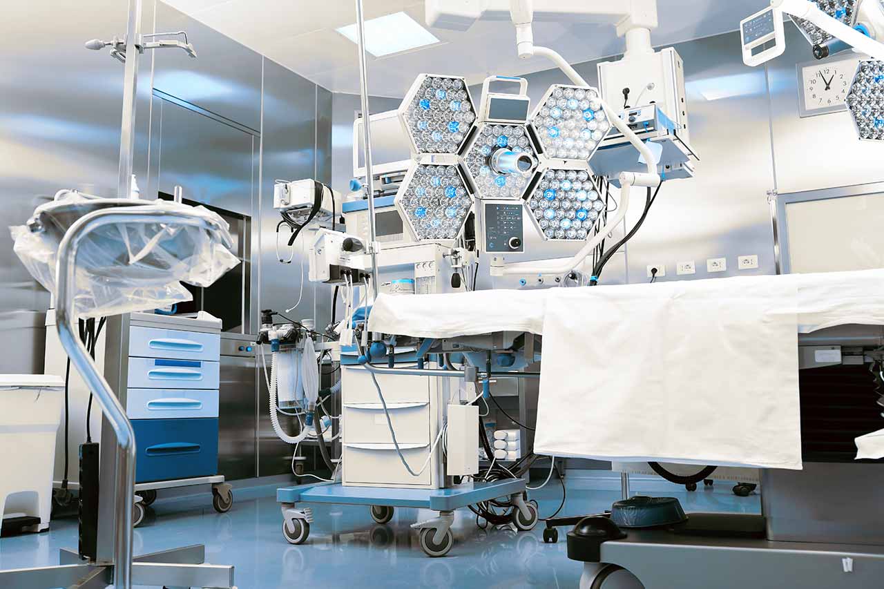
The hospital has created a wonderful atmosphere, which contributes to the rapid recovery of patients. All diagnostic and therapeutic rooms, operating rooms, as well as patient rooms are designed taking into account modern standards of European medicine in order to ensure maximum comfort of each patient. All employees working in the hospital provide the patient with understanding and respect, as well as support him in every possible way during the entire therapeutic process.
The hospital successfully implements a quality management system. It uses its own quality management system implemented by the medical Gesundheit Nordhessen Holding, as well as the IQM (Initiative Qualitätsmedizin) monitoring system. As part of healthcare quality management, the hospital annually clearly provides reports on its clinical activities, the success of diagnostics, treatment, level of patient care, etc. Thus, the hospital stands for maximum openness in its work and makes every effort to maintain the highest level of quality of medical care.
Photo: (с) depositphotos
Accommodation in hospital
Patients rooms
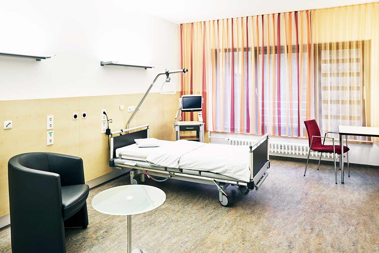
The patients of the Hospital Kassel live in comfortable single, double and triple rooms. The patient rooms are made in a modern design and pastel colors. A standard patient room includes an automatically adjustable bed, a bedside table, a wardrobe, a table and chairs for receiving visitors, a TV and a telephone. The patient rooms have Wi-Fi. Each room has an ensuite bathroom with shower and toilet.
The hospital also offers enhanced-comfort patient rooms. Most of these rooms have a balcony. The bathroom additionally includes a hairdryer, towels and toiletries.
Meals and Menus
The patient and the accompanying person are offered tasty and balanced three meals a day. If for some reason you do not eat all foods, you will be offered an individual menu. Please inform the medical staff about your food preferences prior to treatment. The patients staying in enhanced-comfort rooms are provided with an individual menu every day.
The hospital also has several cafes where one can have a cup of tea or coffee, taste delicious pastries, salads, main hot dishes, pizza, etc.
Further details
Standard rooms include:
Religion
The religious services are available upon request.
Accompanying person
During the inpatient program, the accompanying person can live with the patient in a patient room or a hotel of his choice. Our managers will help you choose the most suitable option.
Hotel
During an outpatient program, the patient can stay at the hotel of his choice. Our managers will help you choose the most suitable option.


