The program includes:
- Initial presentation in the hospital
- Clinical history taking
- Review of available medical records
- Physical examination
- Laboratory tests:
- Complete blood count
- General urine analysis
- Biochemical analysis of blood
- Tumor markers
- Inflammation indicators (CRP, ESR)
- Coagulogram
- Ultrasound scan
- CT scan / MRI
- Preoperative care
- Embolization or chemoembolization, 2 procedures
- Symptomatic treatment
- Cost of essential medicines
- Nursing services
- Elaboration of further recommendations
How program is carried out
During the first visit, the doctor will conduct a clinical examination and go through the results of the available diagnostic tests. After that, you will undergo the necessary additional examination, such as the assessment of liver and kidney function, ultrasound scan, CT scan and MRI. This will allow the doctor to determine which vessels are feeding the tumor and its metastases, as well as determine how well you will tolerate the procedure.
Chemoembolization begins with local anesthesia and catheterization of the femoral artery. The thin catheter is inserted through a few centimeters long incision of the blood vessel. The doctor gradually moves the catheter to the vessel feeding the primary tumor or its metastases. The procedure is carried out under visual control, an angiographic device is used for this. The vascular bed and the position of the catheter in it are displayed on the screen of the angiograph.
When the catheter reaches a suspected artery, a contrast agent is injected through it. Due to the introduction of the contrast agent, the doctor clearly sees the smallest vessels of the tumor and the surrounding healthy tissues on the screen of the angiograph. After that, he injects emboli into the tumor vessels through the same catheter.
Emboli are the spirals or the liquid microspheres. The type of embolus is selected individually, taking into account the diameter of the target vessel. When carrying out chemoembolization, a solution of a chemotherapy drug is additionally injected into the tumor vessel. Due to the subsequent closure of the vessel lumen with an embolus, the chemotherapy drug influences the tumor for a long time. In addition, the drug does not enter the systemic circulation, which allows doctors to use high doses of chemotherapeutic agents without the development of serious side effects. Chemoembolization leads to the destruction of the tumor or slowing down its progression.
After that, the catheter is removed from the artery. The doctor puts a vascular suture on the femoral artery and closes it with a sterile dressing. During chemoembolization, you will be awake. General anesthesia is not used, which significantly reduces the risks of the procedure and allows performing it on an outpatient basis, avoiding long hospital stay.
After the first procedure, you will stay under the supervision of an interventional oncologist and general practitioner. If necessary, you will receive symptomatic treatment. As a rule, a second chemoembolization procedure is performed in 3-5 days after the first one in order to consolidate the therapeutic effect. After that, you will receive recommendations for further follow-up and treatment.
Required documents
- Medical records
- Esophagogastroduodenoscopy (EGD), MRI/CT scan (not older than 3 months)
- Biopsy results (if available)
Service
You may also book:
 BookingHealth Price from:
About the department
The Department of Adult and Pediatric Interventional Radiology at the University Hospital of Ludwig Maximilian University of Munich provides the full range of medical services in the area of its specialization. The department offers modern diagnostic rooms with advanced equipment for imaging examinations for patients with suspected cancer, diseases of the cardiovascular system, thorax, abdominal organs, musculoskeletal system, and breast diseases. The department's diagnostic systems are adapted for examining children of different age groups. The medical facility also offers many therapeutic services, the most popular of which include MRI-guided treatment of uterine fibroids with high-intensity focused ultrasound (HIFU) and uterine artery embolization, prostate adenoma treatment with prostate artery embolization, tumor treatment with brachytherapy, percutaneous radiofrequency ablation, percutaneous microwave ablation, and transarterial chemoembolization. The doctors working at the medical facility also have rich experience in the interventional treatment of vascular pathologies and arteriovenous malformations. The department has an excellent reputation and regularly diagnoses and treats patients not only from Germany but also from other countries in the world. The department is headed by Prof. Dr. med. Jens Ricke.
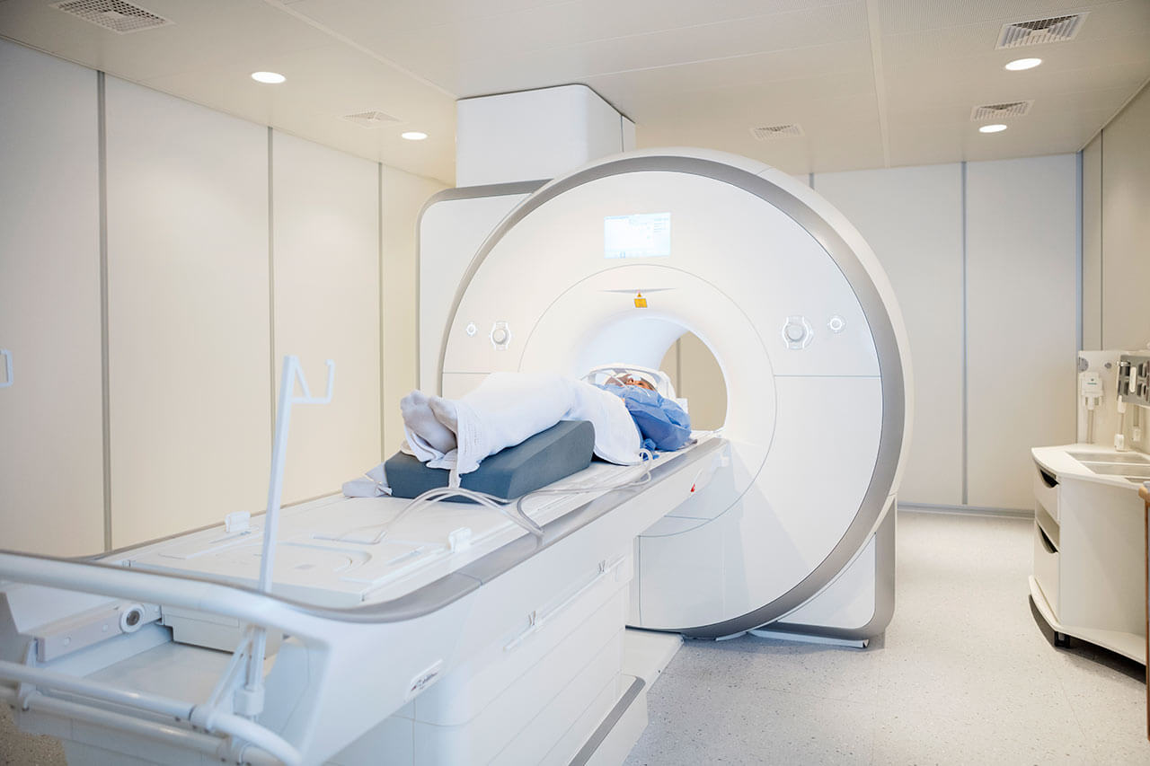
The department's team of doctors often treats patients with uterine fibroids, the most common benign neoplasm in women of childbearing age. Uterine fibroids rarely cause any symptoms, so they are often detected incidentally during a gynecological examination. Uterine fibroids cause infertility, miscarriage, and other pathological conditions. The department's doctors have two effective and sparing therapeutic procedures for uterine fibroid treatment: uterine artery embolization and MRI-guided high-intensity focused ultrasound (HIFU) therapy. In the first case, the therapeutic effect is achieved by introducing emboli into the blood vessels supplying the myoma, due to which the size of the uterine fibroid gradually decreases. As of today, MRI-guided high-intensity focused ultrasound (HIFU) therapy is the most modern treatment for uterine fibroids. The procedure does not require any tissue incisions and is usually performed on an outpatient basis. During the manipulation, the patient is located on the couch of the MRI scanner, which creates 3D images of uterine fibroids using magnetic fields. These images are used as the basis for targeting the neoplasm with HIFU ultrasonic waves, which locally heat the tissues up to 60-80°C. Myoma cells are destroyed under the influence of high temperatures, after which they are excreted naturally. A woman can return to her normal life within 1-2 days after the manipulation.
A fairly common pathology is benign prostatic hyperplasia. The department's doctors successfully carry out prostatic artery embolization to treat the condition. The therapeutic manipulation is performed using a narrow catheter, which is delivered to the blood vessels supplying the prostate gland through the inguinal artery under local anesthesia and imaging guidance. Embolizing material is administered into the blood vessels with a catheter, which contributes to the cessation of blood flow and, consequently, a decrease in the size of the prostate gland.
The department's specialists offer many effective interventional treatment methods for malignant diseases, including brachytherapy, selective internal radiation therapy (SIRT), percutaneous radiofrequency ablation, percutaneous microwave ablation, and transarterial chemoembolization. Brachytherapy is performed under CT or MRI guidance. The department performs exactly that therapeutic manipulation, which was developed in 2004 by radiologists and radiation therapists from the Ludwig Maximilian University of Munich and the Charite University Hospital Berlin. The department's specialists most often perform brachytherapy in patients with primary or secondary liver and lung malignancies. The essence of brachytherapy is to place a radiation source directly into a malignant neoplasm so that doctors can direct the highest possible dose of radiation into it without damaging healthy adjacent tissues. Brachytherapy under CT or MRI guidance contributes to the highest accuracy of treatment.
The department's therapeutic offer is complemented by image-guided interventional treatment of vascular pathologies. The doctors working at the medical facility regularly perform interventional procedures under CT and MRI guidance to eliminate stenosis and obstruction of the blood vessels in the pelvis and lower limbs due to occlusive peripheral artery disease, treat renal artery stenosis due to arterial hypertension and kidney failure, treat abdominal and thoracic aortic aneurysms, repair arteriovenous and venous malformations, including in children.
The department's range of medical services includes the following options:
- Diagnostic examinations in adults and children
- Ultrasound scans, including those with contrast enhancement
- Classic X-ray scans
- Fluoroscopy
- Computed tomography (CT)
- Magnetic resonance imaging (MRI)
- Diagnostics of breast pathologies: mammography, galactography, ultrasound scans and MRI scans
- Image-guided interventional therapeutic procedures in adults and children
- Treatment of uterine fibroids
- Uterine artery embolization
- MRI-guided high-intensity focused ultrasound (HIFU) therapy
- Treatment of benign prostatic hyperplasia
- Prostatic artery embolization
- Treatment of hemorrhoids
- Hemorrhoidal artery embolization
- Treatment of malignant tumors
- Brachytherapy (most commonly used to treat liver and lung tumors)
- Selective internal radiation therapy (SIRT) for liver and lung tumors
- Percutaneous radiofrequency ablation
- Percutaneous microwave ablation
- Transarterial chemoembolization (TACE)
- Treatment of vascular diseases
- Elimination of vascular stenosis and occlusion in the pelvis and lower limbs due to occlusive peripheral arterial disease
- Elimination of renal artery stenosis due to arterial hypertension and kidney failure
- Treatment of abdominal and thoracic aortic aneurysms
- Treatment of vascular tumors and vascular malformations in adults and children
- Treatment of osteoid osteomas
- High-intensity focused ultrasound (HIFU)
- Treatment of portal hypertension
- Transjugular intrahepatic portosystemic shunting (TIPS)
- Implantation of venous catheters and port systems (for example, for dialysis, chemotherapy, etc.)
- Pain management for chronic pain syndromes (for example, in the back)
- Treatment of uterine fibroids
- Other diagnostic and treatment methods
Curriculum vitae
Higher Education and Professional Career
- 1985 - 1992 Human Medicine studies, Faculty of Medicine, Heinrich Heine University of Duesseldorf.
- 1991 Steuben Schurz Society Scholarship, UC Davis School of Medicine, USA.
- 1992 Internship, Department of Surgery, District Hospital Grevenbroich.
- 1993 - 2000 Intern and Research Fellow, Department of Radiation Therapy, Charite University Hospital Berlin.
- 1993 Thesis defense, Heinrich Heine University of Duesseldorf. Subject: "The results of palliative surgery for esophageal cancer".
- 1999 Board certification in Radiology.
- 1999 - 2006 Head of the Working Group on Digital Radiology, including Telemedicine and Picture Archiving and Communication Systems, Department of Radiation Therapy, Charite University Hospital Berlin.
- 2001 Habilitation and Venia Legendi, Charite University Hospital Berlin. Subject: "Optimization of the discrete wavelet transform algorithm for digital X-ray image compression to prevent the loss of its quality".
- 2001 - 2006 Managing Senior Physician, Department of Radiation Therapy, Charite University Hospital Berlin.
- 2004 - 2006 C3 Professorship for Interventional Radiology, Charite University Hospital Berlin.
- 2005 Invitation to the Department of Radiology, Otto von Guericke University Magdeburg.
- 2006 - 2017 Head of the Department of Radiology, Otto von Guericke University Magdeburg; Head Physician of the Department of Radiology and Nuclear Medicine at the University Hospital Magdeburg.
- Since 2011 Founding President of the German Academy for Microtherapy.
- Since 2017 Head of the Department of Radiology and Head Physician of the Department of Adult and Pediatric Interventional Radiology at the University Hospital of Ludwig Maximilian University of Munich.
Research Interests
- Interventional radiology with a special focus on minimally invasive cancer treatment.
- Interventional magnetic resonance imaging.
- Image-guided hybrid therapeutic procedures.
- Image-guided radiation therapy.
- Development of tools for image-guided interventions.
- Contrast-enhanced hepatobiliary MRI scanning.
Photo of the doctor: (c) LMU Klinikum
About hospital
According to the Focus magazine, the University Hospital of Ludwig Maximilian University of Munich is regularly ranked among the best medical institutions in Germany!
The hospital is the largest multidisciplinary medical facility, as well as a leading research and training center in Germany and Europe. The hospital is proud of its bicentenary history and tirelessly confirms its primacy at the national and international levels. The outstanding quality of medical care is complemented by highly productive research activities, thanks to which many effective diagnostic and therapeutic methods, saving people’s lives, have been presented in medical practice.
The medical facility includes two main buildings, Grosshadern and Innenstadt. The hospital has 29 specialized departments, 53 interdisciplinary centers, 11 institutes, and many sections. More than 500,000 patients are treated here every year, which indicates the hospital's excellent reputation. A large and highly professional medical team, consisting of 1,800 doctors and 3,300 nursing staff, works for the benefit of patients. The hospital has 2,000 beds to accommodate patients.
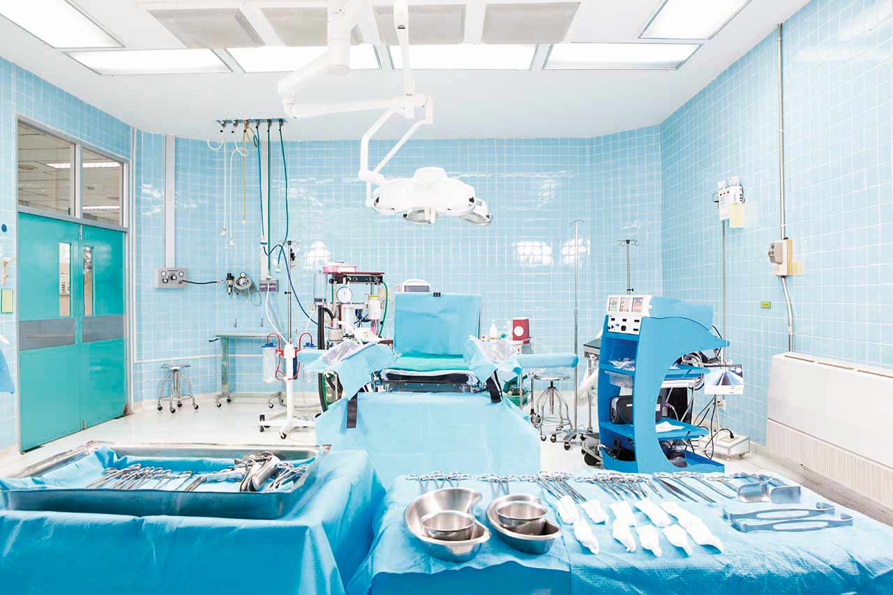
The hospital's infrastructure deserves special attention: advanced diagnostic equipment that allows doctors to detect the slightest pathological changes in the human body, the latest operating rooms with highly efficient monitoring systems, robot-assisted surgical systems that facilitate sparing operations, and proper postoperative care.
Excellent technical resources and highly professional medical staff are undoubtedly the hospital's pride, but the medical facility also pays attention to the patient's comfort and to a humane attitude toward their life situation. When providing the necessary medical care, doctors and nursing staff always show a friendly attitude, inform patients in detail about the upcoming diagnostic and therapeutic procedures, gladly answer all questions of interest to patients, and provide moral support during the therapeutic process.
The hospital has many prestigious quality certificates, including a DIN EN ISO 9001 certificate, an IQM certificate, an endoCert certificate, certificates from the German Cancer Society (DKG) for treating various types of cancer, the German Cardiac Society (DGK), the German Society for Orthopedics and Trauma Surgery (DGOU), etc. Thus, patients can count on the best possible treatment outcome due to the use of the most effective and, at the same time, sparing therapeutic techniques.
Photo: (с) depositphotos
Accommodation in hospital
Patients rooms
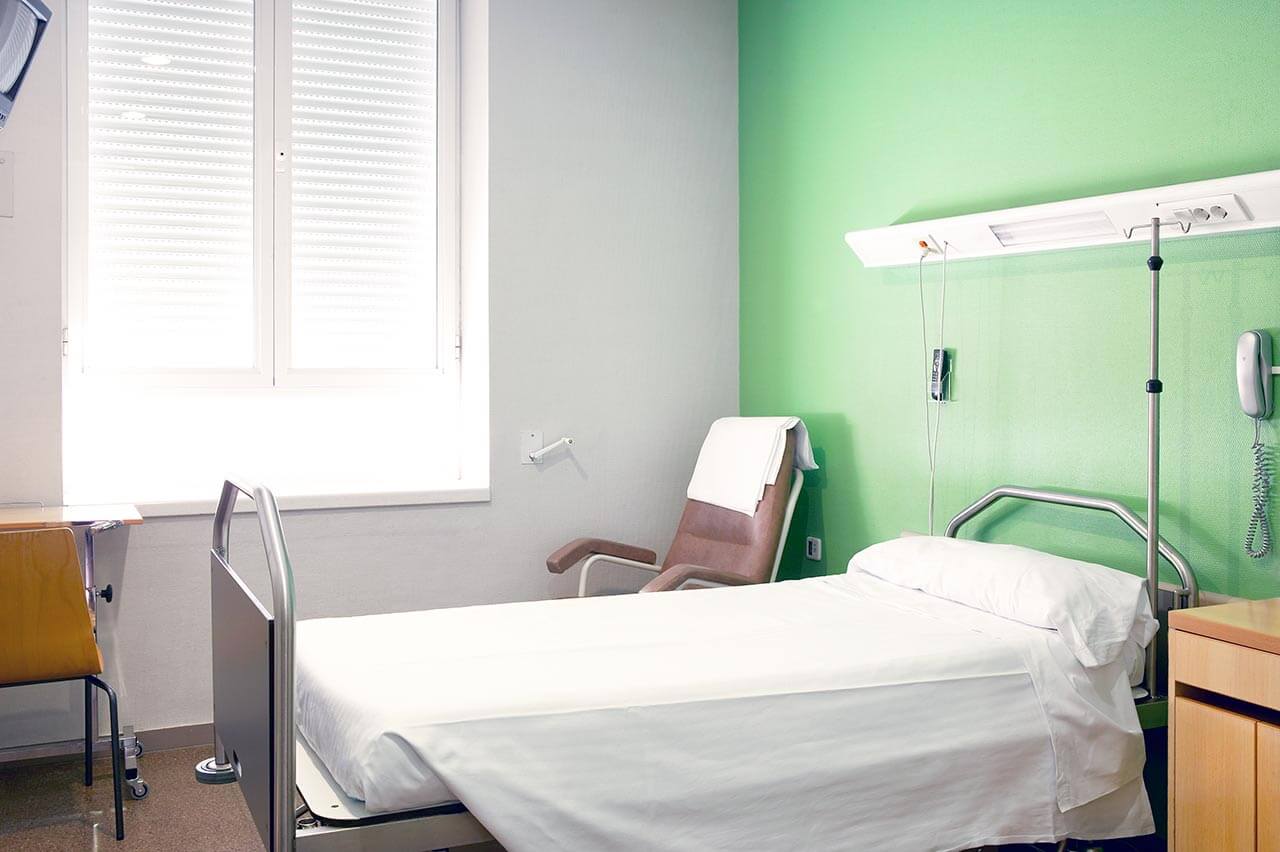
The patients of the University Hospital of Ludwig Maximilian University of Munich live in comfortable, spacious, single and double patient rooms with a modern design. Each room is equipped with an ensuite bathroom with a shower and toilet. The furnishing of a standard patient room includes a comfortable bed, the position of which can be adjusted using the remote control, a locker for storing personal belongings, a TV, and a telephone. Also, if desired, you can connect to the Internet. In addition, patients can opt for enhanced-comfort rooms, with a safe, a fridge, and upholstered furniture.
The hospital has an excellent infrastructure. The medical facility’s area houses a bank, ATMs, a hairdresser, shops with a wide range of food, drinks, newspapers, magazines, and personal hygiene items, play areas for children, and a beautiful garden for walking, etc.
Meals and Menus
The patient and his accompanying person are offered a daily choice of three menus, including a vegetarian one. If you are on a specific diet for any reason, you will be offered an individual menu. Please inform the medical staff about your dietary preferences prior to the treatment.
Further details
Standard rooms include:
Religion
Religious services are available upon request.
Accompanying person
Your accompanying person may stay with you in your room or at a hotel of your choice during the fixed program.
Hotel
You may stay at a hotel of your choice during an outpatient program. Our managers will help you to choose the best option.
The hospital offers a full range of laboratory tests (general, hormonal, tests for infections, antibodies, tumor markers, etc.), genetic tests, various modifications of ultrasound scans, CT scans, MRI and PET/CT, angiography, myelography, biopsies, and other examinations. Treatment with medications, endoscopic and robotic operations, and stereotaxic interventions are carried out here, modern types of radiation therapy are also used. The hospital offers patients all the necessary therapeutic techniques.
- Allogeneic bone marrow transplantation
- Microsurgical transplantation of head and neck tissues
- Microsurgical resection of brain tumors with intraoperative fluorescence
- Minimally invasive treatment of spine pathologies
- Joint replacement with postoperative rehabilitation (fast track program)
Patients with benign and malignant neoplasms of various localizations, pathologies of arteries and veins, herniated discs, osteoporosis, congenital and acquired pathologies of the musculoskeletal system, benign and malignant pathologies of the mammary gland, and other pathologies.
Which specialties of the University Hospital of Ludwig Maximilian University of Munich are the best?
- Interventional and diagnostic neuroradiology
- Vascular surgery
- Cardiac surgery
- Mammalogy
- Gastroenterology and hepatology
Over 1,700 highly qualified doctors work at the hospital.


