Department of Interventional Radiology - WEGE Clinic Bonn
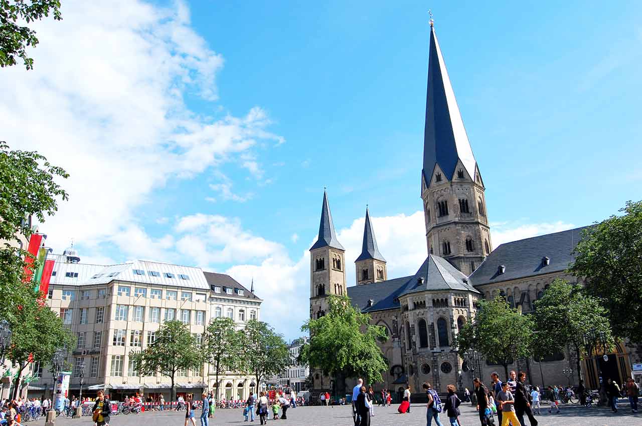
WEGE Clinic Bonn
Bonn, Germany

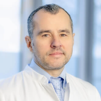
The Department of Interventional Radiology at the WEGE Clinic Bonn offers the full range of modern diagnostic procedures using ionizing radiation, image-guided tissue sampling (biopsy), and minimally invasive therapeutic procedures. Patients may be seen on an inpatient or outpatient basis. To achieve optimal results, the department works closely with specialists in radiation therapy, oncology, neurology, neurosurgery and neuro-oncology, as well as urology, orthopedics, pain management, and palliative care. This collaboration helps ensure an accurate diagnosis and the most effective treatment. The Head Physician of the department is Prof. Dr. med. Attila Kovács.
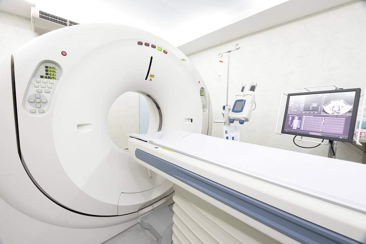
The department performs precise and safe diagnostics using ionizing radiation systems. Key diagnostic options include magnetic resonance imaging, computed tomography, radiography and digital subtraction angiography for vascular imaging. In addition, the department's experts perform all types of biopsies, including vacuum and punch biopsies. All tissue sampling procedures are performed under CT guidance (with the exception of prostate biopsies). Prostate tissue is obtained under MRI guidance. The department also performs systemic chemotherapy and immunotherapy sensitivity testing.
The department offers the widest range of image-guided interventional procedures. One of the most requested interventional procedures is transarterial chemoembolization (TACE). This is a local, minimally invasive therapy used to treat liver cancer and metastases to the kidneys or chest wall. In the early stages of liver cancer, ablation procedures can also achieve good results. However, when the tumor has reached a fairly large size or metastases have spread, the patient is prescribed transarterial chemoembolization. During this procedure, the arteries are embolized with small microspheres, which helps to cut off the supply of oxygen and nutrients to the tumor. At the same time, the microspheres deliver a high dose of a chemotherapy drug to the tumor and release it gradually. Such particles are called drug-coated granules. They allow physicians to target cancer cells more intensively. The department performs two types of transarterial chemoembolization – tumor selective and organ segment selective. Tumor-selective therapy is performed in the presence of a single metastasis in the liver, while segmental chemoembolization is performed in the presence of multiple liver lesions. The advantage of this therapy is the targeted destruction of the tumor and metastases while sparing healthy liver tissue.
The department also offers radiofrequency ablation, which uses heat to destroy the tumor. Variations of this technique include thermal ablation and microwave ablation. However, radiofrequency ablation is the most common method of thermal tumor destruction. It is used to treat malignant neoplasms and metastases in the liver, lungs, kidneys and adrenal glands, retroperitoneal lymph nodes, bones, and soft tissues. The procedure is performed under sedation (anesthesia is not required). Under CT guidance, a special probe is inserted into the body to increase the temperature of the tumor. Radiofrequency ablation allows the physician to heat the tumor cells to a temperature of 60 to 100°C. The heat affects the tumor until it is destroyed. The goal of radiofrequency ablation is to achieve the same result as classical tumor removal. The best results can be achieved with this technique for tumors up to 4 cm in size or liver tumors up to 5 cm in size.
It is worth mentioning that the doctors of the department also have excellent qualifications in kyphoplasty for the treatment of vertebral fractures (minimally invasive procedure). Kyphoplasty helps to relieve pain, and in many cases it can completely eliminate the pain syndrome. The patient lies on his stomach during the procedure. It takes about 20 minutes and is usually performed under general anesthesia. The essence of the therapeutic method is the introduction of a special cannula through the skin (at the level of the affected vertebra), which serves as a working channel for the introduction of the balloon. This balloon is filled with radiopaque fluid and inflated under constant x-ray control. It has two functions: to align the vertebra and to create a bed for the introduction of bone cement, which will help to rebuild the vertebra after the fracture. Kyphoplasty uses a special biocement that mimics real human bone tissue. Kyphoplasty has some risks (for example, damage to the nerve roots or the spinal cord itself), but the high professionalism of the department's doctors allows them to minimize these risks. To relieve symptoms and strengthen the back muscles after the therapeutic manipulation, additional drug therapy may be required.
The department specializes in the diagnostics and treatment of the following diseases:
The department's range of medical services includes:
Higher Education
Clinical and Scientific Career
Qualifications
Memberships in Professional Societies
Photo of the doctor: (c) WEGE Klinik
