



Prof. Dr. Med. Karl Jürgen Oldhafer is a first-class surgeon with extensive clinical experience. He is the Head Physician in the Department of Hepatopancreatobiliary Surgery at the Asklepios Hospital Barmbek Hamburg.
Liver surgery is his life's work. He has performed around 2,500 liver resections, most of which are particularly complex surgical procedures. Prof. Oldhafer is one of the world's most experienced experts in performing liver resections using the innovative ALPPS technique. He also successfully performs robotic liver surgery, which is characterized by excellent precision and the highest safety profile.
In this interview, we discuss new techniques and methods in liver surgery based on harnessing the liver's natural regenerative ability, the latest advances in robotic liver surgery, how personalized 3D modeling helps, innovations in liver cancer treatment, and future possibilities for non-surgical therapies.
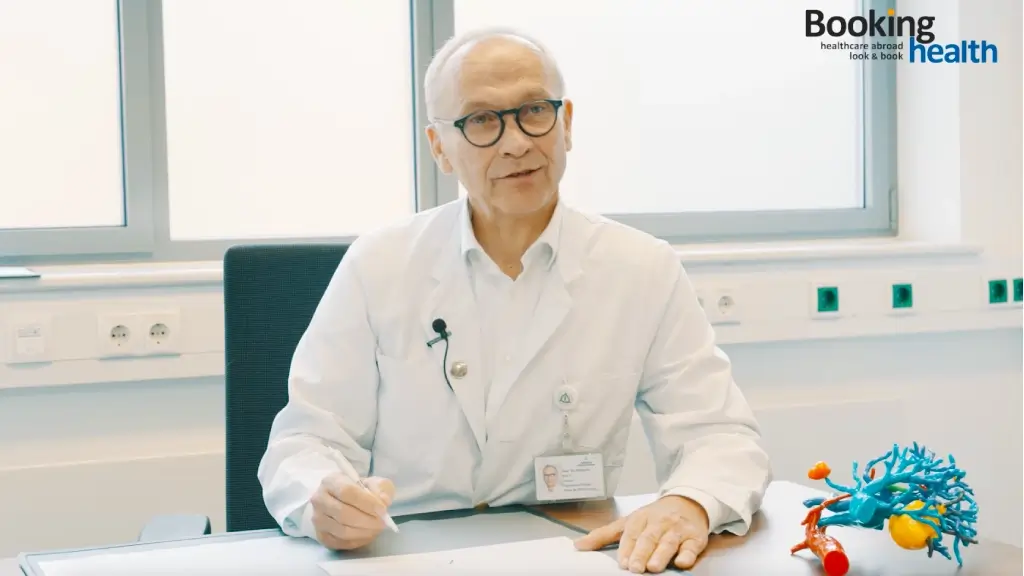
Hello, Prof. Oldhafer! Thank you for taking the time to give us an interview today. Could you please introduce yourself?
Liver surgery is my life's work. Liver surgery caught my interest at the very beginning of my professional career. I studied medicine in Goettingen, and after my graduation from the university, I started working at the Hannover Medical School. At that time, I did not yet know whether I wanted my specialty to be surgery, internal medicine, or something else. I had not yet decided on my future specialty.
I was relatively young. I was 24 years old, and at that time I started doing research in immunology. Immunology research was devoted to studying the mechanisms of the immune system work and the specifics of T-cells. At that period of time I was very interested in these questions, so I fully focused my attention on immunology. It was through my work in immunology that I met doctors from the Department of Surgery at the Hannover Medical School. At that time, this department was headed by Prof. Rudolf Pichlmayr. He was a pioneer in the field of liver transplantation and was looking for a young person to join his team who would be interested in the immunological aspects of these surgical procedures, such as transplant rejection reactions, engraftment of the donor organ, and immunological tolerance, since transplantology deals with these issues. This is how my work in immunology led me to surgery, and my first steps in this field were related to liver transplantation.
After that, I worked with abdominal surgery, but liver surgery was always present in my clinical practice. For all these years, liver surgery has been a kind of red thread for me. During my clinical training, the treatment of liver pathologies was a priority. Of course, I am also a classical abdominal surgeon and have operated on many patients with diseases of other organs, such as the esophagus, and continue to do so now, but liver surgery remains the main focus of my work.
The liver is an amazing organ. I must say that this organ is of particular interest to me personally. It is the largest gland in the human body. Amazing, isn't it? We can replace the function of almost all organs in one way or another: a person can live with one kidney, dialysis can be done, or a kidney transplant can be performed. However, a person cannot live without the liver. There are no methods to replace the function of the liver. The only option is transplant surgery.
So I have been interested in liver surgery for a long time. From the very beginning of my professional career, it has been a kind of red thread for me. Here in Hamburg, I restructured our department, which allowed me to focus exclusively on my specialty – liver and pancreatic surgery. I devote almost all of my time to this area of surgery. This gives me the opportunity to keep up with everything that is happening in the field and to implement modern technologies. It is a kind of innovative approach to work, and it is very exciting.
I have personally performed about 2,500 liver resections and assisted other surgeons in hundreds of operations. This is a tremendous experience gained over the years.
Could you please tell us about the Department of Hepatopancreatobiliary Surgery?
We treat over 200 patients annually and perform approximately 200-220 liver resections. Of course, more patients seek our medical help, but we cannot admit them all for treatment. We receive about 300-350 requests per year.
Our team consists of four specialists: the head physician and three senior physicians. They are highly specialized abdominal surgeons who exclusively perform liver and pancreatic surgery. This is unique for Germany. This is the future, because it is not easy to achieve such a narrow specialization.
It is a very laborious process, and as a medical field develops, there are more and more nuances, so you have to learn the intricacies of what you do. Nevertheless, it is a good experience for us and our colleagues: there is a sense of security, there is no fear of not knowing enough about your specialty, and there is no need to consult with colleagues. It's great to be able to work in a field you know very well. You feel fully informed, there is no discomfort, and you can make an objective assessment of the clinical case. Patients also understand on some unconscious level that they are dealing with professionals. The doctor speaks to the patient in a language he understands and explains the points of interest. And, of course, in such a case, a patient really receives the most effective treatment.
People often ask me: "Does it have to be this way?" Yes, decentralization is a long way and takes a long time. But in my opinion, it is worth it. After all, when you plan a vacation or want to buy a new car, you are looking for something special, right? You consider different options, you visit cities, and you look for something that suits you personally. So when it comes to your health, I believe you can and should go out of your way to find a better alternative.
This was an important step for me that allowed me to fully focus on liver surgery and taking care of the health of this organ.
What are some of the innovative methods and techniques that are currently available in liver surgery?
Now there are two really important and relevant directions. One of them is the method used to stimulate liver hypertrophy. The concept of "hypertrophy" implies that the liver has the ability to regenerate. When part of the liver is removed, the remaining parenchyma is able to grow. This has been known for many years. Here we can cite the example of Prometheus, who, according to legend, was chained to a rock in the Caucasus Mountains thousands of years ago; an eagle flew to him and pecked out part of his liver, but each time it regenerated. This is a story from Greek mythology, but the ability of the liver to regenerate is well known to us, and we use it.
We can stimulate liver growth because there is one problem in liver surgery: a patient undergoes resection of a liver tumor, the operation is a success, but there is little liver parenchyma left after the surgery. This is where the problem arises: the liver is unable to perform its functions, and the patient can only survive if its volume is sufficiently preserved. There is critical liver mass, after which the reduction in organ volume is unacceptable.
When that happens, the patient is at risk of dying from liver failure. We know that. We now have methods that allow us to provide compensatory hypertrophy of the liver parenchyma and stimulate its regeneration. We can do portal vein cross-section – the liver is supplied with blood from the portal vein and the hepatic artery. Unilateral cross-section or ligation of the portal vein helps stimulate the regeneration of the other lobe of the liver.
There are many ways to do this, particularly radiosurgical techniques, especially embolization or surgery. These techniques have allowed us to operate on more patients who were previously considered inoperable due to insufficient liver volume. More approaches and techniques continue to emerge. And we are very grateful to our radiologist colleagues who are increasingly performing interventional procedures before surgery.
We call these surgical interventions liver parenchyma hypertrophy stimulation procedures. This is an important area, it is developing, and we have excellent competence in this field here in Hamburg. Our clinic uses a treatment method called ALPPS, which is a surgical procedure. We have the most extensive experience in the world in performing this surgery. More than one hundred ALPPS operations have been successfully performed in our department. We are the leader in Germany in terms of the number of such surgical procedures and also one of the leading medical centers in the world in this field.
This is the first direction when we talk about new methods in liver surgery. The second is robotics. Of course, robotics is now used in all areas of surgery, and liver surgery is no exception. Robotics is very promising in this field, it is the future.
How is the ALPPS procedure performed? (a method of liver parenchyma hypertrophy stimulation)
Let's say this is the liver. This is the right lobe, and this is the left lobe. The tumor is in the right lobe of the liver, so the liver resection should be done here. If the volume of this part of the organ is insufficient, it needs to be increased. With the help of imaging diagnostics, we can accurately determine the volume of this part of the liver, for example, it is 30% or 40%. If it is too small, we do the following: the portal vein is located here; during a radiosurgical or surgical procedure, the right lobe of the liver is deprived of its blood supply, after which the blood flows from the portal vein to the left lobe and its volume increases.
As a result, this liver lobe moves to the leftmost position, increases in volume, and we can now resect this part of the liver. These operations are referred to as liver parenchyma hypertrophy stimulation or two-stage resection: first we stimulate the growth of the liver volume, and then we remove the tumor in the second stage of the surgical procedure.
We have managed to operate on many patients who were previously considered inoperable, and liver tumor resection is an important part of the therapeutic process in many cases. I cannot say that we can cure everyone and always, but we can certainly improve the prognosis and give years of life. For some patients, these operations allow them to avoid subsequent chemotherapy, and for others, they even allow for a complete recovery.
After four weeks, the liver volume increases by 90% of the initial volume – this is incredibly fast, and the remaining 10% regenerate over the following months. Typically, the waiting period for tumor resection is 4-6 weeks. This is simply unbelievable! The regenerative potential of the liver is impressive. It is truly an amazing organ. So the ALPPS procedure is a wonderful development that offers the opportunity to help more and more patients.
What has changed in liver surgery over the past ten years?
The concept of liver parenchyma hypertrophy stimulation was an important step that took many years of work, and this is not the final step. The next changes will be the use of robotics in liver surgery and minimally invasive techniques in general. These are the innovative approaches that we now have at our disposal. They have had a positive impact on the development of liver surgery. Thanks to the use of minimally invasive techniques, it is possible to provide effective medical care to patients who previously required long hospital stays. Minimally invasive surgery has many advantages, especially for patients with cirrhosis.
What is the role of robotics in liver surgery? What innovations are currently being used in robotic liver surgery?
The robotic system is a true technological miracle for the surgeon. Just imagine: the position of the camera in the robot's manipulator arms exactly matches the specified parameters; the magnification of the surgical field is tenfold, tenfold, just imagine. This means that the surgeon can really see all the anatomical structures, as if under magnifying glass, and perform the necessary manipulations.
When it comes to a liver transplant, I have been operating on patients with a surgical loupe since the beginning of my clinical practice, and this has always been the case. Pediatric liver transplants are performed on young patients who may weigh less than 10 kg. In addition, an anastomosis must be done. Since then, I have always operated with a surgical loupe. It provides 2.5 times magnification. That is already a lot, but now there is tenfold magnification. It allows us to see anatomical structures that we could not see before.
I am impressed by this because now I can see with my own eyes the small lymphatic vessels and nerve endings in the area of the hepatic portal vein. Another advantage of the robot is the secure fixation of the camera in its manipulator arm. When the surgeon holds the camera and you look at the image at tenfold magnification, the slightest tremor is noticeable, but the camera is securely fixed in the robotic arm. That's the first thing.
During the procedure, it is like diving deeper and deeper into the anatomical structures of the liver. This can be compared to diving in a submarine. In the first stage of the dive, you see the liver, then you go deeper and you see the vena cava, and then you see the hepatic veins. And it is very real because the robotic arms hold the camera in such a way that the surgeon has a completely new view of the anatomical structures of the liver. That is another thing that impresses me. It also speaks to the great future of robotic surgery.
Next, the robot is a telemanipulator. This is not like in the automotive industry, where you put a sheet of metal into an automated machine to make a car fender, and the robot does it all by itself. That's not the case here. The robot is actually a manipulator, or rather a telemanipulator, because it is following the surgeon's commands, so the term "robot" is somewhat misleading in this case.
Importantly, the robot is equipped with four manipulator arms. On top of that, the device has imaging capabilities: you look through the camera and can use navigation. We use the fluorescence technique. During surgery, a fluorescent substance is injected, and the camera has a function to detect it. The way it works is similar to a thermal imager.
So by staining the liver tissue with a fluorescent substance, the surgeon can see the things that he couldn't see before. So for surgeons, this is probably like navigation for an aircraft pilot at night. For us, navigation is a real achievement, and the field is just beginning to develop. All of the ITs in imaging and scan processing are evolving. This will also have an impact on robotic surgery.
Today, it is already possible to use artificial intelligence to recognize anatomical structures by analyzing scans. For example, it is possible to color code or color label a blood vessel by staining it white. We are still at the very beginning of the journey. I believe that this technology is very promising in liver surgery as well as advanced medical imaging techniques combined with robotics.
Could you please tell us about 3D printing technology in liver surgery and its importance in planning surgical procedures for complex liver tumors?
This is a model of the liver of the patient we will be operating on tomorrow. This model was created based on a data set, an analysis of CT and MRI scans. In other words, it's a physical model of the patient's liver. Red is the abdominal aorta, blue is the vena cava, and yellow is the tumor that we will remove during surgery tomorrow.
These are colorectal cancer metastases. Thanks to 3D printing technology, we have the opportunity to prepare for the operation, including together with the patient: everything is clearly visible on this model, and the patient understands exactly what the problem is. The model is also useful for discussing the clinical case with colleagues. For example, here is the right hepatic vein, which in this case we will need to remove tomorrow, but there is another vein coming out of the vena cava below that needs to be preserved for local tumor resection. Typically, removing the right hepatic vein means blocking the blood supply to the entire right lobe of the liver, so you have to remove it completely because it has no blood supply.
However, this example shows that part of the right lobe of the liver is supplied by the middle hepatic vein. This means that with the help of 3D technology we were able to develop a personalized treatment regimen for this patient and will put it into practice tomorrow. This is another advantage of this technology. So I am very impressed with 3D technology. Even after many years of working in this field and performing a huge number of liver surgeries, this technology is still an important achievement for me. Advanced technology really opens up great opportunities for us.
We send the data to the institute, and it takes about 3-4 days to make the liver model. You can even take it with you to the operating room. The model is sterilized and placed on the operating table, and the surgeon can check it again. This is really good technology. At first glance, it may seem like a toy, just some colorful thing. The first time I saw such a model, I thought so too, but after using it for the first time, I realized that it is a really useful thing.
Using a model allows us to increase the safety of the surgery because we can better prepare for it. Of course, it is not needed to remove simple tumors. If the tumor is superficial and we have to remove it here, in this case we are talking about a cyst, we do not need such a model. However, if we are dealing with complex tumors, where we have to take into account the location of blood vessels, reconstruct them, or remove them, then the model plays a very important role.
In such complex cases, the operation lasts 4-5 hours. The duration of the operation is shortened by the fact that with the help of the model it is possible to think about the course of the operation in advance and to start it immediately, instead of assessing the situation after the inspection of the surgical field.
In addition to the ability to prepare for surgery, 3D printing technology has the advantage of allowing the surgeon to assess whether it is advisable to perform the operation at all. In addition, the model helps during surgery if there are problems with orientation in the surgical field. So its use certainly affects the duration of the surgery and the preparation for it in general, because you already have a specific plan in mind and you can start working right away.
What is the current state of preoperative planning and simulation modeling programs in liver surgery?
I have been using simulation modeling software for many years, also in cooperation with other medical facilities. The programs and algorithms are becoming more and more reliable and efficient. Previously, many things were not so automated, so you still had to do a lot of things yourself. Modern simulation modeling software is getting better and better. It is now possible to simulate the course of an operation on the computer with the calculation of the necessary and possible volume of resection.
With which physicians from related medical specialties do you work in interdisciplinary collaboration to provide comprehensive medical care to patients?
A Liver Surgery Center like ours needs to have an advanced Department of Radiology because the field is actively developing now and new CT and MRI technologies are emerging. In fact, medical imaging is also a basic condition for creating such models. The better the quality of the imaging, the better the quality and accuracy of the liver model. The clarity of the scans plays a key role in the quality of the 3D printing.
So the Department of Radiology is a very important partner for us. Embolization, which is the interventional procedure that we talked about earlier in the context of liver hypertrophy stimulation techniques, is also a radiological intervention. We work with the Department of Radiology for other therapeutic tasks for the treatment of liver tumors, for example, to do embolization or chemoembolization. These procedures should be available in a center like ours.
Gastroenterology and endoscopy are also important: these are endoscopic procedures on the liver, ERCP, and stenting. Sometimes there can be complications. Unfortunately, no one is immune to this, even when an experienced specialist is working with the patient, but it is not a big deal if the complications can be managed quickly. The development of complications is not a catastrophe. If they do occur, they only complicate the therapeutic process a little. In such cases, patients need medical care from a specialized doctor. This is an important point in establishing centers like ours. Therefore, an important partner of the hepatobiliary surgeon is a gastroenterologist specializing in endoscopy. Our center takes all these aspects into account. I am very proud of our clinic because I and my colleagues from related medical specialties work hand in hand. The treatment of liver diseases is not the exclusive domain of our surgical department, but radiologists and therapists are involved in the process. So there are many specialties involved in the treatment of liver pathologies.
We also use methods of postoperative treatment, especially for patients with oncological diseases. We provide postoperative care, but many patients come to us from other regions of the country, so, of course, we support cooperation with interregional centers.
What are the new trends in liver cancer treatment?
In addition to purely surgical developments in robotics and new methods of scan processing, incredible progress is also being made in oncology. Immunotherapy is a very important area where outstanding successes have been achieved.
Take lung cancer, for example. It has been known for many years that lung cancer, once considered a difficult-to-treat oncology, is now well treatable with modern immunotherapy, with encouraging long-term results.
As for liver tumors, there are two main types of neoplasms: the first is HCC (hepatocellular carcinoma) and the second is CCC (cholangiocellular carcinoma). HCC is, so to speak, a "new lung cancer" or the same oncology as lung cancer, but with abdominal organ involvement. Many immunotherapy methods have been developed to fight it. These tumors respond better to immunotherapy and with good results. This changes the treatment approaches for the better and also gives a chance to improve the long-term prognosis. So immunotherapy is a very important part of the treatment in these cases. It also changes the approach to the surgical treatment because there is an opportunity to operate on more patients or to ensure a good long-term outcome by combining it with other cancer treatments. This is why immunotherapy is such an important discovery for the future of medicine.
Are patients receiving more effective treatment as a result of advances in personalized medicine?
Each tumor has its own peculiarities, so it is necessary to determine the specific type of neoplasm and its characteristics and then prescribe modern treatment based on it. This also applies to liver tumors, including HCC.
The more we know about the tumor, the more accurate the diagnosis will be, and the more effective and personalized the treatment will be.
Take, for example, a liver adenoma, a benign tumor that is most common in women. It is now possible to divide it into subtypes based on genetic testing data and to assess the risk of its degeneration into a malignant tumor. This allows us to develop a personalized treatment regimen.
What do you think about the future of gene therapy?
Many pathological changes can develop in the liver. These also include metabolic disorders that affect the liver.
Gene therapy is not my specialty, but it is an important area to explore in order to be able to treat metabolic diseases based on their underlying causes. I hope to see the success of gene therapy in this field. It is certainly an interesting field, because there are so many metabolic diseases, such as Wilson's disease, and many others. We know exactly how they develop and progress and what pathological changes they cause. I hope that in the future gene therapy methods will be developed to treat such diseases.
What is your vision for the future of liver surgery and the treatment of liver disease?
I believe that immunotherapy opens great prospects for us. There is more and more information about the possibility of treating various abdominal tumors with immunotherapy. On the other hand, we now have better technical capabilities at our disposal and can perform complex surgical procedures.
Two weeks ago I was asked to give a lecture on "Liver surgery yesterday, today, and tomorrow". It was a general lecture. I divided the development of liver surgery into eras.
The first era was from the origins until the 17th century. At that time, liver surgery was not performed but only the anatomy of the organ was studied: the structure of the liver, its anatomical and physiological features, and the bile ducts. This era was called "Liver surgery without liver surgery".
Then came the era of practical application of knowledge. In 1890, the first liver resection was performed, and these operations were performed repeatedly thereafter to evaluate their efficacy. This continued until 1915. This era was called the "Era of surgical techniques". Its legacy is all the liver operations available today.
The pinnacle of liver surgery development is ex situ extracorporeal resection: the surgeon operates on the liver inside the patient's body, but there are problematic, hard-to-reach areas, time is needed for reconstructive measures; the liver is then removed from the patient's body and placed on a special operating table, after which the operation continues outside the patient's body; this provides an approach to anatomical structures from below and above, and vascular reconstruction can be performed.
This operation uses the same principle as transplantation surgery: the liver is cooled with saline solution to gain time, because it must be reimplanted after resection. This operation is called autotransplantation. This is the apogee of liver surgery, the limit of surgical possibilities in this field.
The first such resection was performed by Prof. Pichlmayr, my director, in 1988. I was still a young assistant physician at the time and just observed the course of the surgical procedure. And since then, we have robotics, artificial intelligence, and medical imaging. Now we are living in an era where we are trying to somehow simplify the existing capabilities, to make new technologies simpler, and to make them as accessible as possible for patients, without causing serious complications. This is the time we live in.
What will the next era be like? Where are we heading? In my opinion, from 2100 onwards, there will be an era of the abandonment of surgery, a kind of collapse of the current system, so to speak. Until then, other treatments, such as immunotherapy and gene therapy, will be so advanced and effective that the services of liver surgeons may not be needed at all. But we are still a long way off from that. There is no doubt that patients will need our help for the next 30 to 40 years. I am sure of it.
Dear Prof. Oldhafer, thank you very much for the informative interview!