The program includes:
- Initial presentation in the hospital
- Clinical history taking
- Review of available medical records
- Physical examination
- Laboratory tests:
- Complete blood count
- General urine analysis
- Biochemical analysis of blood
- Tumor markers
- Inflammation indicators (CRP, ESR)
- Coagulogram
- Ultrasound scan
- CT scan / MRI
- Preoperative care
- Embolization or chemoembolization, 2 procedures
- Symptomatic treatment
- Cost of essential medicines
- Nursing services
- Elaboration of further recommendations
How program is carried out
During the first visit, the doctor will conduct a clinical examination and go through the results of the available diagnostic tests. After that, you will undergo the necessary additional examination, such as the assessment of liver and kidney function, ultrasound scan, CT scan and MRI. This will allow the doctor to determine which vessels are feeding the tumor and its metastases, as well as determine how well you will tolerate the procedure.
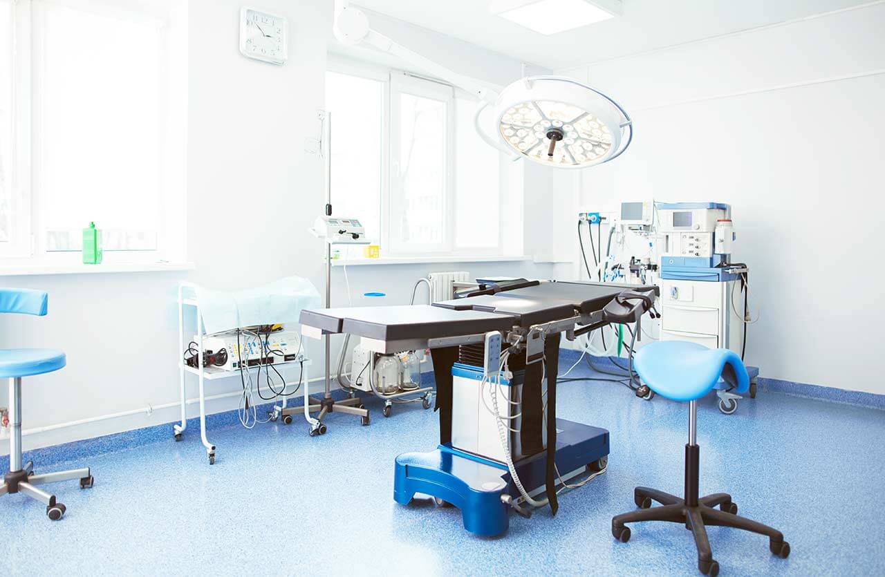
Chemoembolization begins with local anesthesia and catheterization of the femoral artery. The thin catheter is inserted through a few centimeters long incision of the blood vessel. The doctor gradually moves the catheter to the vessel feeding the primary tumor or its metastases. The procedure is carried out under visual control, an angiographic device is used for this. The vascular bed and the position of the catheter in it are displayed on the screen of the angiograph.
When the catheter reaches a suspected artery, a contrast agent is injected through it. Due to the introduction of the contrast agent, the doctor clearly sees the smallest vessels of the tumor and the surrounding healthy tissues on the screen of the angiograph. After that, he injects emboli into the tumor vessels through the same catheter.
Emboli are the spirals or the liquid microspheres. The type of embolus is selected individually, taking into account the diameter of the target vessel. When carrying out chemoembolization, a solution of a chemotherapy drug is additionally injected into the tumor vessel. Due to the subsequent closure of the vessel lumen with an embolus, the chemotherapy drug influences the tumor for a long time. In addition, the drug does not enter the systemic circulation, which allows doctors to use high doses of chemotherapeutic agents without the development of serious side effects. Chemoembolization leads to the destruction of the tumor or slowing down its progression.
After that, the catheter is removed from the artery. The doctor puts a vascular suture on the femoral artery and closes it with a sterile dressing. During chemoembolization, you will be awake. General anesthesia is not used, which significantly reduces the risks of the procedure and allows performing it on an outpatient basis, avoiding long hospital stay.
After the first procedure, you will stay under the supervision of an interventional oncologist and general practitioner. If necessary, you will receive symptomatic treatment. As a rule, a second chemoembolization procedure is performed in 3-5 days after the first one in order to consolidate the therapeutic effect. After that, you will receive recommendations for further follow-up and treatment.
Service
You may also book:
 BookingHealth Price from:
About the department
The Department of Interventional Radiology and Neuroradiology at the Schlosspark Hospital Berlin provides all the options of modern imaging diagnostics and also specializes in image-guided therapeutic interventional procedures, including those for treating vascular diseases of the nervous system. The department's diagnostic rooms are equipped with state-of-the-art systems for digital X-ray, fluoroscopy, computed tomography, magnetic resonance imaging, and angiography. The department performs numerous highly effective and minimally invasive interventional procedures under imaging guidance for treating vascular stenosis and occlusions in all locations, internal bleeding, tumors, brain aneurysms, brain arteriovenous malformations, strokes, etc. Patients with neurovascular diseases are treated at a specialized center that is part of the department. Neuroradiologists, neurologists, and neurosurgeons cooperate closely here to provide patients with the best possible therapy recommended by a multidisciplinary team specializing in the treatment of neurological disorders. The department's patients will benefit from a modern and comfortable infrastructure as well as a highly professional and caring medical team. The Head Physician of the department is Dr. med. Annette Förschler.
In the field of interventional radiology, the department most often performs percutaneous transluminal balloon angioplasty. This minimally invasive procedure can restore vascular patency in cases of stenosis or occlusion. The procedure is widely used to dilate the coronary arteries, carotid arteries, renal arteries, femoral artery, and abdominal aorta. At the initial stage of the intervention, the doctor makes a skin puncture in the groin and inserts a special guidewire with a balloon attached to it into the vascular system. The guidewire is delivered to the area of arterial narrowing or occlusion under imaging guidance, after which a balloon is placed. The inflating balloon restores the patency of the artery, due to which the blood flow is normalized. The final stage of the endovascular procedure is stent implantation to prevent recurrent vascular occlusion. Percutaneous transluminal balloon angioplasty is performed under local anesthesia. Thus, the interventional procedure is minimally traumatic but highly effective and poses almost no risks to the patient's health.
The department's interventional radiologists have exceptional professional skills in performing embolization for benign and malignant tumors. Embolization is a minimally invasive procedure that involves the cessation of blood supply to the tumor by injecting blocking embolic agents into the blood vessels that supply the tumor. Patients with malignant tumors are also offered transarterial chemoembolization. This procedure is similar to classic embolization but involves the use of emboli combined with a chemotherapy agent. The tumor is therefore deprived of a blood supply and exposed to chemotherapy drugs. Embolization can be used as a stand-alone treatment or as a complement to a surgical procedure. For example, embolization prior to surgery allows surgeons to remove the entire large neoplasm by shrinking it beforehand. In some cases, embolization can also be used as a palliative measure. The procedure is performed under angiographic guidance and local anesthesia. In most cases, the targeted blood vessel is approached through a puncture in the femoral artery. Embolization is indicated for patients with liver, kidney, lung, uterine, prostate, and head and neck tumors.
The department also performs many interventional procedures for neurovascular diseases, including thrombolysis and mechanical thrombectomy for stroke, carotid balloon angioplasty with stenting, clipping for aneurysms, treatment of vascular malformations, and removal of angiomas, angiolipomas, and cavernomas. The department's specialists also offer interventional pain management procedures for chronic back pain, such as periradicular therapy and facet joint block. All interventional procedures are performed under imaging guidance, so the patient can be confident in the safety of the treatment. The optimal treatment for a neurovascular disease is determined by a special board involving neuroradiologists, neurologists, and neurosurgeons. The specialists cooperatively study the patient's clinical case and develop the most effective course of treatment.
The department's range of therapeutic services includes:
- Interventional radiology
- Therapeutic procedures
- Percutaneous transluminal balloon angioplasty followed by stent implantation
- Tumor embolization
- Embolization for internal bleeding
- Transarterial chemoembolization
- Percutaneous transhepatic biliary drainage
- Radiofrequency catheter ablation
- Therapeutic procedures
- Interventional neuroradiology
- Therapeutic procedures
- Thrombolysis and mechanical thrombectomy for stroke
- Carotid balloon angioplasty with stenting
- Clipping for aneurysms
- Endovascular repair for vascular malformations
- Endovascular removal of angiomas, angioblastomas, and cavernomas
- Image-guided interventional treatment for chronic back pain
- Periradicular therapy
- Facet joint block
- Therapeutic procedures
- Other therapeutic options
Curriculum vitae
Higher Education and Postgraduate Training
- 1994 - 2001 Medical studies at the Humboldt University of Berlin.
- 2003 Admission to medical practice.
- 2003 - 2007 Advanced training, Department of Diagnostic and Interventional Radiology, University Hospital Leipzig.
- 2008 Board certification in Radiology.
- 2009 Doctoral thesis defense, Technical University of Munich. Subject: "Research of the role of a new diagnostic method for brain tumors using functional magnetic resonance imaging with BOLD contrast enhancement".
- 2011 Specialization in Neuroradiology.
- 2014 Certified Instructor of the German Society for Interventional Radiology (DeGIR) and the German Society for Neuroradiology (DGNR).
Professional Career
- 2002 - 2003 Internship, Department of Radiology and Neuroradiology, Charite University Hospital Berlin.
- 2007 Scientific paper in the Fraunhofer Institute for Cell Therapy and Immunology, Leipzig.
- Since 01.04.2008 Senior Physician, Department of Neuroradiology, University Hospital Rechts der Isar Munich.
- 2010 - 2012 Managing Senior Physician, Department of Neuroradiology (focus on image-guided interventions for neurovascular diseases and functional imaging in neuro-oncology), University Hospital Rechts der Isar Munich.
- 2013 - 2016 Deputy Head Physician, Department of Interventional Neuroradiology (focus on image-guided interventions for neurovascular diseases and functional imaging in neurooncology), Vivantes Neukölln Hospital.
- Since 2016 Head Physician, Department of Interventional Radiology and Neuroradiology at the Schlosspark Hospital Berlin.
Research Focuses
- Multimodal imaging in neuro-oncology.
- Interventional treatment for stroke.
Memberships in Professional Societies
- German Society for Interventional Radiology (DeGIR).
- German Society for Neuroradiology (DGNR).
- German Radiological Society (DRG).
- European Society of Neuroradiology (ESNR).
- European Society of Radiology (ESR).
Photo of the doctor: (c) Schlosspark-Klinik GmbH
About hospital
The Schlosspark Hospital Berlin began its work in 1970 and, during this time, has gained an excellent reputation not only in Germany but also in the international medical arena. The Schlosspark Hospital Berlin is an academic hospital of the Charite University Hospital Berlin, which is one of the best medical centers in Europe and throughout the world. The successful clinical practice of the medical facility is based on an advanced medical and technical base, access to the very latest and most effective treatment methods, and the exceptional competence and experience of medical personnel. The hospital is located in the picturesque Charlottenburg Park, away from the hustle and bustle of the city, which contributes to the peace of mind of patients.
The hospital's bed fund includes 340 beds. Structurally, the hospital consists of 12 specialized departments with narrowly focused centers integrated into them, which are responsible for the treatment of a particular group of diseases. More than 14,000 patients are treated annually at the hospital. One of the priorities of the medical facility is emergency medical care. With an annual number of more than 10,000 surgical interventions, including operations of high complexity, the department is particularly interested in surgical treatment.
The hospital's medical team consists of more than 900 employees. The departments at the hospital are headed by professors who are well-known in the medical community and have impressive clinical experience and outstanding professional achievements. In the course of diagnostics and treatment, an individual approach is used for each patient, and his needs and wishes are also taken into account. The hospital pays due attention to patient care during the therapeutic process. Despite state-of-the-art medical equipment and the automation of many processes, a humane and respectful attitude towards the patient is the first priority for the doctors at the hospital. The specialists are always open to dialogue and do their best to achieve the best treatment outcomes.
Photo: (с) depositphotos
Accommodation in hospital
Patients rooms
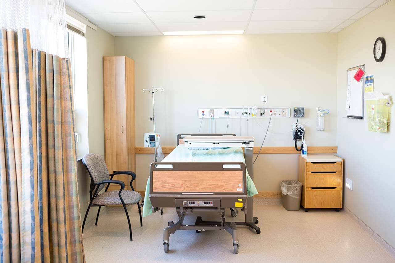
The patients of the Schlosspark Hospital Berlin live in comfortable rooms with light colors. Each patient room has an ensuite bathroom with a shower and a toilet. The standard patient room includes a comfortable automatically adjustable bed, a bedside cabinet with a pull-out table, a wardrobe, a table and chairs for receiving visitors, a telephone, and a TV. Wi-Fi is available on the territory of the hospital. The hospital also offers enhanced-comfort rooms with a more exquisite design.
Meals and Menus
The patient and their accompanying person are offered three meals a day: breakfast, lunch, and dinner. A new menu is prepared for patients each week. Only high-quality and fresh products are used for cooking meals. The menu includes dietary and vegetarian dishes. The patient will be offered an individual menu if necessary.
There is a cozy cafe on the ground floor of the hospital that serves full breakfasts, a wide range of hot and cold drinks, delicious desserts, and snacks.
Further details
Standard rooms include:
Accompanying person
The accompanying person may stay with you in the patient room or at the hotel of your choice during the inpatient program.
Hotel
You may stay at the hotel of your choice during the outpatient program. Our manager will help you choose the best option.


