About the Department of Interventional Radiology at Specialized Pulmonary Clinic Asklepios Munich-Gauting
The Department of Interventional Radiology at the Specialized Pulmonary Clinic Asklepios Munich-Gauting offers the full range of medical services in the area of its specialization. The department successfully performs modern imaging examinations of the thoracic cavity, abdominal cavity, head and neck, and spine. The key focus in the medical facility is on computed tomography (CT), including CT angiography, high-resolution CT, multiphase CT, and dynamic CT. The department has state-of-the-art equipment for chest X-rays. The medical facility also performs interventional procedures under imaging guidance, such as CT-guided lung puncture for suspected malignant processes; CT-guided puncture for detecting metastases in the liver, kidneys, and adrenal glands; CT-guided pleural puncture for pleurisy; CT-guided drainage for removing air or fluid from the pleural cavities or pericardium. All diagnostic and therapeutic image-guided procedures are carried out in strict accordance with the requirements of professional medical societies and in compliance with radiation protection standards. The department's specialists guarantee accurate diagnostics for each patient, which is the key to developing an optimal treatment regimen and achieving the desired therapeutic result. The Head Physician of the department is Prof. Dr. med. Julien Dinkel.
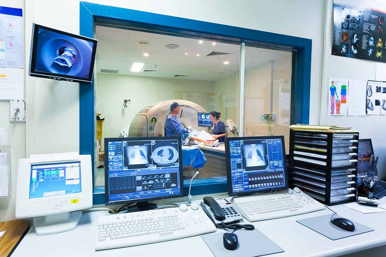
The team of the department's doctors cooperates closely with pulmonologists and thoracic surgeons at the Specialized Pulmonary Clinic Asklepios Munich-Gauting, providing high-precision examinations of the chest organs. The department has state-of-the-art equipment for X-ray diagnostics. Computed tomography (CT) is of particular interest. CT scanning is a highly informative diagnostic method based on the use of X-rays. With the help of CT scans, the department's specialists obtain 3D images of the respiratory organs, which are impossible with conventional X-rays. The department's modern multislice CT system allows doctors to perform high-resolution computed tomography (HRCT) of the lungs to examine and assess therapeutic results in patients with interstitial lung diseases, computed tomography angiography to visualize the blood vessels in the head, neck, chest, and abdomen, multiphase computed tomography to detect lung and airway tumors, dynamic computed tomography to diagnose spontaneous pneumothorax, etc. Whenever required, PET-CT scanning can be used. This is a diagnostic method that combines positron emission tomography and computed tomography. The results of the performed imaging tests are stored in a special picture archiving and communication system (PACS), so that all doctors can quickly access the necessary diagnostic data.
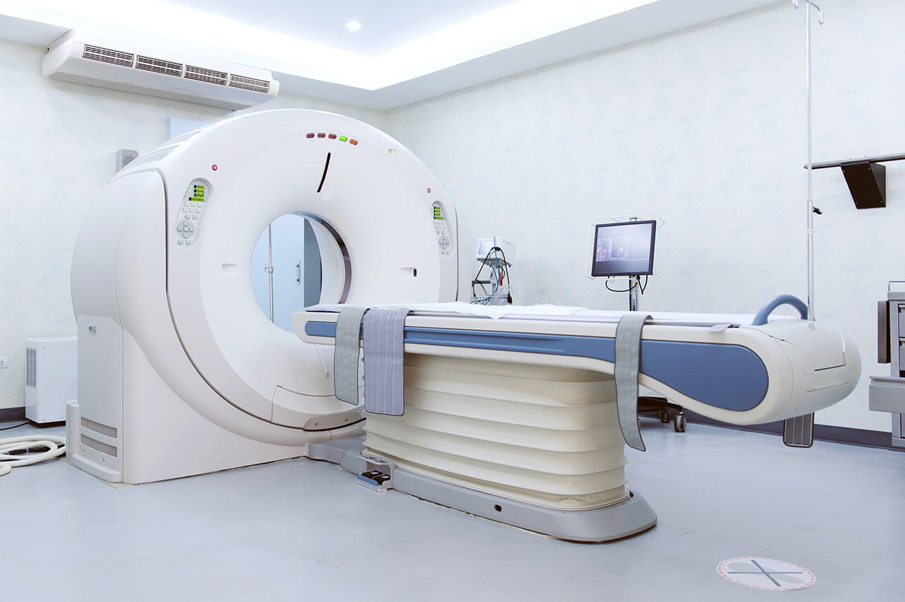
A CT scan is a painless procedure that only takes a few minutes. Before the scan, a patient takes a prone position on the table of the CT machine, after which a doctor gives him the necessary instructions. As a rule, during the examination of the respiratory system, a patient needs to hold his breath several times. The department uses an open-type CT device, which contributes to the comfort of patients with claustrophobia (fear of enclosed spaces).
The department also regularly performs CT-guided interventional diagnostic procedures, including CT-guided lung puncture for suspected malignancy, CT-guided puncture for detecting metastases in the liver, kidneys, and adrenal glands, CT-guided pleural puncture for pleurisy, etc. Such procedures are usually indicated in cases where a bronchoscopic examination does not ensure reliable data necessary for making an accurate diagnosis. Punctures are performed under local anesthesia, so they are completely painless. During the procedure, a thin biopsy needle is used, which is advanced by an interventional radiologist under imaging guidance to the pathological focus to obtain a tissue sample. The use of CT imaging during the puncture guarantees high accuracy of the procedure, minimizing the risk of damage to the adjacent organs.
Image-guided therapeutic procedures are also available in the department. The most popular of these is CT-guided drainage to remove air or fluid from the pleural cavities. For example, this interventional treatment is indicated for patients with pneumothorax, in which air enters the pleural cavity, which subsequently leads to partial or total lung collapse. The essence of the procedure is to place a drainage pleural tube through which air is removed from the pleural cavity. The tube is inserted under local anesthesia. The use of CT imaging allows the department's doctors to carry out the placement as accurately as possible as well as control the process of removing air from the pleural cavity in real time. As a rule, the procedure gives good results because, following it, patients do not experience any respiratory disorders and associated discomfort.
The department's range of medical services includes:
- Diagnostics
- Non-invasive examinations
- X-ray
- Classical computed tomography
- High-resolution computed tomography (HRCT) of the lungs
- Computed tomography angiography
- Multiphase computed tomography
- Dynamic computed tomography
- Positron emission tomography combined with computed tomography (PET-CT)
- Interventional examinations
- CT-guided lung puncture for suspected malignant tumors
- CT-guided puncture for detecting metastases in the liver, kidney, and adrenal glands
- CT-guided pleural puncture for pleurisy
- Preoperative labeling of pathological foci
- Non-invasive examinations
- Treatment
- CT-guided drainage for removing air or fluid from the pleural cavities
- Other diagnostic and treatment methods
Curriculum vitae
Higher Education and Postgraduate Training
- 10.1997 - 10.2003 Medical School, Louis Pasteur University, Strasbourg, France.
- 09.1999 - 07.2003 Master, Louis Pasteur University, Strasbourg, France.
- 01.2010 Thesis defense with honors, University of Heidelberg, Germany. Subject: "4D multislice CT scans of the lungs: qualitative comparison and reproducibility of small volumes in an ex vivo model".
- Since 10.2011 Research habilitation program, Medical School, University of Heidelberg, Germany.
- Since 10.2014 Full Professor for Thoracic Imaging, Ludwig Maximilian University of Munich.
Professional Career
- 01.2004 - 06.2005 Residency, Department of Radiation Oncology, University Hospital Heidelberg.
- 01.07.2005 Admission to medical practice, Stuttgart, Germany.
- 07.2005 - 08.2006 Residency, Department of Radiology, University Hospital Heidelberg.
- 09.2006 - 07.2011 Residency, German Cancer Research Center, Heidelberg.
- 08.2011 Board certification in Radiology, Germany.
- 08-10.2011 Radiologist, German Cancer Research Center, Heidelberg.
- 11.2011 - 02.2013 Postdoctoral Research Fellowship, Department of Radiology, Massachusetts General Hospital, Harvard Medical School, Boston, USA.
- 03.2013 - 09.2013 Radiologist, Department of Radiology, University Hospital Heidelberg.
- 10.2013 - 09.2014 Radiologist, Department of Radiology (focus on Thoracic Surgery), University Hospital Heidelberg.
- Since 10.2014 Full Professor for Thoracic Imaging, Radiologist, Department of Radiology, University Hospital of Ludwig Maximilian University of Munich.
- Since 01.2018 Head Physician of the Department of Interventional Radiology at the Specialized Pulmonary Clinic Asklepios Munich-Gauting.
- Since 12.2021 Head of the Section of Cardiovascular Imaging, Department of Radiology, University Hospital of Ludwig Maximilian University of Munich.
Awards and Honors
- 2007 Poster Award, German Society for Radiation Oncology (DEGRO).
- 2008 Poster Award from the Alumni Association of the German Cancer Research Center.
- 2009 Poster Award, Journées françaises de radiologie.
- 2010 Research Award, Experimental Radiology.
Clinical Interests
- Chest imaging.
- Oncologic imaging.
- CT and MR imaging protocols; ultrafast imaging.
- Functional imaging: DTI, DWI, PWI; motion assessment, DECT.
- Phase-contrast imaging.
Photo of the doctor: (c) Asklepios Fachkliniken München-Gauting


