The program includes:
- Initial presentation in the clinic
- clinical history taking
- review of medical records
- physical examination
- laboratory tests:
- complete blood count
- general urine analysis
- biochemical analysis of blood
- TSH-basal, fT3, fT4
- tumor markers (AFP, CEA, СА-19-9)
- inflammation indicators (CRP, ESR)
- indicators of blood coagulation
- abdominal ultrasound scan
- CT/MRI of abdomen
- preoperative care
- percutaneous embolization (coiling) or chemoembolization
- symptomatic treatment
- cost of essential medicines
- nursing services
- elaboration of further recommendations
Indications
- Inoperable liver metastases
- Poor response to systemic chemotherapy
Treatment is not indicated in:
- Presence of extrahepatic metastases
- Affection of more than 70% of the liver
How program is carried out
During the first visit, the doctor will conduct a clinical examination and go through the results of the available diagnostic tests. After that, you will undergo the necessary additional examination, such as the assessment of liver and kidney function, ultrasound scan, CT scan and MRI. This will allow the doctor to determine which vessels are feeding the tumor and its metastases, as well as determine how well you will tolerate the procedure.
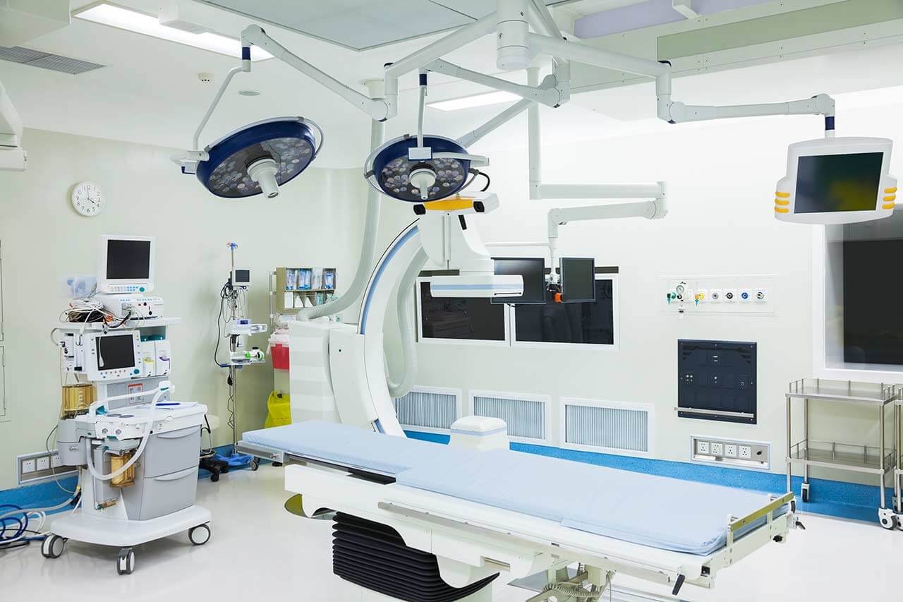
Chemoembolization begins with local anesthesia and catheterization of the femoral artery. The thin catheter is inserted through a few centimeters long incision of the blood vessel. The doctor gradually moves the catheter to the vessel feeding the primary tumor or its metastases. The procedure is carried out under visual control, an angiographic device is used for this. The vascular bed and the position of the catheter in it are displayed on the screen of the angiograph.
When the catheter reaches a suspected artery, a contrast agent is injected through it. Due to the introduction of the contrast agent, the doctor clearly sees the smallest vessels of the tumor and the surrounding healthy tissues on the screen of the angiograph. After that, he injects emboli into the tumor vessels through the same catheter.
Emboli are the spirals or the liquid microspheres. The type of embolus is selected individually, taking into account the diameter of the target vessel. When carrying out chemoembolization, a solution of a chemotherapy drug is additionally injected into the tumor vessel. Due to the subsequent closure of the vessel lumen with an embolus, the chemotherapy drug influences the tumor for a long time. In addition, the drug does not enter the systemic circulation, which allows doctors to use high doses of chemotherapeutic agents without the development of serious side effects. Chemoembolization leads to the destruction of the tumor or slowing down its progression.
After that, the catheter is removed from the artery. The doctor puts a vascular suture on the femoral artery and closes it with a sterile dressing. During chemoembolization, you will be awake. General anesthesia is not used, which significantly reduces the risks of the procedure and allows performing it on an outpatient basis, avoiding long hospital stay.
After the first procedure, you will stay under the supervision of an interventional oncologist and general practitioner. If necessary, you will receive symptomatic treatment. As a rule, a second chemoembolization procedure is performed in 3-5 days after the first one in order to consolidate the therapeutic effect. After that, you will receive recommendations for further follow-up and treatment.
Required documents
- Medical records
- Abdominal ultrasound (if available)
- MRI/CT scan of the abdomen (if available)
- Biopsy results (if available)
Service
You may also book:
 BookingHealth Price from:
About the department
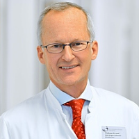
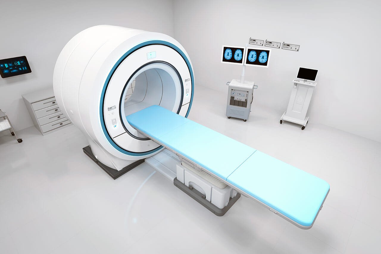
The Department of Interventional Radiology at the ViDia Hospital Karlsruhe offers the full range of medical services in the areas of its specialization. The department's team of doctors performs imaging diagnostics and image-guided therapeutic interventional procedures. The department has state-of-the-art equipment, which allows the doctors to perform the full range of imaging tests, such as X-ray, sonography, mammography, angiography, computed tomography, and magnetic resonance imaging, as well as PET-CT and SPECT-CT (in collaboration with the nuclear medicine specialists). As for the department's therapeutic offer, the focus is on endovascular treatment of vascular diseases and cancer. The department has an emergency service that provides 24-hour care for patients with acute bleeding, vascular obstructions, and other urgent conditions. Diagnostic examinations and most therapeutic procedures are performed on an outpatient basis without mandatory hospitalization. The department's doctors are always open to dialog with the patient and tell them in detail about the upcoming procedure and its expected results. To store the results of imaging tests, the department uses a modern picture archiving and communication system (PACS), which allows for unhindered access to images for doctors in all departments of the hospital. The Head Physician of the department is Prof. Dr. med. Karl-Jürgen Lehmann.
The department successfully performs image-guided endovascular interventions for the treatment of vascular diseases. Doctors regularly perform percutaneous transluminal balloon angioplasty with follow-up stent implantation for the elimination of stenoses of the coronary arteries, carotid arteries, and lower limb arteries. During this procedure, a balloon catheter is percutaneously introduced into the vascular system through the groin, positioned at the area of the narrowing, and inflated to expand the artery. All the manipulations are done under angiography or computed tomography guidance. Then a stent, resembling a metal frame, is placed in the area of the narrowed blood vessel. The stent prevents recurrent stenosis. Percutaneous transluminal balloon angioplasty is performed under local anesthesia and does not require any skin or soft tissue incisions, so patients quickly recover after treatment.
Lysis therapy for stroke is regularly performed in the department's operating rooms. This treatment involves targeted catheter-based drug injections to dissolve blood clots that impede normal blood supply to the brain.
Doctors also have vast experience in endovascular repair of abdominal aortic aneurysms (EVAR). The essence of the method is the implantation of a special stent graft to strengthen the walls of the blood vessel in the area of the aneurysm, preventing its rupture and related consequences. The EVAR procedure is performed through a vascular approach in the groin area with the use of local anesthesia under angiography guidance. This method of aneurysm repair is a good alternative to open surgery, which potentially carries high risks for the patient.
The department's team of interventional radiologists performs many effective therapeutic procedures for patients with cancer. The most popular procedures in this area include chemoembolization and radiofrequency ablation. Chemoembolization involves localized chemotherapy for the treatment of malignant tumors of various localizations. The essence of the therapeutic manipulation is to close the lumen of the artery supplying the tumor with embolization microspheres containing chemotherapy drugs. As a result, the tumor is deprived of blood supply, and the local effect of chemotherapeutic agents leads to the destruction of the neoplasm. The procedure is performed under local anesthesia and is relatively well tolerated.
Radiofrequency ablation is most often performed for primary and secondary malignant liver tumors. When performing this therapeutic procedure, the specialists introduce electrodes into the area affected by cancer, causing a local temperature increase to 60-110°C. The extreme heat applied to the tumor leads to its necrosis. It is important that this treatment method affects the cancer focus directly without harming healthy tissues. Doctors can repeat radiofrequency ablation if required.
The department's range of medical services includes:
- Diagnostics
- Imaging tests
- X-ray
- Sonography
- Mammography
- Angiography
- Computed tomography
- Magnetic resonance imaging
- PET-CT and SPECT-CT (in collaboration with the nuclear medicine specialists)
- Lymphography
- Image-guided interventional tests
- Bronchial arteriography
- Image-guided biopsy
- Imaging tests
- Treatment
- Image-guided interventional therapeutic procedures
- Percutaneous transluminal balloon angioplasty followed by stent implantation
- Lysis therapy
- Atherectomy
- Thrombectomy
- Endovascular repair of abdominal aortic aneurysm (EVAR)
- Endovascular repair of arteriovenous malformations
- Embolization for arresting bleeding
- Embolization for uterine myomas
- Sclerotherapy for varicocele
- Chemoembolization, including transarterial chemoembolization (TACE)
- Radiofrequency ablation for liver tumors
- Portal vein embolization
- Percutaneous transhepatic cholangiography
- Preoperative tumor blood vessel embolization
- Percutaneous nephrostomy
- Image-guided pain management
- Endovascular interventions for urgent conditions (for example, massive hemorrhage or vascular occlusion)
- Image-guided interventional therapeutic procedures
- Other diagnostic and treatment methods
Curriculum vitae
Since November 1999, Prof. Dr. med. Karl-Jürgen Lehmann has been the Head of the Department of Interventional Radiology at the ViDia Hospital Karlsruhe. The specialist also served as Medical Director of the ViDia Hospital Karlsruhe for more than 13 years, where he made significant contributions to the development of the medical facility. Additionally, from July 2015 to November 2019, the professor was a member of the Supervisory Board of the ViDia Hospital Karlsruhe.
Photo of the doctor: (c) ViDia Kliniken Karlsruhe
About hospital
The ViDia Hospital Karlsruhe is a modern medical facility with a rich history and traditions. The medical complex is an academic hospital of the University of Freiburg, granting patients access to advanced university medicine and the very latest therapeutic developments. The hospital first opened its doors in 1851 and, since then, has maintained a leading position in the European medical arena. The health facility offers a state-of-the-art technical base, comfortable infrastructure, and highly qualified doctors. All this allows the hospital to provide patients with top-class healthcare in accordance with modern standards. In addition, the hospital's team honors Christian traditions, emphasizing a humane attitude towards the patient and striving to provide understanding and support.
The hospital employs a large team of specialists, consisting of over 3,200 staff members, including 400 highly qualified physicians. The medical team admits more than 50,000 inpatients every year, and about 100,000 patients are diagnosed and treated on an outpatient basis or in a day hospital. More than 3,000 babies are born in the maternity rooms of the Department of Obstetrics every year. More and more patients, including those from abroad, come to the hospital for medical care annually.
The hospital has 24 specialized departments with 25 highly certified, narrowly focused centers integrated into them. A large Cancer Center certified according to the German Cancer Society (DKG) standards also operates here. Thus, one of the main clinical focuses of the medical complex is cancer treatment. The hospital also excels in other specialties, such as general surgery, abdominal surgery, thoracic surgery, orthopedics, cardiology, endocrinology, otolaryngology, pulmonology, gastroenterology, and others. There are 37 operating rooms available here for surgical treatment, the equipment of which corresponds to the highest technical level. Priority is given to performing minimally traumatic operations using minimally invasive, endoscopic, arthroscopic, and endovascular techniques.
The ViDia Hospital Karlsruhe enjoys a high reputation in Germany and far beyond its borders. The health facility successfully combines innovative medicine with Christian values, thanks to which the patient receives not only effective treatment but also care, understanding, and support.
Photo: (с) depositphotos
Accommodation in hospital
Patients rooms
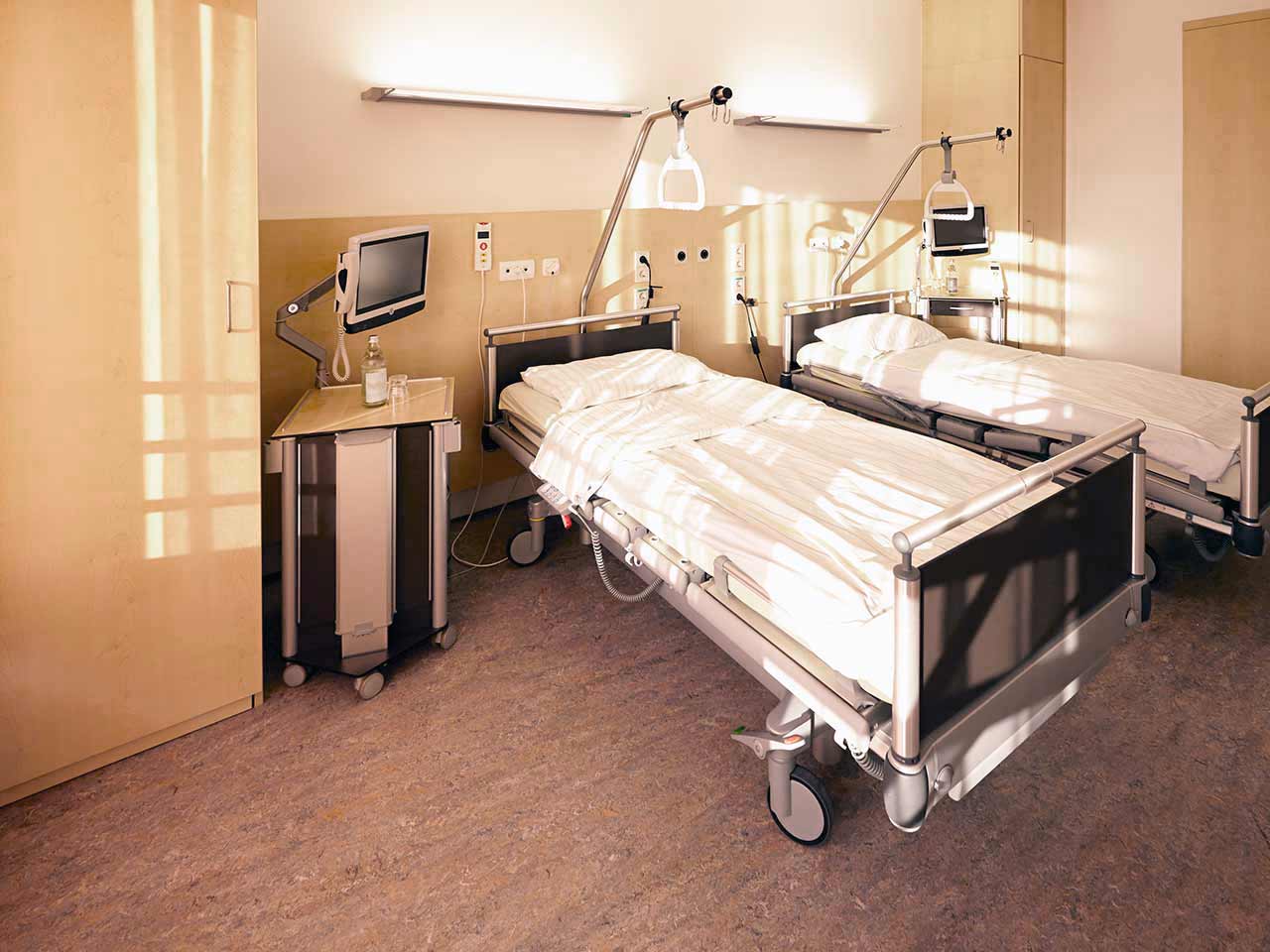
The patients of the ViDia Hospital Karlsruhe stay in comfortable single and double rooms with modern design. Each patient room has an ensuite bathroom with a shower and a toilet. The standard room furnishings include an automatically adjustable bed, a bedside table, a table and chairs, a wardrobe, a telephone, a TV, and a radio. Wi-Fi access is also available in the patient rooms.
Patients can also be accommodated in enhanced comfort rooms. These rooms are very spacious and are additionally equipped with a safe, a mini-fridge, and upholstered furniture.
Meals and Menus
The patients of the hospital are offered three tasty meals a day: breakfast is served buffet-style, and there are several set menus to choose from for lunch and dinner.
If, for some reason, you do not eat all of the foods, you will be offered an individual menu. Please inform the medical staff about your dietary preferences prior to treatment.
Further details
Standard rooms include:
![]() Toilet
Toilet
![]() Shower
Shower
![]() Wi-Fi
Wi-Fi
![]() TV
TV
Religion
There are several chapels on the territory of the hospital. Regular Catholic and evangelical services are held here. Patients can also visit one of the chapels at any time to find a quiet place to pray, if desired.
Accompanying person
Your accompanying person may stay with you in your patient room or at the hotel of your choice during the inpatient program.
Hotel
You may stay at the hotel of your choice during the outpatient program. Our managers will support you for selecting the best option.


