Treatment of liver cancer with embolization or chemoembolization in University Hospital Carl Gustav Carus Dresden
University Hospital Carl Gustav Carus Dresden
Dresden, Germany
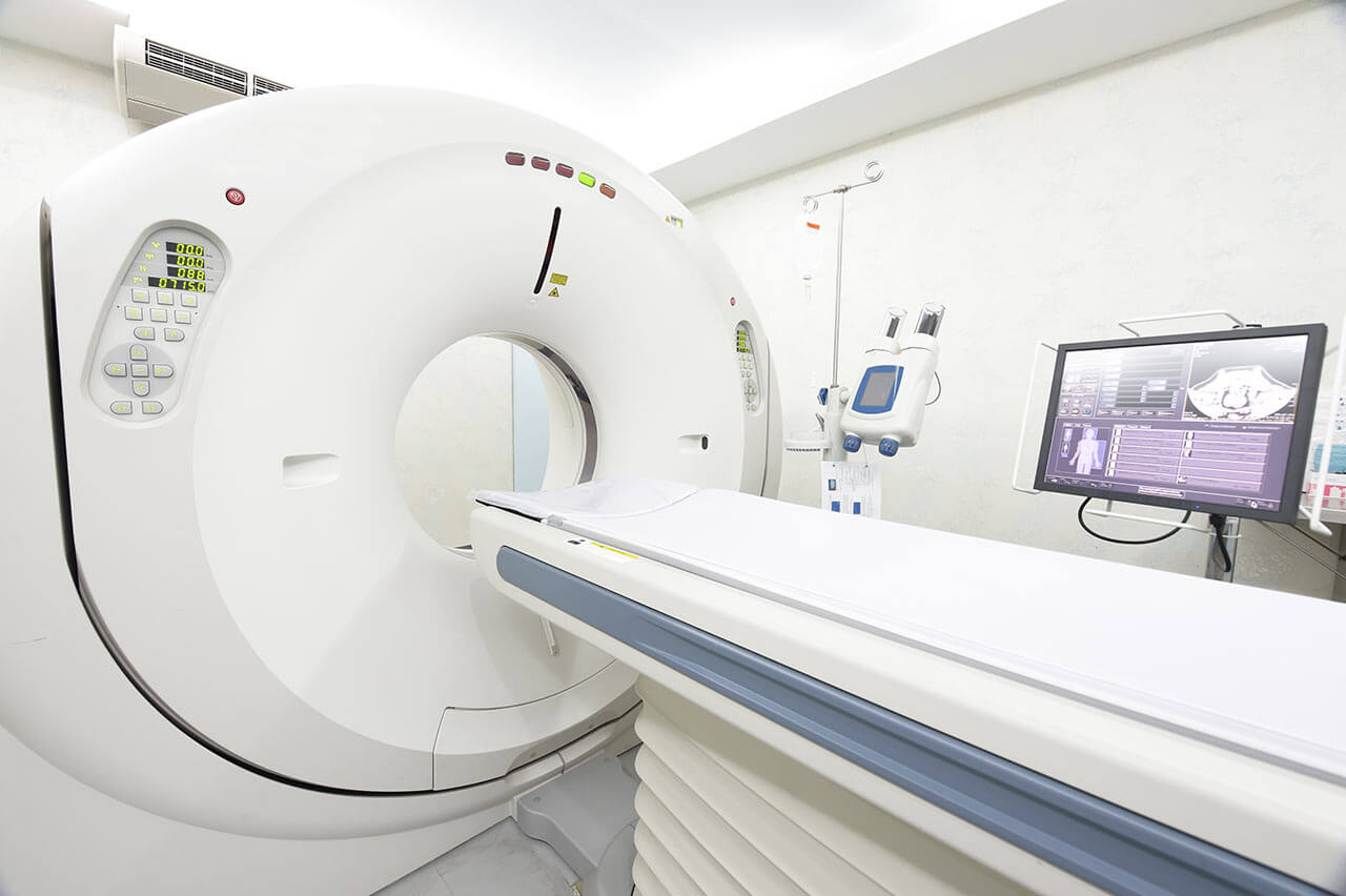


During the first visit, the doctor will conduct a clinical examination and go through the results of the available diagnostic tests. After that, you will undergo the necessary additional examination, such as the assessment of liver and kidney function, ultrasound scan, CT scan and MRI. This will allow the doctor to determine which vessels are feeding the tumor and its metastases, as well as determine how well you will tolerate the procedure.
Chemoembolization begins with local anesthesia and catheterization of the femoral artery. The thin catheter is inserted through a few centimeters long incision of the blood vessel. The doctor gradually moves the catheter to the vessel feeding the primary tumor or its metastases. The procedure is carried out under visual control, an angiographic device is used for this. The vascular bed and the position of the catheter in it are displayed on the screen of the angiograph.
When the catheter reaches a suspected artery, a contrast agent is injected through it. Due to the introduction of the contrast agent, the doctor clearly sees the smallest vessels of the tumor and the surrounding healthy tissues on the screen of the angiograph. After that, he injects emboli into the tumor vessels through the same catheter.
Emboli are the spirals or the liquid microspheres. The type of embolus is selected individually, taking into account the diameter of the target vessel. When carrying out chemoembolization, a solution of a chemotherapy drug is additionally injected into the tumor vessel. Due to the subsequent closure of the vessel lumen with an embolus, the chemotherapy drug influences the tumor for a long time. In addition, the drug does not enter the systemic circulation, which allows doctors to use high doses of chemotherapeutic agents without the development of serious side effects. Chemoembolization leads to the destruction of the tumor or slowing down its progression.
After that, the catheter is removed from the artery. The doctor puts a vascular suture on the femoral artery and closes it with a sterile dressing. During chemoembolization, you will be awake. General anesthesia is not used, which significantly reduces the risks of the procedure and allows performing it on an outpatient basis, avoiding long hospital stay.
After the first procedure, you will stay under the supervision of an interventional oncologist and general practitioner. If necessary, you will receive symptomatic treatment. As a rule, a second chemoembolization procedure is performed in 3-5 days after the first one in order to consolidate the therapeutic effect. After that, you will receive recommendations for further follow-up and treatment.
 BookingHealth Price from:
The Department of Neuroradiology at the University Hospital Carl Gustav Carus Dresden offers the full range of imaging diagnostics and minimally invasive image-guided therapy for patients with nervous system diseases. The department's medical team specializes in care for pathologies of the brain, spinal cord, peripheral nerves and muscles. The department has state-of-the-art systems for computed tomography (CT) and magnetic resonance imaging (MRI), which provide doctors with information about the structure and functioning of the nervous system, as well as its metabolic processes. The department's therapeutic options include catheter-based procedures for the treatment of acute and chronic pathologies of the vessels of the brain and spinal cord. The department's specialists cooperate closely with neurologists and neurosurgeons. In especially complex clinical cases, the doctors of these medical fields gather for a board to develop the most effective, but at the same time sparing treatment regimen. The department is certified in accordance with the requirements of the German Society of Neuroradiology (DGNR), so patients can count on top-class medical care with the use of the very latest medical advances. The Head Physician of the department is Prof. Dr. med. Jennifer Linn.
It is no secret that accurate diagnostics is the key to successful treatment of any disease. The department has modern diagnostic rooms for comprehensive diagnostics of the nervous system. Patients can undergo the full range of imaging tests here: computed tomography, magnetic resonance imaging, perfusion computed tomography, functional magnetic resonance imaging, magnetic resonance spectroscopy, catheter angiography, myelography, classical radiography and other tests. The necessary test or their set is prescribed by the doctor, based on the patient's anamnesis and his complaints. Prior to any of the above mentioned tests, the patient receives recommendations for preparation so that the result is as informative as possible.
In addition to diagnostics, the department performs a variety of image-guided interventional therapeutic procedures. Such treatment is an excellent alternative to neurosurgical intervention, after which a long recovery is required. One of the primary focuses of the department's specialists is mechanical thrombectomy for stroke treatment. The procedure is an endovascular removal of a blood clot that disrupts cerebral blood flow. The best results with mechanical thrombectomy are achieved within the first 6 hours after symptom onset. The procedure is performed using anesthesia or sedation. A special catheter is inserted into the femoral artery and then directed to the carotid artery through the aorta. At this stage, the doctor performs the necessary therapeutic manipulations to remove the blood clot. Before mechanical thrombectomy, CT or MRI scanning is mandatory in order to assess the patient's condition and the feasibility of this therapeutic manipulation.
The department also successfully deals with the treatment of brain aneurysms. In some cases, there is no need for any therapeutic procedures to remove the aneurysm, and the patient is recommended to be monitored over time. If the patient still requires brain aneurysm repair, the department's specialists consider the option of performing endovascular coiling. The procedure is performed under general anesthesia and with angiography guidance. A flexible catheter is inserted through the femoral artery in the groin and directed to one of the arteries in the neck that lead to the brain. A special dye is injected into the cerebral vessels to imagine them on the screen. After the catheter reaches the aneurysm, a thin platinum coil is inserted into the aneurysm, which fills the aneurysm and reduces the risk of its rupture. The coil stays inside the aneurysm permanently. After the procedure, the patient remains under monitoring in the intensive care unit for several days, after which he is discharged.
Another popular therapeutic manipulation is endovascular repair of carotid artery stenosis that may provoke a stroke. The essence of the procedure is the placement of a special stent into the carotid artery, which helps the patients to restore normal blood flow and significantly reduce the risk of developing cerebral hemorrhage. The endovascular procedure is also performed under image guidance.
The department's range of medical services includes:
Prof. Dr. med. Jennifer Linn studied human medicine at the Saarland University and the Free University of Brussels. Jennifer Linn defended her doctoral thesis at the Institute of Neurology of the Technical University of Munich. Habilitation for hemorrhagic stroke followed in 2010 at Ludwig Maximilian University of Munich.
Since October 9, 2014, the Professor has been heading the Department of Neuroradiology at the University Hospital Carl Gustav Carus Dresden. Prior to that, the Professor held the position of Senior Physician in the Department of Neuroradiology and the position of Head of the Research Unit at the University Hospital of Ludwig Maximilian University of Munich. Dr. Linn specializes in the early diagnostics of various forms of stroke. Of particular interest to her are microangiopathies – lesions of the small vessels of the brain, which in many cases can provoke stroke in the elderly, causing hemorrhages and acute cerebrovascular accidents. The head of the department has repeatedly received scholarships from the German Research Foundation. Her accomplishments also include the Kurt Decker Award of the German Society of Neuroradiology and the title of Invited Professor in the Department of Neuroradiology at Johns Hopkins University in Baltimore.
Photo: (с) depositphotos
According to the reputable Focus magazine, the University Hospital Carl Gustav Carus Dresden ranks among the top five German hospitals!
The hospital is the benchmark for modern high-quality medicine. Positioning itself as a maximum care medical facility, the hospital represents all medical fields. There are 26 specialized departments, 6 institutes and 17 interdisciplinary centers, which cooperate closely with the clinical and scientific facilities of the Faculty of Medicine. The basis of successful practice is excellent equipment, which is regularly updated, as well as highly qualified, experienced medical personnel: world famous doctors and professors work here for the benefit of patients.
In addition to its main goal of caring for patients, the hospital is also active in training and professional development of medical personnel, as well as in the field of public health care. The priority focus of the work is research activity, which allows the doctors to introduce the innovative diagnostic and therapeutic techniques into clinical practice.
A special feature of the hospital is also the diagnostics and treatment of rare diseases. State-of-the-art equipment and well-coordinated work of doctors of various medical specialties make it possible to timely recognize pathologies rarely encountered in medical practice and select the most effective therapy. Specialization in rare diseases include neurology, endocrinology, hematology/oncology, and rare autoimmune diseases.
The hospital has 1,410 beds for patient hospitalization. About 55,900 inpatients and more than 233,975 outpatients undergo treatment here annually. A large medical team, consisting of about 1,000 highly qualified doctors, as well as over 2,000 nursing staff take care of the patients' health. Each patient is guaranteed an individual approach and the most effective treatment in accordance with current clinical protocols.
It should be noted that the university hospital enjoys an impeccable reputation not only in Germany, but also far beyond its borders, including Arab countries, post-Soviet states, Great Britain and the United States. Patients from different parts of the world come here for high-quality treatment for diseases of any severity. The highest credit of patient confidence is the main indicator of the fruitful work of doctors.
Photo: (с) depositphotos
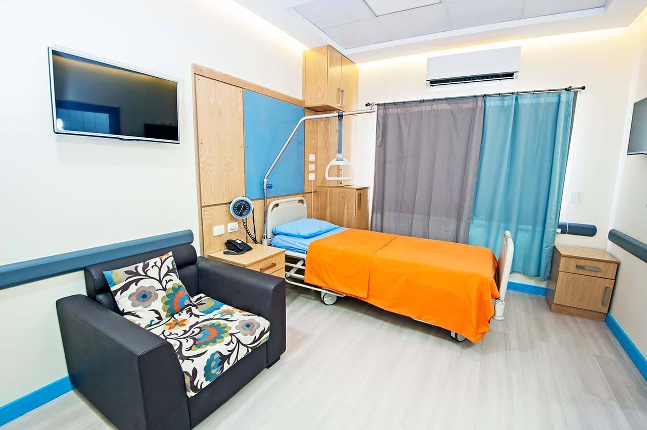
The patients of the University Hospital Carl Gustav Carus Dresden live in comfortable rooms made in bright colors and equipped with everything necessary. The standard patient room includes an automatically adjustable bed, a bedside table with a sliding table, a wardrobe, a telephone and a TV. There is also Wi-Fi (free) in the patient rooms.
If desired, patients may live in enhanced comfort patient rooms. These patient rooms have a more sophisticated design, upholstered furniture and a safe for storing valuables.
The patients of the hospital are offered a tasty, healthy and varied three meals a day. The menu is based on local cuisine and seasonal food. If you for some reason do not eat certain products, please inform the medical staff of the hospital in advance, and you will be offered an individual menu. The nutrition provided in the hospital is certified in accordance with the quality standards of the German Nutrition Society (DGE) for catering in German hospitals.
Standard rooms include:
The religious services are available upon request.
Your accompanying person may stay with you in your patient room or at the hotel of your choice during the inpatient program.
You may stay at the hotel of your choice during the outpatient program. Our managers will support you for selecting the best option.
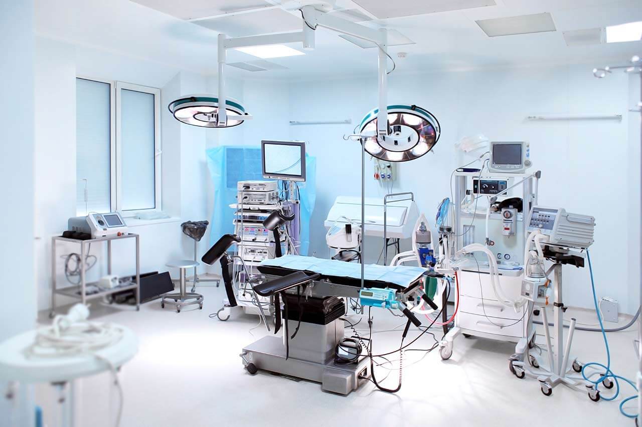
The hospital offers a full range of laboratory diagnostic procedures (general, hormonal, tests for tumor markers, infections, antibodies, etc.), genetic tests, various modifications of ultrasound scans, CT scans, MRI and PET / CT, angiography, myelography, biopsy and other examinations. Treatment with medications, endoscopic and robotic operations, stereotaxic interventions is carried out here, modern types of radiation therapy are also used. The hospital offers patients all the necessary therapeutic techniques.
These are head and neck tumors, hearloss, amyotrophic lateral sclerosis, epilepsy, Parkinson disease, infertility, malignant tumors of the reproductive system, congenital anomalies of the genital organs and the urinary system, urinary incontinence, blood clotting disorders, leukemia and other pathologies.
About 1,000 highly qualified doctors work at the hospital.
