{"translation_price":"50","translation_doc_price":"40","child_coefficient":"1.1","transfer_price":"2.00","transfer_price_vip":"5.00","constant_transfer_price_vip":350,"constant_transfer_price":150,"constant_transfer_distanse":60,"type":"treatment","program_full_story":"<ul>\n\t<li>Initial presentation in the hospital<\/li>\n\t<li>Clinical history taking<\/li>\n\t<li>Review of available medical records<\/li>\n\t<li>Physical examination<\/li>\n\t<li>Laboratory tests:\n\t<ul>\n\t\t<li>Complete blood count<\/li>\n\t\t<li>General urine analysis<\/li>\n\t\t<li>Biochemical analysis of blood<\/li>\n\t\t<li>Tumor markers<\/li>\n\t\t<li>Inflammation indicators (CRP, ESR)<\/li>\n\t\t<li>Coagulogram<\/li>\n\t<\/ul>\n\t<\/li>\n\t<li>Ultrasound\u200b scan<\/li>\n\t<li>CT scan \/ MRI<\/li>\n\t<li>Preoperative care<\/li>\n\t<li>Embolization or chemoembolization, 2 procedures<\/li>\n\t<li>Symptomatic treatment<\/li>\n\t<li>Cost of essential medicines<\/li>\n\t<li>Nursing services<\/li>\n\t<li>Elaboration of further recommendations<\/li>\n<\/ul>\n<div class=\"program_how_program_going mt-4\"><h4>How program is carried out<\/h4><p style=\"text-align: justify;\"><strong>During the first visit<\/strong>, the doctor will conduct a clinical examination and go through the results of the available diagnostic tests. After that, you will undergo the necessary additional examination, such as the assessment of liver and kidney function, ultrasound scan, CT scan and MRI. This will allow the doctor to determine which vessels are feeding the tumor and its metastases, as well as determine how well you will tolerate the procedure.<\/p>\n\n<p style=\"text-align: justify;\"><strong>Chemoembolization <\/strong>begins with local anesthesia and catheterization of the femoral artery. The thin catheter is inserted through a few centimeters long incision of the blood vessel. The doctor gradually moves the catheter to the vessel feeding the primary tumor or its metastases. The procedure is carried out under visual control, an angiographic device is used for this. The vascular bed and the position of the catheter in it are displayed on the screen of the angiograph.<\/p>\n\n<p style=\"text-align: justify;\">When the catheter reaches a suspected artery, a contrast agent is injected through it. Due to the introduction of the contrast agent, the doctor clearly sees the smallest vessels of the tumor and the surrounding healthy tissues on the screen of the angiograph. After that, he injects emboli into the tumor vessels through the same catheter.<\/p>\n\n<p style=\"text-align: justify;\">Emboli are the spirals or the liquid microspheres. The type of embolus is selected individually, taking into account the diameter of the target vessel. When carrying out chemoembolization, a solution of a chemotherapy drug is additionally injected into the tumor vessel. Due to the subsequent closure of the vessel lumen with an embolus, the chemotherapy drug influences the tumor for a long time. In addition, the drug does not enter the systemic circulation, which allows doctors to use high doses of chemotherapeutic agents without the development of serious side effects. Chemoembolization leads to the destruction of the tumor or slowing down its progression.<\/p>\n\n<p style=\"text-align: justify;\">After that, the catheter is removed from the artery. The doctor puts a vascular suture on the femoral artery and closes it with a sterile dressing. During chemoembolization, you will be awake. General anesthesia is not used, which significantly reduces the risks of the procedure and allows performing it on an outpatient basis, avoiding long hospital stay.<\/p>\n\n<p style=\"text-align: justify;\"><strong>After the first procedure<\/strong>, you will stay under the supervision of an interventional oncologist and general practitioner. If necessary, you will receive symptomatic treatment. As a rule, a second chemoembolization procedure is performed in 3-5 days after the first one in order to consolidate the therapeutic effect. After that, you will receive recommendations for further follow-up and treatment.<\/p>\n<\/div><div class=\"program_required_documents mt-4\"><h4>Required documents<\/h4><ul>\n\t<li style=\"text-align: justify;\">Medical records<\/li>\n\t<li style=\"text-align: justify;\">MRI\/CT scan (not older than 3 months)<\/li>\n\t<li style=\"text-align: justify;\">Biopsy results (if available)<\/li>\n<\/ul>\n<\/div>","program_full_story_crm":"<ul>\n\t<li>Initial presentation in the hospital<\/li>\n\t<li>Clinical history taking<\/li>\n\t<li>Review of available medical records<\/li>\n\t<li>Physical examination<\/li>\n\t<li>Laboratory tests:\n\t<ul>\n\t\t<li>Complete blood count<\/li>\n\t\t<li>General urine analysis<\/li>\n\t\t<li>Biochemical analysis of blood<\/li>\n\t\t<li>Tumor markers<\/li>\n\t\t<li>Inflammation indicators (CRP, ESR)<\/li>\n\t\t<li>Coagulogram<\/li>\n\t<\/ul>\n\t<\/li>\n\t<li>Ultrasound\u200b scan<\/li>\n\t<li>CT scan \/ MRI<\/li>\n\t<li>Preoperative care<\/li>\n\t<li>Embolization or chemoembolization, 2 procedures<\/li>\n\t<li>Symptomatic treatment<\/li>\n\t<li>Cost of essential medicines<\/li>\n\t<li>Nursing services<\/li>\n\t<li>Elaboration of further recommendations<\/li>\n<\/ul>\n<div class=\"program_how_program_going mt-4\"><h4>How program is carried out<\/h4><p style=\"text-align: justify;\"><strong>During the first visit<\/strong>, the doctor will conduct a clinical examination and go through the results of the available diagnostic tests. After that, you will undergo the necessary additional examination, such as the assessment of liver and kidney function, ultrasound scan, CT scan and MRI. This will allow the doctor to determine which vessels are feeding the tumor and its metastases, as well as determine how well you will tolerate the procedure.<\/p>\n\n<p style=\"text-align: justify;\"><strong>Chemoembolization <\/strong>begins with local anesthesia and catheterization of the femoral artery. The thin catheter is inserted through a few centimeters long incision of the blood vessel. The doctor gradually moves the catheter to the vessel feeding the primary tumor or its metastases. The procedure is carried out under visual control, an angiographic device is used for this. The vascular bed and the position of the catheter in it are displayed on the screen of the angiograph.<\/p>\n\n<p style=\"text-align: justify;\">When the catheter reaches a suspected artery, a contrast agent is injected through it. Due to the introduction of the contrast agent, the doctor clearly sees the smallest vessels of the tumor and the surrounding healthy tissues on the screen of the angiograph. After that, he injects emboli into the tumor vessels through the same catheter.<\/p>\n\n<p style=\"text-align: justify;\">Emboli are the spirals or the liquid microspheres. The type of embolus is selected individually, taking into account the diameter of the target vessel. When carrying out chemoembolization, a solution of a chemotherapy drug is additionally injected into the tumor vessel. Due to the subsequent closure of the vessel lumen with an embolus, the chemotherapy drug influences the tumor for a long time. In addition, the drug does not enter the systemic circulation, which allows doctors to use high doses of chemotherapeutic agents without the development of serious side effects. Chemoembolization leads to the destruction of the tumor or slowing down its progression.<\/p>\n\n<p style=\"text-align: justify;\">After that, the catheter is removed from the artery. The doctor puts a vascular suture on the femoral artery and closes it with a sterile dressing. During chemoembolization, you will be awake. General anesthesia is not used, which significantly reduces the risks of the procedure and allows performing it on an outpatient basis, avoiding long hospital stay.<\/p>\n\n<p style=\"text-align: justify;\"><strong>After the first procedure<\/strong>, you will stay under the supervision of an interventional oncologist and general practitioner. If necessary, you will receive symptomatic treatment. As a rule, a second chemoembolization procedure is performed in 3-5 days after the first one in order to consolidate the therapeutic effect. After that, you will receive recommendations for further follow-up and treatment.<\/p>\n<\/div>","is_ambulant":"1","bh_fee":"0","only_for_children":"0","no_service":"0","with_prepayment":"1","show_calculator":"1","paket_type":"1","btn_type":"0","clinic_icon":"60018c63b444d.jpg","city":"Berlin","clinic_site":"http:\/\/www.charite.de\/","department_recommend":"1","country":"Germany","country_id":"1","clinic_name":"Charite University Hospital Berlin","cinic_name":"Charite University Hospital Berlin","department_id":"2747","duration":"7","direction":"Interventional radiology","min_duration":0,"clinic_id":"254","paketPrice":7800,"paket":"<ul>\n <li>Interpreter up to 17 hours<\/li>\n <li>Translation up to 10 pages<\/li>\n <li>Visa support<\/li>\n\n \n<\/ul>","title":"Treatment of lung cancer with embolization or chemoembolization","price":{"val":41043.05,"type":"val"},"price_surcharge":0,"price_surcharge_clear":0,"extra_service_clinic":[],"extra_service":[],"translation_hours":"0","translation_doc_count":null,"roads":[{"id":"22","distance":"20","airport_title":"Berlin-Tegel"},{"id":"20","distance":"172","airport_title":"Leipzig"},{"id":"3","distance":"288","airport_title":"Hannover"}],"pakets":[],"lang":{"day":"Day","days":"days","ambulatory":"Outpatient","stationaryProgram":"Inpatient"}}
Treatment of lung cancer with embolization or chemoembolization in Charite University Hospital Berlin
During the first visit, the doctor will conduct a clinical examination and go through the results of the available diagnostic tests. After that, you will undergo the necessary additional examination, such as the assessment of liver and kidney function, ultrasound scan, CT scan and MRI. This will allow the doctor to determine which vessels are feeding the tumor and its metastases, as well as determine how well you will tolerate the procedure.
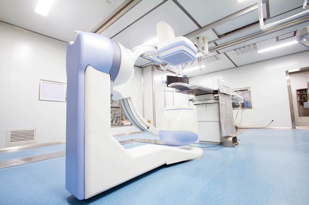
Chemoembolization begins with local anesthesia and catheterization of the femoral artery. The thin catheter is inserted through a few centimeters long incision of the blood vessel. The doctor gradually moves the catheter to the vessel feeding the primary tumor or its metastases. The procedure is carried out under visual control, an angiographic device is used for this. The vascular bed and the position of the catheter in it are displayed on the screen of the angiograph.
When the catheter reaches a suspected artery, a contrast agent is injected through it. Due to the introduction of the contrast agent, the doctor clearly sees the smallest vessels of the tumor and the surrounding healthy tissues on the screen of the angiograph. After that, he injects emboli into the tumor vessels through the same catheter.
Emboli are the spirals or the liquid microspheres. The type of embolus is selected individually, taking into account the diameter of the target vessel. When carrying out chemoembolization, a solution of a chemotherapy drug is additionally injected into the tumor vessel. Due to the subsequent closure of the vessel lumen with an embolus, the chemotherapy drug influences the tumor for a long time. In addition, the drug does not enter the systemic circulation, which allows doctors to use high doses of chemotherapeutic agents without the development of serious side effects. Chemoembolization leads to the destruction of the tumor or slowing down its progression.
After that, the catheter is removed from the artery. The doctor puts a vascular suture on the femoral artery and closes it with a sterile dressing. During chemoembolization, you will be awake. General anesthesia is not used, which significantly reduces the risks of the procedure and allows performing it on an outpatient basis, avoiding long hospital stay.
After the first procedure, you will stay under the supervision of an interventional oncologist and general practitioner. If necessary, you will receive symptomatic treatment. As a rule, a second chemoembolization procedure is performed in 3-5 days after the first one in order to consolidate the therapeutic effect. After that, you will receive recommendations for further follow-up and treatment.
Required documents
Medical records
MRI/CT scan (not older than 3 months)
Biopsy results (if available)
Service
Price from:
Type of program :
Price for 1 day:
Expected duration of the program:
The minimum duration of the program:
Select
You may also book:
Hospital directly:
Saving:
BookingHealth Price from:
About the department
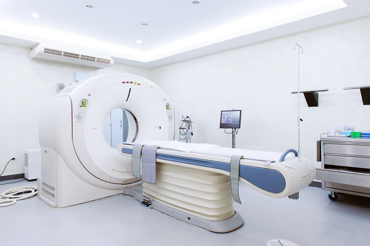
The Department of Adult and Pediatric Diagnostic, Interventional Radiology at the Charite University Hospital Berlin offers the full range of medical services in these fields. In order to ensure the most accurate and reliable diagnostics, the department has state-of-the-art systems for CT, MRI, PET, ultrasound and X-ray examinations, as well as PET-CT and SPECT-CT hybrid examinations. The department specializes in interventional radiology, namely, imaging-guided therapy. It performs all vascular (for example, vascular recanalization) and non-vascular imaging-guided interventions (for example, tumor ablation). The department is one of the largest and leading medical facilities of this kind in Europe. The Chief Physician of the department is Prof. Dr. med. Bernd Hamm.
In the field of diagnostic radiology, all modern imaging examinations are carried out. The great advantage of the department is the availability of specially adapted diagnostic systems for the diagnostics of children, for example, ultrasound, X-ray, CT, etc. The cutting-edge medical equipment guarantees an optimal radiation protection, so the diagnostic tests do not harm the health of either children or adults.
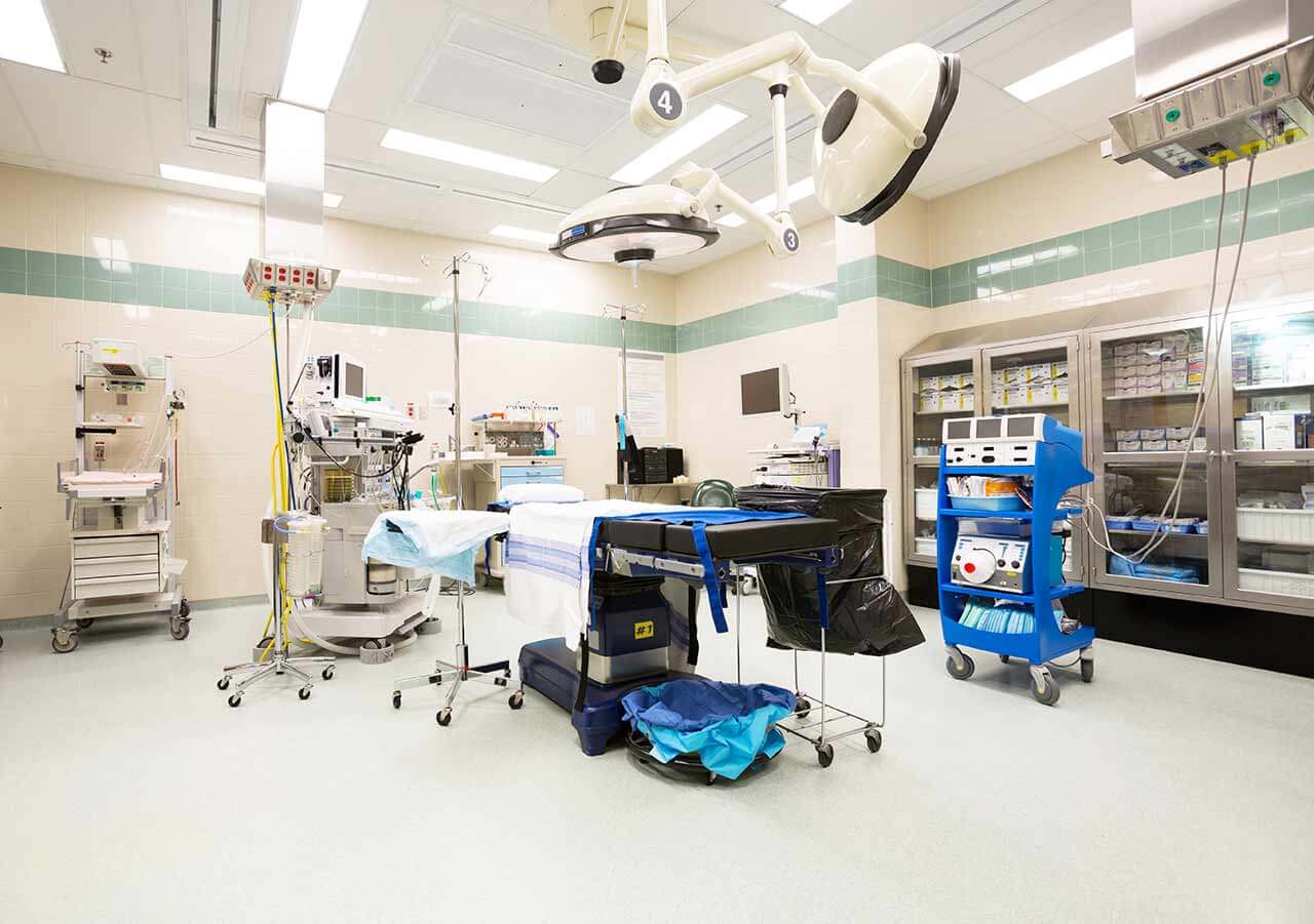
It should be noted that the department is a recognized Center for Minimally Invasive Surgery. In the case of minimally invasive interventions, such radiological methods as X-ray, computed tomography (CT) and magnetic resonance imaging (MRI) serve as imaging-guided tools. This ensures accurate, targeted surgery. In most cases, local anesthesia is used. An exception is the treatment of osteoid osteoma, which is performed under general anesthesia. Minimally invasive procedures include tissue sampling for diagnostic tests, blockade of nerve endings during pain therapy, use of laser techniques for the treatment of tumors and spinal disc herniation, restoration of blood vessel patency in thrombosis, arrest of arterial hemorrhages, blockade of tumor blood supply, etc.
The department's specialists always prefer an individual approach to treatment. Many minimally invasive techniques have been developed or improved by the department's doctors, which guarantees the highest treatment quality. The department annually performs more than 2,000 minimally invasive interventions (from tissue sampling to tumor treatment). Prior to any type of treatment, the department's doctors discuss in detail the therapeutic possibilities with the patient, including therapy risks and alternative treatment methods.
The service range of the department includes:
Diagnostic radiology
Magnetic resonance imaging (MRI)
MRI of organs and anatomical structures of the neck (for example, salivary glands, larynx, trachea)
Breast MRI
Abdominal MRI
Pelvic MRI
Musculoskeletal MRI
Vascular MRI (MR angiography)
MRI of the nervous system
Ultrasound examinations
Radiodiagnostics in inflammatory rheumatic and degenerative joint diseases
Breast imaging
Cancer screening
Breast MRI
Vacuum-assisted breast biopsy
Mammography
Breast ultrasound examination
Tomosynthesis
MRI diagnostics of the pelvic organs in women
Prostate imaging
Imaging examinations in children (ultrasound, X-ray, CT, MRI)
Interventional radiology
Treatment of vascular diseases
Dialysis access formation
Implantation of central venous port systems
Transjugular intrahepatic portosystemic shunting
Treatment of peripheral arterial occlusive disease
X-ray-, CT- and MRI-guided infiltration of nerve roots, vertebral joints, sacroiliac joints and sympathetic trunk
Periradicular injection therapy
Injection therapy of facet and sacroiliac joints
Treatment of uterine fibroids
Uterine fibroid embolization
Other medical services
Curriculum vitae
1972 - 1978 Study of Human Medicine at the Free University of Berlin.
1978 - 1986 Scientific Assistant at the Institute of Pathology, Department of Radiation Therapy, Department of Radiology and Nuclear Medicine at the Steglitz Clinic of the Free University of Berlin.
07.07.1982 Doctorate at the Faculty of Medicine of the Free University of Berlin, "magna cum laude".
16.05.1986 Specialist examination and recognition as a Medical Specialist in Radiology.
02.1989 Habilitation and Venia legendi by the Faculty of Medicine at the Free University of Berlin in Radiology.
1990 - 1993 Head of the Department of Radiology and Nuclear Medicine at the Steglitz Clinic of the Free University of Berlin.
04.1993 Appointment as C3 Professor for Clinical Radiology at the Free University of Berlin.
1993 Call for the C4 Professorship for Diagnostic Radiology at the Medical University of Essen (primo loco).
1994 Received and accepted the call for C4 Professorship for X-ray Diagnostics at the Charité, Faculty of Medicine at the Humboldt University of Berlin (primo loco).
1998 Received the call for C4 Professorship for Diagnostic Radiology at the Goethe University Frankfurt (primo loco).
01.2006 - 04.2009 Acting Head of the Department of Nuclear Medicine at the Charité.
Since 2006 Head of the CharitéCenter 6 (Diagnostic and Interventional Radiology/Nuclear Medicine/Medical Physics) and taking responsibility for budget of the mentioned areas, Charité.
10.2006 Appointment as the Head of the Department of Clinical Radiology and Director of the merged Radiology at the Charité – Campus Mitte and Campus Virchow Clinic.
Since 2004/2006 Director of four Medical Care Centers (MVZ) for Radiology and Nuclear Medicine at the Charité, as well as a Research and Clinical Director of three Centers for Imaging Diagnostics in Berlin.
10.2010 Appointment as the Head of the Department of Radiology and Head of three merged Departments of Radiology at the Charité – Campus Mitte, Campus Virchow and Campus Benjamin Franklin.
2012 Honorary Professorship at the China-Japan Friendship Hospitals, Peking.
Photo of the doctor: (c) Charité – Universitätsmedizin Berlin
About hospital
According to the reputable Focus magazine, the Charite University Hospital Berlin ranks 1st among the best healthcare facilities in Germany!
The hospital is one of the largest and leading university medical complexes in Europe, and also consistently holds leading positions in the international medical arena. The Charite operates on the basis of the Faculty of Medicine of the Free University of Berlin and the Humboldt University of Berlin. Patients are offered modern diagnostics and treatment with the very latest methods, many of which were developed by professors and scientists of the medical complex. More than half of all German Nobel Prize winners in medicine and physiology, such as Emil von Behring, Robert Koch, and Paul Ehrlich, studied and worked at the Charite University Hospital Berlin. The medical complex includes more than 100 specialized departments and institutes, which helps to ensure that patients receive care in all existing medical specialties. The hospital has exceptional experience in treating complex clinical cases.
Each year, the hospital treats more than 137,800 inpatients and more than 787,700 outpatients. The hospital has a bed capacity of 3,293 beds. A huge medical team consisting of 5,670 scientists and doctors and more than 6,000 nurses work for the benefit of the patients. The main task of all specialists of the medical facility is to restore the patient's health or save his life in critical cases. The hospital has a friendly atmosphere where every patient feels care, respect and empathy.
The Charite University Hospital Berlin is generously funded by the German government, which is why it offers patients the latest generation of excellent equipment and comfortable infrastructure. The Charite medical complex is equipped with da Vinci robotic surgery systems, laser technologies, equipment for endovascular catheter-based interventions, neuronavigation devices, intraoperative monitoring systems, equipment for proton therapy available only in the most advanced medical centers in the world, and many other technologies. All these resources, combined with the experience and professional skills of the hospital's doctors, are the key to providing the most effective and safe treatment in accordance with the highest international medical standards.
The hospital is recognized with a huge number of quality certificates, including DIN EN ISO 9001:2015, certificates from the German Cancer Society (DKG), the German Society for General and Visceral Surgery (DGAV), the German Society for Thoracic Surgery (DGT), the German Hernia Society (DHG), and the ERAS Society.
The Charite University Hospital Berlin is a benchmark in the European healthcare system. Patients therefore receive impeccable medical service, quality care, and personalized service that puts the patient and their individual needs first.
Photo: (с) depositphotos
Accommodation in hospital
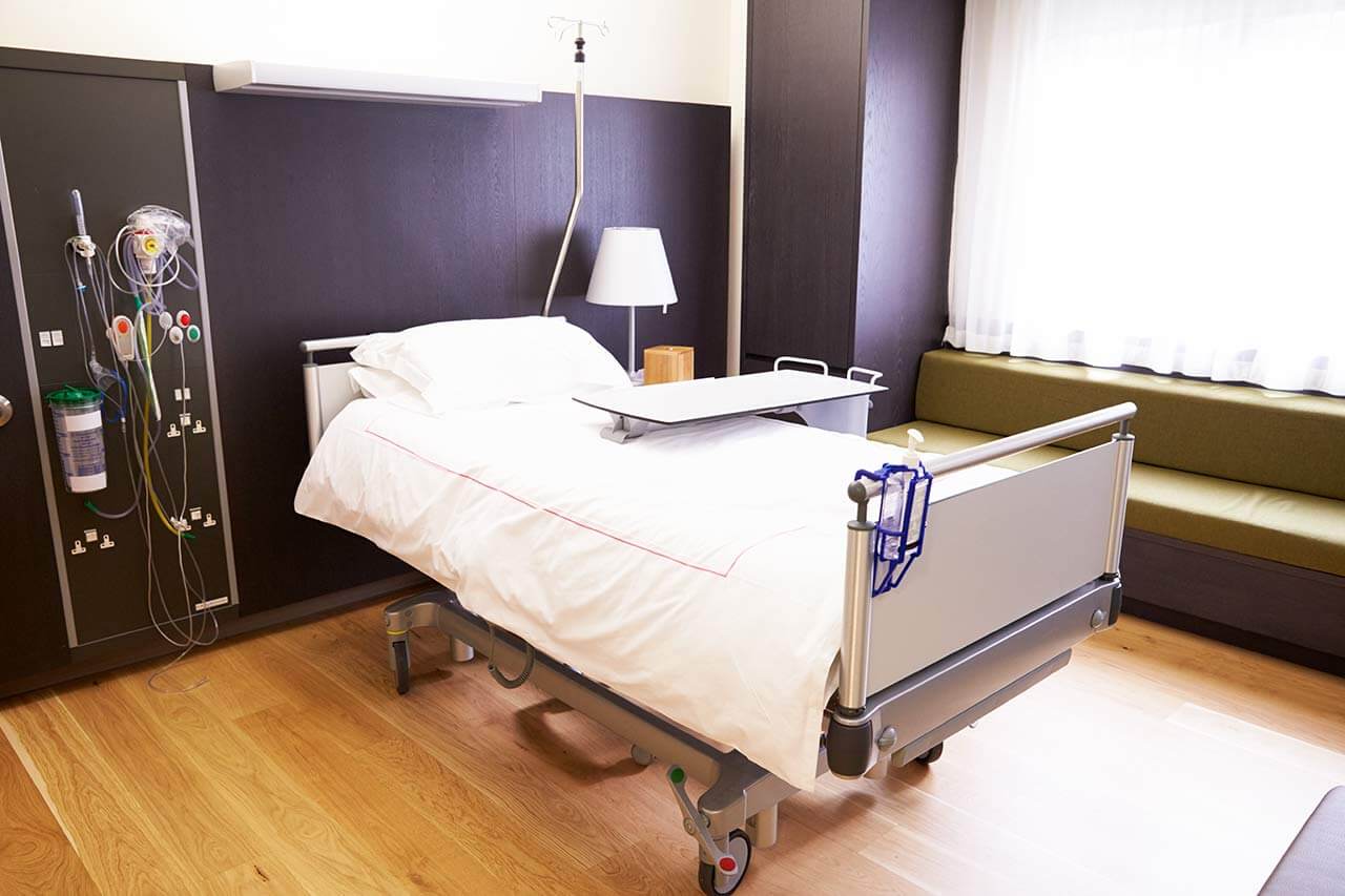
Patients rooms
The patients of the Charite University Hospital Berlin live in comfortable rooms made of modern design. Each room is equipped with an ensuite bathroom with a toilet and a shower. The standard room furnishing includes an automatically adjustable bed, a bedside table, a wardrobe for storing clothes, a table and chairs for receiving visitors, and a TV. If desired, Wi-Fi access can be provided. The hospital also offers enhanced-comfort rooms.
Meals and Menus
The patient and his accompanying person have a daily choice of three menus. If for any reason, you do not like the food, you will be offered an individual menu. Please inform the medical staff about your dietary preferences before the treatment.
Further details
Standard rooms include:
Toilet
Shower
Wi-Fi
TV
Religion
Religious services are available upon request.
Accompanying person
During the inpatient program, an accompanying person may stay with you in a patient room or at the hotel of your choice.
Hotel
During the outpatient program, you can live at a hotel of your choice. Managers will help you to choose the most suitable options.
The hospital offers a full range of laboratory tests (general, hormonal, tests for infections, antibodies, tumor markers, etc.), genetic tests, various modifications of ultrasound scans, CT scans, MRI and PET / CT, angiography, myelography, biopsy and other examinations. Treatment with medications, endoscopic and robotic operations, stereotaxic interventions is carried out here, modern types of radiation therapy are also used. The hospital offers patients all the necessary therapeutic techniques.
These are oncological diseases, benign neoplasms of the brain and spinal cord, heart valve defects, diabetes mellitus and its complications, joint diseases and other pathologies.
thank you for reaching out! One of our medical advisors is currently reviewing your details. You can expect a call from us within one business day to discuss your request.
Note: The incoming call will appear as a German number or your local area code. This call is entirely free of charge.
Booking Health guarantees:
Expert Placement
We use rigorous statistical analysis to ensure you are treated at the best-rated facility for your specific diagnosis.
Total Cost Protection
No hidden fees or unexpected bills. We provide a fixed price guarantee, backed by insurance that covers any additional complications.
12-Month Extended Care
Your recovery is our priority. Benefit from a full year of direct medical support following your procedure.


 BookingHealth Price from:
