The program includes:
- Initial presentation in the clinic
- clinical history taking
- review of medical records
- physical examination
- laboratory tests:
- complete blood count
- biochemical analysis of blood
- inflammation indicators (CRP, ESR)
- indicators of blood coagulation
- blood gas analysis
- chest x-ray examination
- Holter monitoring (24h)
- measurement of arterial blood pressure
- electrocardiogram (ECG)
- pulmonary function test
- echocardiography
- doppler echocardiography
- high-resolution computed tomography (HR-CT)/MRI (on indication 950/1200€)
- CT angiography (on indication 1300€)
- nursing services
- consultation of related specialists
- treatment by chief physician and all leading experts
- explanation of individual treatment plan
(the cost of medicines is not included)
Required documents
- Medical records
- Echocardiography (if available)
Service
You may also book:
 BookingHealth Price from:
About the department
The Department of Pediatric Cardiac Surgery at the University Hospital Erlangen offers the full range of modern surgical interventions for congenital and acquired heart defects in children. The surgical treatment corresponds to the highest level of university medicine. All cardiac surgeons working in the department have excellent qualifications and vast clinical experience required for the successful treatment outcomes in the long term. Of particular importance is the optimal preoperative preparation and postoperative care, as well as communication with young patients aimed at establishing a trusting relationship and overcoming the child's fear. Many young patients receive sparing minimally invasive treatment, which excludes severe pain syndrome, high risks of postoperative complications and a long hospital stay. The department is a leader in the area of its specialization in Germany. In addition, the medical facility regularly demonstrates one of the highest success rates in the treatment of pediatric heart pathologies in Europe. The department is headed by Prof. Dr. med. Oliver Dewald.
The department's team of surgeons successfully treats atrial septal defects – one of the most common congenital heart defects in children. The disorder is the formation of a pathological opening in the interatrial septum. As a result, blood from the left atrium enters the right atrium and overflows the pulmonary vessels. The most common symptoms are fatigue, shortness of breath, and abnormal heart rhythm, but there are also cases when symptoms are absent. Should atrial septal defect be suspected, pediatric cardiac surgeons along with pediatric cardiologists carry out a clinical examination and a complex of laboratory and instrumental tests, including echocardiography, electrocardiography, x-ray, etc. During preliminary diagnostics, doctors determine the exact size of the defect, its hemodynamic characteristics, the presence or absence of comorbidities and other important clinical data, which form the basis for choosing the optimal option for atrial septal defect repair. First of all, experts consider the least invasive technique – image-guided endovascular placement of occluder (without thoracotomy). Such interventions are performed jointly with pediatric cardiologists. However, not all atrial septal defects can be repaired with an occluder. For example, large defects and a combination of the defect with abnormal drainage of the pulmonary veins are contraindications for endovascular procedures. The last-line therapy is classic open surgery with a median sternotomy. During the operation, the patient is connected to a heart-lung machine, which means that the heart temporarily stops, and its defect is sutured or repaired with pericardial tissues. The department's cardiac surgeons regularly perform operations for atrial septal defect repair, so young patients receive safe and highly effective treatment in accordance with high European standards.
The department also specializes in the correction of ventricular septal defects – pathological openings in the interventricular septum. As a result of this anatomical abnormality, blood flows from the left ventricle to the right, which leads to mixing of arterial and venous blood and overflow of the pulmonary circulation. The symptoms include shortness of breath, frequent colds, pneumonia, as well as a delay in physical development. In addition, the ventricular septal defect can contribute to the development of such a serious complication as pulmonary hypertension, which is a contraindication to surgery for heart defect. The main method for diagnosing the defect is echocardiography. In some cases, cardiac catheterization can be additionally performed. When the diagnosis is confirmed, cardiac surgeons begin treatment planning. Whenever possible, preference is given to an endovascular intervention for the placement of an occluder, which completely isolates the ventricles from each other. The endovascular procedure is performed under imaging guidance and is minimally traumatic for the child. Unfortunately, this treatment method is effective only for small and medium-sized ventricular septal defects. If the child has an extensive defect, then open-heart surgery with a heart-lung machine is required. When performing the operation, cardiac surgeons assess the location and size of the defect. If the hole size does not exceed 7-8 mm, it is sutured, otherwise, plastic repair with a patch is carried out. With the timely operation, the child's health condition improves significantly, and he can return to an active lifestyle, including doing sports.
Another priority focus of the department's clinical practice is on the treatment of young patients with tetralogy of Fallot. This pathology is formed even before birth and is an impairment of the development of the right heart. Pulmonary artery stenosis, right ventricular hypertrophy, ventricular septal defect, and aortic dextroposition are typical for tetralogy of Fallot. This disease provokes serious disorders in the circulatory system. The child has cyanosis of the skin and mucous membranes. The disease is characterized by seizures, during which the blood oxygen levels decrease. At the same time, there is a delay in physical development, severe shortness of breath, rapid fatigue during physical exertion and loss of consciousness. Upon admission to the department, doctors conduct comprehensive diagnostics: physical examination, laboratory and instrumental tests, assessment of the blood oxygen levels, blood gas analysis, electrocardiography and chest X-ray, echocardiography, cardiac catheterization and angiocardiography. Based on the diagnostic data obtained, an optimal treatment regimen is developed. As a rule, the department's cardiac surgeons combine drug therapy and surgery, which is the only way to completely cure the defect. During the operation, the patient is connected to a heart-lung machine, after which an incision is made in the middle side of the chest. The surgeon closes the ventricular septal defect with a patch and performs excision of the hypertrophied myocardium, thereby eliminating the obstacle to the outflow of blood from the right ventricle. Thanks to the long experience of the department's specialists and their high professional skills, the risks of developing complications after surgery do not exceed 1%. In the future, the child develops normally, without any restrictions.
The department's main clinical focuses include the following heart surgeries in children:
- Surgical repair of atrial septal defects
- Surgical repair of ventricular septal defects
- Surgical treatment of tetralogy of Fallot
- Surgical repair of double outlet right ventricle
- Surgical repair of partial or total anomalous pulmonary venous drainage
- Surgical repair of the atrioventricular canal
- Tricuspid valve reconstruction for Ebstein's anomaly
- Anatomical repair of transposition of the great vessels, including in case of pulmonary artery stenosis (REV procedure)
- Surgical repair of univentricular defects (Fontan procedure)
- Complex surgical repair and treatment of hypoplastic left heart syndrome (Norwood procedure)
- Ross or Ross-Konno procedure
- Unifocalization for tetralogy of Fallot with pulmonary atresia
- Other heart interventions
Curriculum vitae
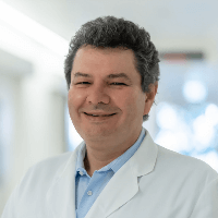
On June 1, 2022, Prof. Dr. med. Oliver Dewald headed the Department of Pediatric Cardiac Surgery at the University Hospital Erlangen. Prior to this, the specialist headed the Department of Cardiac Surgery at the University Hospital Oldenburg. Previously, Prof. Dewald headed the Section for Congenital Heart Disease Surgery in Children and Adults at the Department of Cardiac Surgery at the University Hospital Bonn.
From 1992 to 1999, Prof. Oliver Dewald studied human medicine at Ludwig Maximilian University of Munich, where he received his doctorate. This was followed by his internship in the Department of Cardiac Surgery at the University Hospital Bonn. As part of a research grant from the German Research Foundation, the doctor also conducted research at the Baylor College of Medicine in Houston, Texas, USA (2000 - 2003). In 2008, Prof. Dewald completed his habilitation and board certification in Cardiac Surgery, Department of Cardiac Surgery at the University Hospital Bonn. The doctor then took up the post of a Senior Physician in the Department of Cardiac Surgery at the University Hospital of Bonn. In 2015, Prof. Oliver Dewald became the Head of the Section for Pediatric Cardiac Surgery with a focus on the correction of congenital heart defects in adults. In 2016, he was appointed an Extraordinary Professor. In 2019, the specialist was appointed the Head Physician in the Department of Cardiac Surgery at the University Hospital of Oldenburg.
Dr. Dewald is the recipient of many awards, including the Ernst Derra Prize and the Ulrich Karsten Foundation Science Prize from the German Society for Thoracic and Cardiovascular Surgery. In addition, the specialist is engaged in reviewing activities and is a member of many professional societies.
The priority clinical focuses of the specialist's work include minimally invasive and hybrid interventions on the heart, for example, for heart valve reconstruction, severe heart defect correction, ECMO in newborns, the installation of modern heart support systems and interventions on large blood vessels of the heart.
Photo of the doctor: (c) Universitätsklinikum Erlangen
About hospital
According to the Focus magazine, University Hospital Erlangen ranks among the best medical facilities in Germany!
The hospital is one of the leading healthcare facilities in Bavaria and offers top-class medical care distinguished by the close intertwining of clinical activities with research and training of medical students. The hospital was founded in 1815 and today is proud of its rich traditions, numerous medical achievements and an excellent reputation not only in Germany, but also in the international arena. The hospital has 25 specialized departments, 7 institutes and 41 interdisciplinary centers, whose experts work tirelessly for the benefit of their patients.
The hospital has the status of a maximum care center, and therefore it represents almost all fields of modern medicine. Oncology, transplant medicine, and robot-assisted surgery are among the top priorities of the clinical activities of the medical complex. Oncology is represented by the Comprehensive Cancer Center Erlangen, which is one of 13 centers of excellence in Germany certified by the German Cancer Society. The university hospital has a high-tech center with high success rates for heart, liver, kidney, pancreas, cornea and bone marrow transplants. In addition, the hospital is a leader in the use of robot-assisted surgery. The medical facility has at its disposal innovative robotic technologies, in particular the da Vinci Surgical System, with the help of which surgeons perform many sparing interventions in various medical fields.
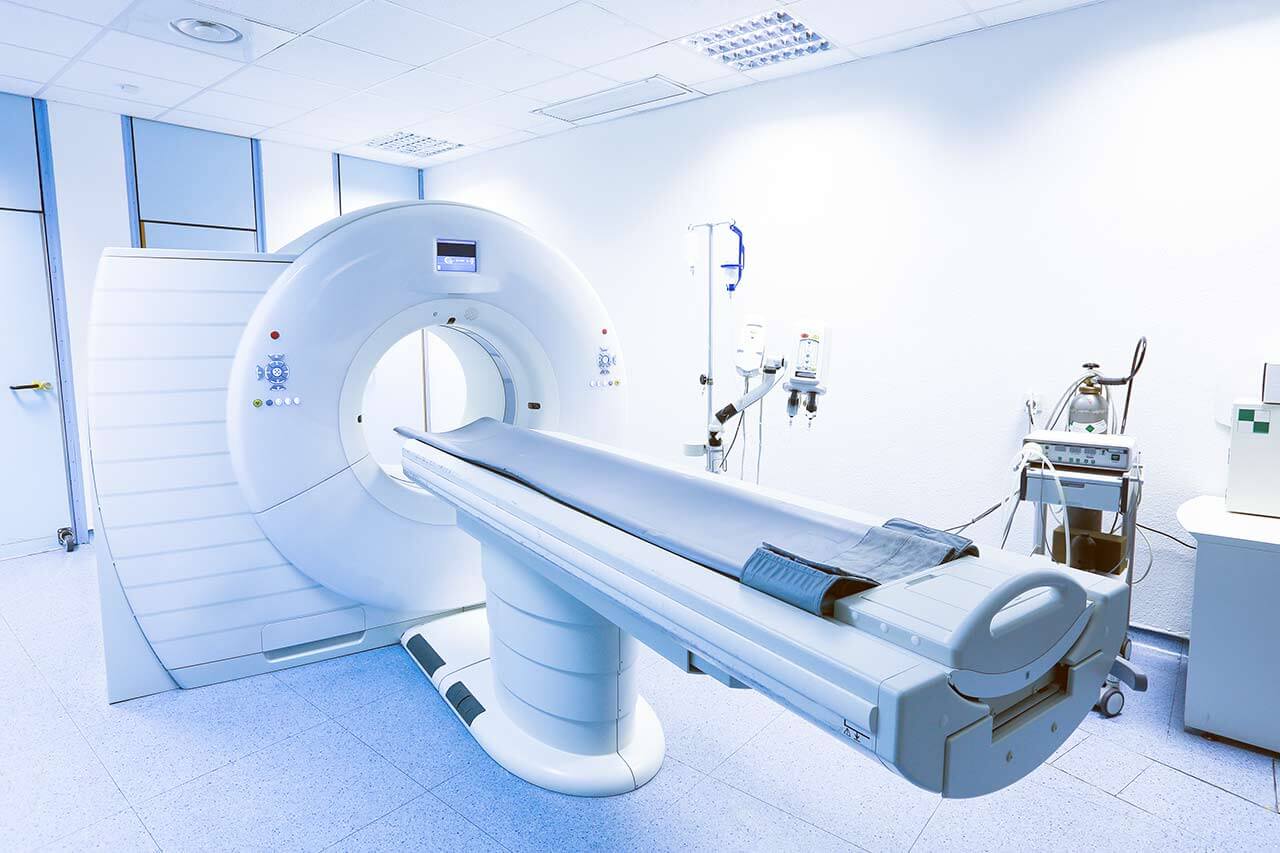
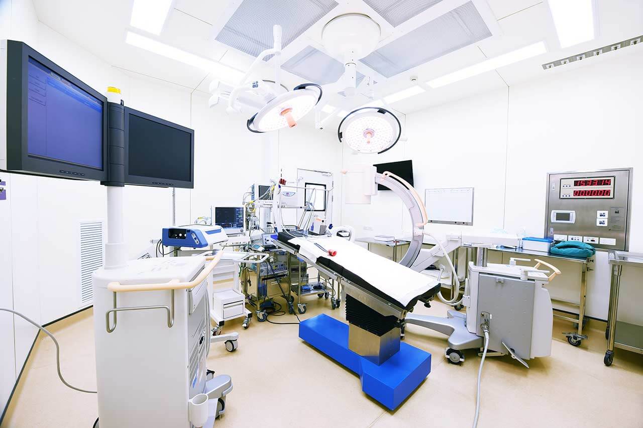
The medical team of the hospital consists of highly professional therapists, surgeons and nursing staff. The focus of their efforts is on the patient, his health and peace of mind, as well as comfort during treatment. The clinical practice of doctors is based on an individual approach to each case, which results in high treatment success rates. State-of-the-art technical equipment also plays an important role in the therapeutic process. The hospital is proud of the most advanced devices for imaging diagnostics (X-ray, ultrasound, CT, MRI, PET-CT, SPECT-CT, etc.), endoscopic examinations, laboratory tests, as well as specially equipped operating rooms for robot-assisted interventions, image-guided therapeutic manipulations, minimally invasive and classical surgeries of any complexity. Thus, the doctors of the university hospital have all the necessary resources to effectively treat the most severe pathologies and save lives.
The combination of high-tech equipment, experienced and highly qualified personnel, as well as strict adherence to the standards of modern medicine, form a solid foundation for the provision of the best medical care at the European level. An undeniable proof of the high prestige of the hospital is the constantly growing number of patients who come here from various regions of Germany and other countries of the world.
Photo: (с) depositphotos
Accommodation in hospital
Patients rooms
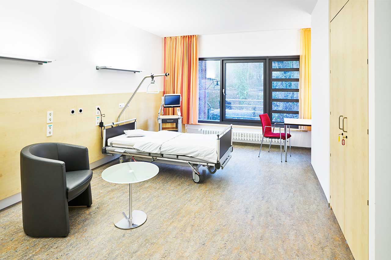
The patients of the University Hospital Erlangen live in comfortable rooms with light colors and modern design. Each patient room has an ensuite bathroom with shower and toilet. The furnishing of the patient room includes an automatically adjustable bed with an orthopedic mattress, a bedside table, a wardrobe, a table and chairs for receiving visitors, a TV, a radio and a telephone. Wi-Fi can be provided upon request. The use of a mobile phone is prohibited in many rooms of the hospital.
Patients can also live in enhanced-comfort rooms with a more sophisticated design. The enhanced-comfort rooms additionally include upholstered furniture, a minifridge and a safe.
Meals and Menus
The hospital offers healthy and tasty food distinguished by many awards, including the 1st place in the prestigious ESSEN PRO GESUNDHEIT competition of the Bavarian State Ministry of the Environment and Consumer Protection.
The patient and the accompanying person have three meals a day. Breakfast is served buffet style: scrambled eggs, boiled eggs, sausage, cheese, bread and buns with butter and jam, cereals, etc. There are three set menus for lunch and dinner to choose from: a classic menu featuring local cuisine dishes, a Mediterranean menu and a vegetarian menu.
If for some reason you do not eat all the foods, you will be offered an individual menu. Please inform the medical staff about your dietary preferences prior to the treatment.
The hospital also houses many cafeterias, which will delight with a wide range of delicious dishes and drinks.
Further details
Standard rooms include:
Religion
The hospital regularly hosts catholic and evangelical devine services. The services of representatives of other religions are available upon request.
Accompanying person
During an inpatient program, an accompanying person can stay with you in the patient room or in a hotel of your choice.
Hotel
During an outpatient program, you can stay in a hotel of your choice. The managers will help you choose the most suitable options.


