{"translation_price":"50","translation_doc_price":"40","child_coefficient":"1.1","transfer_price":"2.00","transfer_price_vip":"5.00","constant_transfer_price_vip":350,"constant_transfer_price":150,"constant_transfer_distanse":60,"type":"treatment","program_full_story":"<ul>\n\t<li>Initial presentation in the clinic<\/li>\n\t<li>clinical history taking<\/li>\n\t<li>review of medical records<\/li>\n\t<li>physical examination<\/li>\n\t<li>laboratory tests:\n\t<ul>\n\t\t<li>complete blood count<\/li>\n\t\t<li>general urine analysis<\/li>\n\t\t<li>biochemical analysis of blood<\/li>\n\t\t<li>inflammation indicators (CRP, ESR)<\/li>\n\t\t<li>indicators of blood coagulation<\/li>\n\t<\/ul>\n\t<\/li>\n\t<li>neurological examination<\/li>\n\t<li>CT\/MRI scan<\/li>\n\t<li>neuropsychological tests (if indicated):\n\t<ul>\n\t\t<li>ENMG (electroneuromyography)<\/li>\n\t\t<li>EEG (electroencephalography)<\/li>\n\t\t<li>SEPs (somatosensory evoked potentials)<\/li>\n\t\t<li>VEPs (visually evoked potentials)<\/li>\n\t\t<li>BAEP tests (brainstem auditory evoked potentials)<\/li>\n\t<\/ul>\n\t<\/li>\n\t<li>preoperative care<\/li>\n\t<li>MRI-guided laser ablation of glioblastoma<\/li>\n\t<li>postoperative MRI control<\/li>\n\t<li>symptomatic treatment<\/li>\n\t<li>control examinations<\/li>\n\t<li>the cost of essential medicines and materials<\/li>\n\t<li>nursing services<\/li>\n\t<li>full hospital accommodation<\/li>\n\t<li>developing of further guidance by specialists<br \/>\n\tin oncology and radiotherapy<\/li>\n<\/ul>\n<div class=\"program_how_program_going mt-4\"><h4>How program is carried out<\/h4><p style=\"text-align:justify\"><strong>During the first visit<\/strong>, the physician will conduct a clinical examination and neurological examination, and go through the results of the available diagnostic tests. After that, you will undergo the necessary additional examination, such as the laboratory blood and urine tests, MRI scan with obtaining 3D images and creating volumetric images of the whole brain. Based on the results of an additional examination, the physician will clarify the localization of glioblastoma. After that, the preparation according to the preoperative standard will begin.<\/p>\n\n<p style=\"text-align:justify\"><strong>Laser ablation of glioblastoma<\/strong> begins with the formation of access to the brain. When using a catheter with a laser fiber, a hole with a diameter of 3.2 mm is sufficient for this. The miniature size and flexibility of the catheter help preserve healthy brain tissue.<\/p>\n\n<p style=\"text-align:justify\">When the catheter reaches glioblastoma, energy is delivered through the laser fiber. Under the influence of high temperature, glioblastoma is destroyed. The procedure is guided by real-time MRI. Surgeons also assess the temperature of healthy brain tissues using thermographic images in order to avoid overheating.<\/p>\n\n<p style=\"text-align:justify\"><strong>After the completion of the operation<\/strong>, you will be transferred to the ward, under the round-the-clock supervision of physicians and nurses. In several days, the attending physician will evaluate the results of the postoperative MRI scans, schedule the date of discharge from the hospital and give you detailed recommendations for further follow-up and treatment.<\/p>\n<\/div><div class=\"program_required_documents mt-4\"><h4>Required documents<\/h4><ul>\n\t<li style=\"text-align: justify;\">Medical records<\/li>\n\t<li style=\"text-align: justify;\">MRI\/CT scan (not older than 3 months)<\/li>\n\t<li style=\"text-align: justify;\">Biopsy results (if available)<\/li>\n<\/ul>\n<\/div>","program_full_story_crm":"<ul>\n\t<li>Initial presentation in the clinic<\/li>\n\t<li>clinical history taking<\/li>\n\t<li>review of medical records<\/li>\n\t<li>physical examination<\/li>\n\t<li>laboratory tests:\n\t<ul>\n\t\t<li>complete blood count<\/li>\n\t\t<li>general urine analysis<\/li>\n\t\t<li>biochemical analysis of blood<\/li>\n\t\t<li>inflammation indicators (CRP, ESR)<\/li>\n\t\t<li>indicators of blood coagulation<\/li>\n\t<\/ul>\n\t<\/li>\n\t<li>neurological examination<\/li>\n\t<li>CT\/MRI scan<\/li>\n\t<li>neuropsychological tests (if indicated):\n\t<ul>\n\t\t<li>ENMG (electroneuromyography)<\/li>\n\t\t<li>EEG (electroencephalography)<\/li>\n\t\t<li>SEPs (somatosensory evoked potentials)<\/li>\n\t\t<li>VEPs (visually evoked potentials)<\/li>\n\t\t<li>BAEP tests (brainstem auditory evoked potentials)<\/li>\n\t<\/ul>\n\t<\/li>\n\t<li>preoperative care<\/li>\n\t<li>MRI-guided laser ablation of glioblastoma<\/li>\n\t<li>postoperative MRI control<\/li>\n\t<li>symptomatic treatment<\/li>\n\t<li>control examinations<\/li>\n\t<li>the cost of essential medicines and materials<\/li>\n\t<li>nursing services<\/li>\n\t<li>full hospital accommodation<\/li>\n\t<li>developing of further guidance by specialists<br \/>\n\tin oncology and radiotherapy<\/li>\n<\/ul>\n<div class=\"program_how_program_going mt-4\"><h4>How program is carried out<\/h4><p style=\"text-align:justify\"><strong>During the first visit<\/strong>, the physician will conduct a clinical examination and neurological examination, and go through the results of the available diagnostic tests. After that, you will undergo the necessary additional examination, such as the laboratory blood and urine tests, MRI scan with obtaining 3D images and creating volumetric images of the whole brain. Based on the results of an additional examination, the physician will clarify the localization of glioblastoma. After that, the preparation according to the preoperative standard will begin.<\/p>\n\n<p style=\"text-align:justify\"><strong>Laser ablation of glioblastoma<\/strong> begins with the formation of access to the brain. When using a catheter with a laser fiber, a hole with a diameter of 3.2 mm is sufficient for this. The miniature size and flexibility of the catheter help preserve healthy brain tissue.<\/p>\n\n<p style=\"text-align:justify\">When the catheter reaches glioblastoma, energy is delivered through the laser fiber. Under the influence of high temperature, glioblastoma is destroyed. The procedure is guided by real-time MRI. Surgeons also assess the temperature of healthy brain tissues using thermographic images in order to avoid overheating.<\/p>\n\n<p style=\"text-align:justify\"><strong>After the completion of the operation<\/strong>, you will be transferred to the ward, under the round-the-clock supervision of physicians and nurses. In several days, the attending physician will evaluate the results of the postoperative MRI scans, schedule the date of discharge from the hospital and give you detailed recommendations for further follow-up and treatment.<\/p>\n<\/div>","is_ambulant":"0","bh_fee":"0","only_for_children":"0","no_service":"0","with_prepayment":"1","show_calculator":"1","paket_type":"1","btn_type":"2","clinic_icon":"5ffc48d6204db.jpg","city":"Mainz","clinic_site":"http:\/\/www.unimedizin-mainz.de\/","department_recommend":"0","country":"Germany","country_id":"1","clinic_name":"University Hospital Mainz","cinic_name":"University Hospital Mainz","department_id":"3032","duration":"5","direction":"Neurosurgery","min_duration":0,"clinic_id":"1699","paketPrice":13800,"paket":"<ul>\n <li>Interpreter up to 30 hours<\/li>\n <li>Translation up to 15 pages<\/li>\n <li>Visa support<\/li>\n\n<\/ul>","title":"Glioblastoma treatment with MRI-guided laser ablation","price":{"val":70953.78,"type":"val"},"price_surcharge":0,"price_surcharge_clear":0,"extra_service_clinic":[{"id":"need_to_use_head_doctor","title":"Treatment by leading experts","type":"val","val":"13824.00","req":0},{"id":"1_person_room","title":" Single room (whole period)","parent":"duration","type":"count_val","val":"76.00"},{"id":"2_person_room","title":" double room (whole period)","parent":"duration","type":"count_val","val":"27.00"},{"id":"attendant_place","title":"Accommodation for the accompanying person ","parent":"duration","type":"count_val","val":"90.00"}],"extra_service":[],"translation_hours":"0","translation_doc_count":null,"roads":[{"id":"6","distance":"37","airport_title":"Frankfurt a.M."},{"id":"7","distance":"168","airport_title":"Cologne-Bonn"},{"id":"19","distance":"211","airport_title":"Stuttgart"}],"pakets":[],"lang":{"day":"Day","days":"days","ambulatory":"Outpatient","stationaryProgram":"Inpatient"}}
Glioblastoma treatment with MRI-guided laser ablation in University Hospital Mainz
developing of further guidance by specialists in oncology and radiotherapy
How program is carried out
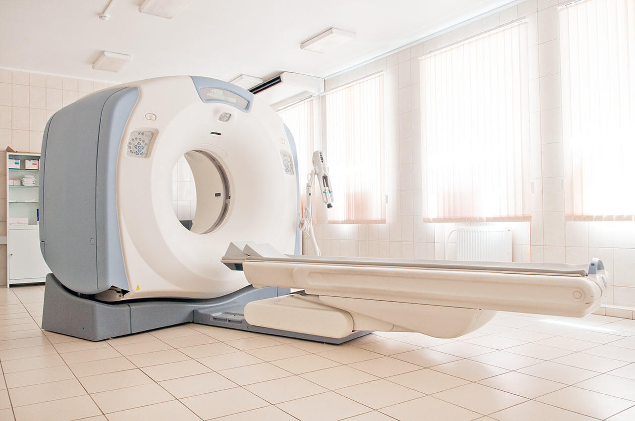
During the first visit, the physician will conduct a clinical examination and neurological examination, and go through the results of the available diagnostic tests. After that, you will undergo the necessary additional examination, such as the laboratory blood and urine tests, MRI scan with obtaining 3D images and creating volumetric images of the whole brain. Based on the results of an additional examination, the physician will clarify the localization of glioblastoma. After that, the preparation according to the preoperative standard will begin.
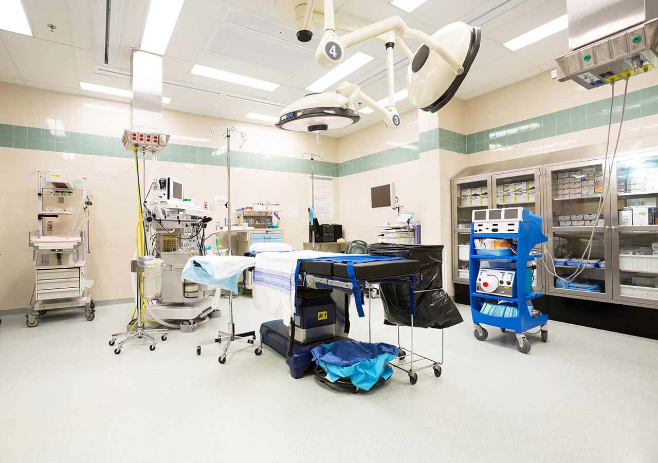
Laser ablation of glioblastoma begins with the formation of access to the brain. When using a catheter with a laser fiber, a hole with a diameter of 3.2 mm is sufficient for this. The miniature size and flexibility of the catheter help preserve healthy brain tissue.
When the catheter reaches glioblastoma, energy is delivered through the laser fiber. Under the influence of high temperature, glioblastoma is destroyed. The procedure is guided by real-time MRI. Surgeons also assess the temperature of healthy brain tissues using thermographic images in order to avoid overheating.
After the completion of the operation, you will be transferred to the ward, under the round-the-clock supervision of physicians and nurses. In several days, the attending physician will evaluate the results of the postoperative MRI scans, schedule the date of discharge from the hospital and give you detailed recommendations for further follow-up and treatment.
Required documents
Medical records
MRI/CT scan (not older than 3 months)
Biopsy results (if available)
Service
Price from:
Type of program :
Price for 1 day:
Expected duration of the program:
The minimum duration of the program:
Select
You may also book:
Hospital directly:
Saving:
BookingHealth Price from:
About the department
The Department of Adult and Pediatric Neurosurgery at the University Hospital Mainz offers the full range of services in this field. The department specializes in the surgical treatment of diseases of the brain, spine and peripheral nerves in adults and children. During the treatment, the department uses the very latest surgical techniques and technical equipment, for example, robotic spinal surgery. The department is one of the leading neurosurgical medical institutions in Germany and Europe. The Chief Physician of the department is Prof. Dr. med. Florian Ringel.
It should be noted that one of the most important focuses of the department is the treatment of cancer of the nervous system. In this field, the department is certified as the Neuro-Oncology Center by the German Cancer Society.
In addition, the department specializes in early rehabilitation (phase B), which plays a key role in the success of therapy. For this purpose, an interdisciplinary team of professionals, such as highly specialized doctors, therapists, nurses and social workers, work here.
The main clinical focuses of the department include:
Surgical treatment of benign meningiomas
Surgical treatment of pituitary tumors (in most cases benign ones)
Surgical treatment of benign peripheral nerve tumors
Surgical treatment of gliomas and glioblastomas
Surgical treatment of the spinal diseases – robotic surgery using the Renaissance™ Mini Roboter system (the system was used here in Europe for the first time)
Functional interventions for the treatment of chronic pain and movement disorders
Pediatric neurosurgery
Surgical treatment of craniosynostosis
Surgical treatment of scaphocephaly/dolichocephaly
Surgical treatment of trigonocephaly
Surgical treatment of hydrocephalus
Psycho-oncological care
Ergotherapy
Speech therapy
Physiotherapy
Self-help groups
Intensive care
Other medical services
Curriculum vitae
1991 - 1998 Study of Human Medicine at the Ludwig Maximilian University of Munich.
1999 - 2000 Intern at the Institute for Surgical Research at the University of Munich (Head: Prof. Dr. med. Dr. h.c. K. Messmer).
July 2000 Admission to medical practice.
2000 - 2001 Research Assistant at the Institute for Surgical Research at the University of Munich (Head: Prof. Dr. med. Dr. h.c. K. Messmer).
2001 - 2006 Research Assistant, Department of Neurosurgery, University Hospital Bonn (Head: Prof. Dr. med. J. Schramm).
2004 Doctoral thesis defense (Doctor of Medicine). Subject: "Acidosis-induced edema and intracellular acidosis of glial cells – the value of anions and calcium".
2006 Medical Specialist in Neurosurgery (Medical Association of North Rhine-Westphalia).
Since 2006 Senior Physician in the Department of Neurosurgery, University Hospital Rechts der Isar Munich (Head: Prof. Dr. med. B. Meyer).
2009 Habilitation (Neurosurgery) and Venia Legendi.
Photo of the doctor: (c) Universitätsmedizin der Johannes Gutenberg-Universität Mainz
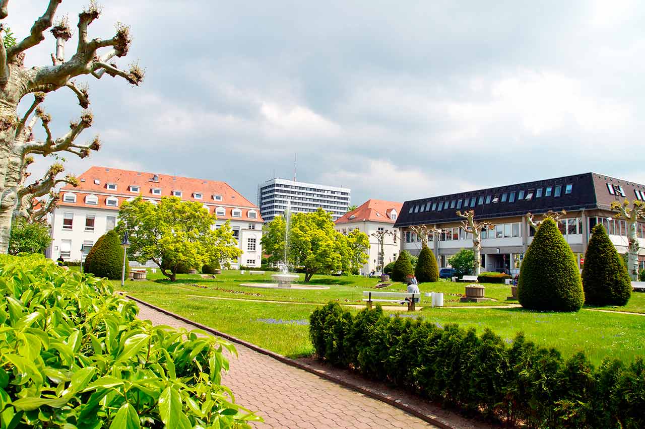
About hospital
The University Hospital Mainz is one of the best maximum care medical facilities in Germany and an internationally recognized scientific center. There are more than 60 departments and institutes, which represent all fields of modern medicine. The hospital serves more than 68,000 inpatients and more than 273,000 outpatients annually, which testifies to the excellent reputation of this medical institution.
The key to the successful clinical practice is also a highly qualified medical staff, which consists of 7.800 employees from various fields. The doctors of the hospital are convinced that each clinical case requires an individual approach, therefore, they devote much time to consultations and communication with patients. The main goal of all hospital employees is to provide an optimal medical care based on the use of the state-of-art diagnostic and therapeutic measures, as well as the introduction of the latest scientific achievements into the medical practice.
The best interns and assistant physicians are trained here. The world-famous leading physicians of the hospital share their long experience and professional skills. Naturally, an integral part of the university hospital work is research, thanks to which many innovative possibilities in the field of diagnostics and therapy have been developed.
Photo: (c) depositphotos
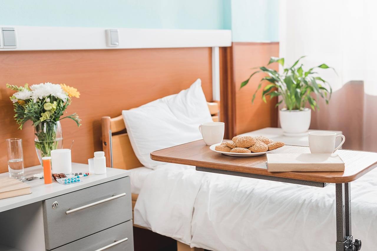
Accommodation in hospital
Patients rooms
The patients of the University Hospital Mainz live in bright, spacious and comfortable rooms. The patient rooms are equipped with modern electronically adjustable beds, which at the touch of a button automatically assume a specified position. Also, there is a TV and a telephone in the patient rooms. To use them, it is necessary to get a prepaid card (in vending machines at the entrance to the hospital). The use of a TV is free, but the patient will need headphones. Telephone calls are made for a fixed fee, which will be withdrawn from the prepaid card at discharge from the hospital. Also, each patient room is equipped with an ensuite bathroom with shower, washbasin and toilet.
Meals and Menus
The patients of the hospital are offered a tasty and balanced three meals a day: breakfast, lunch and dinner. The menu also includes vegetarian and dietary dishes. In addition, for both patients and their visitors there are available cafeterias and bakeries, where one can buy baked goods, snacks, hot and cold drinks.
Further details
Standard rooms include:
Shower
Toilet
TV
Select your currency:
Your guarantee
We are the only TÜV-certified company with an ISO 9001:2015 certificate
thank you for reaching out! One of our medical advisors is currently reviewing your details. You can expect a call from us within one business day to discuss your request.
Note: The incoming call will appear as a German number or your local area code. This call is entirely free of charge.
Booking Health guarantees:
Expert Placement
We use rigorous statistical analysis to ensure you are treated at the best-rated facility for your specific diagnosis.
Total Cost Protection
No hidden fees or unexpected bills. We provide a fixed price guarantee, backed by insurance that covers any additional complications.
12-Month Extended Care
Your recovery is our priority. Benefit from a full year of direct medical support following your procedure.

 BookingHealth Price from:
