{"translation_price":"50","translation_doc_price":"40","child_coefficient":"1.1","transfer_price":"2.00","transfer_price_vip":"5.00","constant_transfer_price_vip":350,"constant_transfer_price":150,"constant_transfer_distanse":60,"type":"treatment","program_full_story":"<ul>\n\t<li>Initial presentation in the hospital<\/li>\n\t<li>Clinical history taking<\/li>\n\t<li>Review of available medical records<\/li>\n\t<li>Physical examination<\/li>\n\t<li>Laboratory tests:\n\t<ul>\n\t\t<li>Complete blood count<\/li>\n\t\t<li>General urine analysis<\/li>\n\t\t<li>Biochemical analysis of blood<\/li>\n\t\t<li>Tumor markers<\/li>\n\t\t<li>Inflammation indicators (CRP, ESR)<\/li>\n\t\t<li>Coagulogram<\/li>\n\t<\/ul>\n\t<\/li>\n\t<li>Ultrasound\u200b scan<\/li>\n\t<li>CT scan \/ MRI<\/li>\n\t<li>Preoperative care<\/li>\n\t<li>Embolization or chemoembolization, 2 procedures<\/li>\n\t<li>Symptomatic treatment<\/li>\n\t<li>Cost of essential medicines<\/li>\n\t<li>Nursing services<\/li>\n\t<li>Elaboration of further recommendations<\/li>\n<\/ul>\n<div class=\"program_how_program_going mt-4\"><h4>How program is carried out<\/h4><p style=\"text-align: justify;\"><strong>During the first visit<\/strong>, the doctor will conduct a clinical examination and go through the results of the available diagnostic tests. After that, you will undergo the necessary additional examination, such as the assessment of liver and kidney function, ultrasound scan, CT scan and MRI. This will allow the doctor to determine which vessels are feeding the tumor and its metastases, as well as determine how well you will tolerate the procedure.<\/p>\n\n<p style=\"text-align: justify;\"><strong>Chemoembolization <\/strong>begins with local anesthesia and catheterization of the femoral artery. The thin catheter is inserted through a few centimeters long incision of the blood vessel. The doctor gradually moves the catheter to the vessel feeding the primary tumor or its metastases. The procedure is carried out under visual control, an angiographic device is used for this. The vascular bed and the position of the catheter in it are displayed on the screen of the angiograph.<\/p>\n\n<p style=\"text-align: justify;\">When the catheter reaches a suspected artery, a contrast agent is injected through it. Due to the introduction of the contrast agent, the doctor clearly sees the smallest vessels of the tumor and the surrounding healthy tissues on the screen of the angiograph. After that, he injects emboli into the tumor vessels through the same catheter.<\/p>\n\n<p style=\"text-align: justify;\">Emboli are the spirals or the liquid microspheres. The type of embolus is selected individually, taking into account the diameter of the target vessel. When carrying out chemoembolization, a solution of a chemotherapy drug is additionally injected into the tumor vessel. Due to the subsequent closure of the vessel lumen with an embolus, the chemotherapy drug influences the tumor for a long time. In addition, the drug does not enter the systemic circulation, which allows doctors to use high doses of chemotherapeutic agents without the development of serious side effects. Chemoembolization leads to the destruction of the tumor or slowing down its progression.<\/p>\n\n<p style=\"text-align: justify;\">After that, the catheter is removed from the artery. The doctor puts a vascular suture on the femoral artery and closes it with a sterile dressing. During chemoembolization, you will be awake. General anesthesia is not used, which significantly reduces the risks of the procedure and allows performing it on an outpatient basis, avoiding long hospital stay.<\/p>\n\n<p style=\"text-align: justify;\"><strong>After the first procedure<\/strong>, you will stay under the supervision of an interventional oncologist and general practitioner. If necessary, you will receive symptomatic treatment. As a rule, a second chemoembolization procedure is performed in 3-5 days after the first one in order to consolidate the therapeutic effect. After that, you will receive recommendations for further follow-up and treatment.<\/p>\n<\/div><div class=\"program_required_documents mt-4\"><h4>Required documents<\/h4><ul>\n\t<li style=\"text-align: justify;\">Medical records<\/li>\n\t<li style=\"text-align: justify;\">MRI\/CT scan (not older than 3 months)<\/li>\n\t<li style=\"text-align: justify;\">Biopsy results (if available)<\/li>\n<\/ul>\n<\/div>","program_full_story_crm":"<ul>\n\t<li>Initial presentation in the hospital<\/li>\n\t<li>Clinical history taking<\/li>\n\t<li>Review of available medical records<\/li>\n\t<li>Physical examination<\/li>\n\t<li>Laboratory tests:\n\t<ul>\n\t\t<li>Complete blood count<\/li>\n\t\t<li>General urine analysis<\/li>\n\t\t<li>Biochemical analysis of blood<\/li>\n\t\t<li>Tumor markers<\/li>\n\t\t<li>Inflammation indicators (CRP, ESR)<\/li>\n\t\t<li>Coagulogram<\/li>\n\t<\/ul>\n\t<\/li>\n\t<li>Ultrasound\u200b scan<\/li>\n\t<li>CT scan \/ MRI<\/li>\n\t<li>Preoperative care<\/li>\n\t<li>Embolization or chemoembolization, 2 procedures<\/li>\n\t<li>Symptomatic treatment<\/li>\n\t<li>Cost of essential medicines<\/li>\n\t<li>Nursing services<\/li>\n\t<li>Elaboration of further recommendations<\/li>\n<\/ul>\n<div class=\"program_how_program_going mt-4\"><h4>How program is carried out<\/h4><p style=\"text-align: justify;\"><strong>During the first visit<\/strong>, the doctor will conduct a clinical examination and go through the results of the available diagnostic tests. After that, you will undergo the necessary additional examination, such as the assessment of liver and kidney function, ultrasound scan, CT scan and MRI. This will allow the doctor to determine which vessels are feeding the tumor and its metastases, as well as determine how well you will tolerate the procedure.<\/p>\n\n<p style=\"text-align: justify;\"><strong>Chemoembolization <\/strong>begins with local anesthesia and catheterization of the femoral artery. The thin catheter is inserted through a few centimeters long incision of the blood vessel. The doctor gradually moves the catheter to the vessel feeding the primary tumor or its metastases. The procedure is carried out under visual control, an angiographic device is used for this. The vascular bed and the position of the catheter in it are displayed on the screen of the angiograph.<\/p>\n\n<p style=\"text-align: justify;\">When the catheter reaches a suspected artery, a contrast agent is injected through it. Due to the introduction of the contrast agent, the doctor clearly sees the smallest vessels of the tumor and the surrounding healthy tissues on the screen of the angiograph. After that, he injects emboli into the tumor vessels through the same catheter.<\/p>\n\n<p style=\"text-align: justify;\">Emboli are the spirals or the liquid microspheres. The type of embolus is selected individually, taking into account the diameter of the target vessel. When carrying out chemoembolization, a solution of a chemotherapy drug is additionally injected into the tumor vessel. Due to the subsequent closure of the vessel lumen with an embolus, the chemotherapy drug influences the tumor for a long time. In addition, the drug does not enter the systemic circulation, which allows doctors to use high doses of chemotherapeutic agents without the development of serious side effects. Chemoembolization leads to the destruction of the tumor or slowing down its progression.<\/p>\n\n<p style=\"text-align: justify;\">After that, the catheter is removed from the artery. The doctor puts a vascular suture on the femoral artery and closes it with a sterile dressing. During chemoembolization, you will be awake. General anesthesia is not used, which significantly reduces the risks of the procedure and allows performing it on an outpatient basis, avoiding long hospital stay.<\/p>\n\n<p style=\"text-align: justify;\"><strong>After the first procedure<\/strong>, you will stay under the supervision of an interventional oncologist and general practitioner. If necessary, you will receive symptomatic treatment. As a rule, a second chemoembolization procedure is performed in 3-5 days after the first one in order to consolidate the therapeutic effect. After that, you will receive recommendations for further follow-up and treatment.<\/p>\n<\/div>","is_ambulant":"1","bh_fee":"0","only_for_children":"0","no_service":"0","with_prepayment":"1","show_calculator":"1","paket_type":"1","btn_type":"0","clinic_icon":"5ffc48d6204db.jpg","city":"Mainz","clinic_site":"http:\/\/www.unimedizin-mainz.de\/","department_recommend":"0","country":"Germany","country_id":"1","clinic_name":"University Hospital Mainz","cinic_name":"University Hospital Mainz","department_id":"3047","duration":"7","direction":"Interventional radiology","min_duration":0,"clinic_id":"1699","paketPrice":7800,"paket":"<ul>\n <li>Interpreter up to 17 hours<\/li>\n <li>Translation up to 10 pages<\/li>\n <li>Visa support<\/li>\n\n \n<\/ul>","title":"Treatment of lung cancer with embolization or chemoembolization","price":{"val":29426.46,"type":"val"},"price_surcharge":0,"price_surcharge_clear":0,"extra_service_clinic":[],"extra_service":[],"translation_hours":"0","translation_doc_count":null,"roads":[{"id":"6","distance":"37","airport_title":"Frankfurt a.M."},{"id":"7","distance":"168","airport_title":"Cologne-Bonn"},{"id":"19","distance":"211","airport_title":"Stuttgart"}],"pakets":[],"lang":{"day":"Day","days":"days","ambulatory":"Outpatient","stationaryProgram":"Inpatient"}}
Treatment of lung cancer with embolization or chemoembolization in University Hospital Mainz
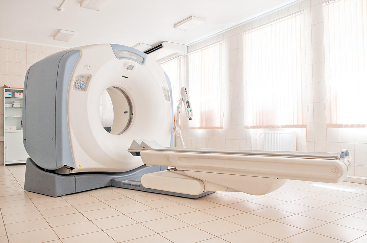
During the first visit, the doctor will conduct a clinical examination and go through the results of the available diagnostic tests. After that, you will undergo the necessary additional examination, such as the assessment of liver and kidney function, ultrasound scan, CT scan and MRI. This will allow the doctor to determine which vessels are feeding the tumor and its metastases, as well as determine how well you will tolerate the procedure.
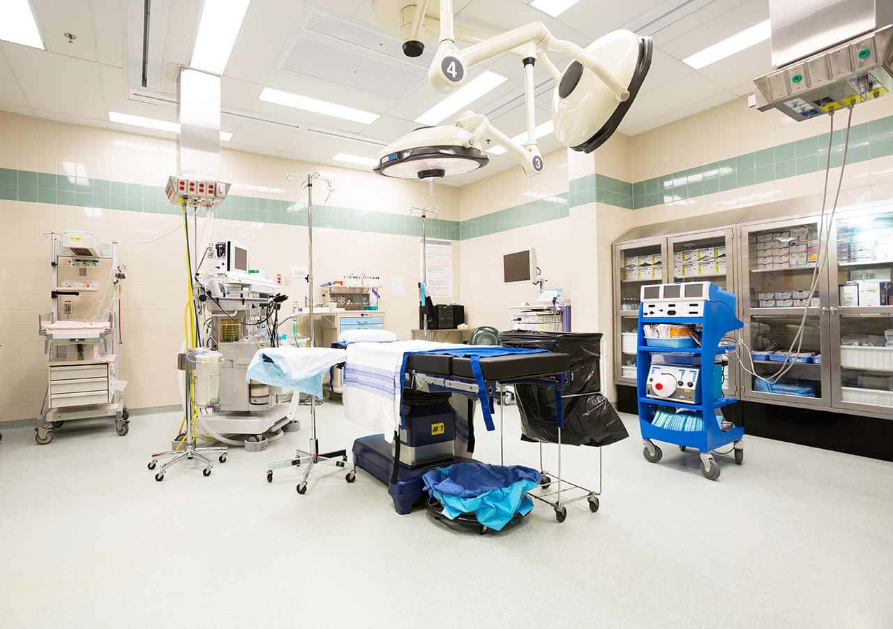
Chemoembolization begins with local anesthesia and catheterization of the femoral artery. The thin catheter is inserted through a few centimeters long incision of the blood vessel. The doctor gradually moves the catheter to the vessel feeding the primary tumor or its metastases. The procedure is carried out under visual control, an angiographic device is used for this. The vascular bed and the position of the catheter in it are displayed on the screen of the angiograph.
When the catheter reaches a suspected artery, a contrast agent is injected through it. Due to the introduction of the contrast agent, the doctor clearly sees the smallest vessels of the tumor and the surrounding healthy tissues on the screen of the angiograph. After that, he injects emboli into the tumor vessels through the same catheter.
Emboli are the spirals or the liquid microspheres. The type of embolus is selected individually, taking into account the diameter of the target vessel. When carrying out chemoembolization, a solution of a chemotherapy drug is additionally injected into the tumor vessel. Due to the subsequent closure of the vessel lumen with an embolus, the chemotherapy drug influences the tumor for a long time. In addition, the drug does not enter the systemic circulation, which allows doctors to use high doses of chemotherapeutic agents without the development of serious side effects. Chemoembolization leads to the destruction of the tumor or slowing down its progression.
After that, the catheter is removed from the artery. The doctor puts a vascular suture on the femoral artery and closes it with a sterile dressing. During chemoembolization, you will be awake. General anesthesia is not used, which significantly reduces the risks of the procedure and allows performing it on an outpatient basis, avoiding long hospital stay.
After the first procedure, you will stay under the supervision of an interventional oncologist and general practitioner. If necessary, you will receive symptomatic treatment. As a rule, a second chemoembolization procedure is performed in 3-5 days after the first one in order to consolidate the therapeutic effect. After that, you will receive recommendations for further follow-up and treatment.
Required documents
Medical records
MRI/CT scan (not older than 3 months)
Biopsy results (if available)
Service
Price from:
Type of program :
Price for 1 day:
Expected duration of the program:
The minimum duration of the program:
Select
You may also book:
Hospital directly:
Saving:
BookingHealth Price from:
About the department
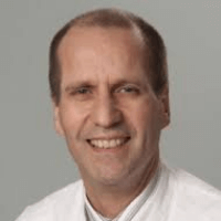
The Department of Adult and Pediatric Diagnostic, Interventional Radiology at the University Hospital Mainz offers the full range of services in these medical fields. Patient care is provided both on an outpatient and inpatient basis. The department has all the modern imaging methods for the most accurate diagnostics and interventional therapy. The highly qualified doctors with extensive clinical experience and high rates of successful treatment take care of patient health. The department is headed by Prof. Dr. med. Christoph Düber.
The service range of the department includes:
Diagnostic radiology
Conventional X-ray studies
Functional diagnostics
Contrast-enhanced examinations of the gastrointestinal tract
Joint examination (arthrography in combination with MRI or CT)
Spinal cord examination (myelography)
Venous examination (phlebography)
Bile duct examination
Ultrasound diagnostics (in accordance with the requirements of the German Society for Ultrasound in Medicine)
Breast imaging diagnostics and minimally invasive biopsy
Full-field digital mammography with a minimum radiation dose
Tomosynthesis
Magnetic resonance imaging (MRI)
High resolution ultrasound with color duplex sonography (3D and 4D)
Needle biopsy and stereotactic vacuum biopsy
Galactography
Preoperative marking and intraoperative radiography
Second opinion and consultation on diagnostics
Computed tomography (CT)
Combined PET-CT method
Magnetic resonance imaging (MRI)
MRI of the musculoskeletal system (joints, ligaments, muscles, tendons, bones)
MRI of the central nervous system (brain and spinal cord)
MRI of the abdominal cavity and retroperitoneal space (liver, spleen, pancreas, kidney)
MRI of the pelvic organs (bladder, rectum, genital organs)
MRI of the heart and blood vessels
Angiography
Interventional radiology
Patency restoration of the narrowed or occluded blood vessels of the legs and arms, head and neck, internal organs (for example, liver, kidneys, intestines or lungs) using a balloon catheter, a stent or thrombolysis
Treatment of thoracic and abdominal aortic aneurysms (EVAR) and aneurysms of other arteries
Treatment of vascular dissections and vascular lesions
Blood vessel closure (embolization) in bleeding and tumors
Uterine artery embolization for the treatment of uterine myomas
Puncture (tissue sampling)
Drainage placement to eliminate abscesses and fluid accumulations
Local ablation therapy of tumors using radiofrequency ablation, irreversible electroporation and microwave ablation (in particular, in tumors of the liver, lungs, bones and other organs)
Percutaneous transhepatic gallbladder drainage
Stenting in esophageal and intestinal stenosis
Installation of feeding tubes in the stomach and intestines (gastrostomy, jejunostomy)
Transjugular intrahepatic portosystemic shunting
Pain therapy (for example, solar plexus block)
Vertebroplasty for the treatment of pathological vertebral fractures
Prostate artery embolization
Transarterial chemotherapy for the treatment of liver cancer and liver metastases
Internal selective radiation therapy and radioembolization for the treatment of liver cancer and liver metastases
Pediatric radiology
Ultrasound examinations
Computed tomography (CT)
Magnetic resonance imaging (MRI)
X-ray examinations
Other diagnostic and therapeutic options in radiology
Curriculum vitae
1976 -1982 Study of Human Medicine at the Johannes Gutenberg University Mainz, Fellow of the German Foundation Studienstiftung des Deutschen Volkes.
02.28.1984 Doctoral dissertation defense (summa cum laude), Johannes Gutenberg University Mainz.
Professional and Academic Training
1982 - 1984 Research Assistant, Institute of Pathology at the University of Mainz (Head: Prof. Dr. V. Thoenes).
1984 - 1989 Research Assistant, Department of Adult and Pediatric Diagnostic, Interventional Radiology, University Hospital Mainz (Head: Prof. Dr. M. Thelen).
06.03.1989 Medical Specialist in Radiology.
1991 - 2000 Senior Physician, Department of Adult and Pediatric Diagnostic, Interventional Radiology, University Hospital Mainz (Head: Prof. Dr. M. Thelen).
04.11.1993 Habilitation in Radiology.
01.11.2000 C4 Professor in Clinical Radiology, Department of Clinical Medicine at the University of Heidelberg, as well as Head of the Institute for Clinical Radiology at the University Hospital Mannheim.
01.05.2005 W3 Professor in Diagnostic and Interventional Radiology at the Johannes Gutenberg University Mainz and Head of the Department of Adult and Pediatric Diagnostic, Interventional Radiology, University Hospital Mainz.
Photo of the doctor: (c) Universitätsmedizin der Johannes Gutenberg-Universität Mainz
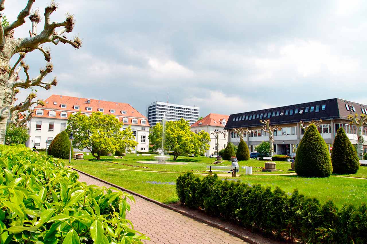
About hospital
The University Hospital Mainz is one of the best maximum care medical facilities in Germany and an internationally recognized scientific center. There are more than 60 departments and institutes, which represent all fields of modern medicine. The hospital serves more than 68,000 inpatients and more than 273,000 outpatients annually, which testifies to the excellent reputation of this medical institution.
The key to the successful clinical practice is also a highly qualified medical staff, which consists of 7.800 employees from various fields. The doctors of the hospital are convinced that each clinical case requires an individual approach, therefore, they devote much time to consultations and communication with patients. The main goal of all hospital employees is to provide an optimal medical care based on the use of the state-of-art diagnostic and therapeutic measures, as well as the introduction of the latest scientific achievements into the medical practice.
The best interns and assistant physicians are trained here. The world-famous leading physicians of the hospital share their long experience and professional skills. Naturally, an integral part of the university hospital work is research, thanks to which many innovative possibilities in the field of diagnostics and therapy have been developed.
Photo: (c) depositphotos
Accommodation in hospital
Patients rooms
The patients of the University Hospital Mainz live in bright, spacious and comfortable rooms. The patient rooms are equipped with modern electronically adjustable beds, which at the touch of a button automatically assume a specified position. Also, there is a TV and a telephone in the patient rooms. To use them, it is necessary to get a prepaid card (in vending machines at the entrance to the hospital). The use of a TV is free, but the patient will need headphones. Telephone calls are made for a fixed fee, which will be withdrawn from the prepaid card at discharge from the hospital. Also, each patient room is equipped with an ensuite bathroom with shower, washbasin and toilet.
Meals and Menus
The patients of the hospital are offered a tasty and balanced three meals a day: breakfast, lunch and dinner. The menu also includes vegetarian and dietary dishes. In addition, for both patients and their visitors there are available cafeterias and bakeries, where one can buy baked goods, snacks, hot and cold drinks.
Further details
Standard rooms include:
Shower
Toilet
TV
Select your currency:
Your guarantee
We are the only TÜV-certified company with an ISO 9001:2015 certificate
thank you for reaching out! One of our medical advisors is currently reviewing your details. You can expect a call from us within one business day to discuss your request.
Note: The incoming call will appear as a German number or your local area code. This call is entirely free of charge.
Booking Health guarantees:
Expert Placement
We use rigorous statistical analysis to ensure you are treated at the best-rated facility for your specific diagnosis.
Total Cost Protection
No hidden fees or unexpected bills. We provide a fixed price guarantee, backed by insurance that covers any additional complications.
12-Month Extended Care
Your recovery is our priority. Benefit from a full year of direct medical support following your procedure.

 BookingHealth Price from:
