{"translation_price":"50","translation_doc_price":"40","child_coefficient":"1.1","transfer_price":"2.00","transfer_price_vip":"5.00","constant_transfer_price_vip":350,"constant_transfer_price":150,"constant_transfer_distanse":60,"type":"treatment","program_full_story":"<ul>\n\t<li>Initial presentation in the hospital<\/li>\n\t<li>Clinical history taking<\/li>\n\t<li>Review of available medical records<\/li>\n\t<li>Physical examination<\/li>\n\t<li>Laboratory tests:\n\t<ul>\n\t\t<li>Complete blood count<\/li>\n\t\t<li>General urine analysis<\/li>\n\t\t<li>Biochemical analysis of blood<\/li>\n\t\t<li>Tumor markers<\/li>\n\t\t<li>Inflammation indicators (CRP, ESR)<\/li>\n\t\t<li>Coagulogram<\/li>\n\t<\/ul>\n\t<\/li>\n\t<li>Ultrasound\u200b scan<\/li>\n\t<li>CT scan \/ MRI<\/li>\n\t<li>Preoperative care<\/li>\n\t<li>Embolization or chemoembolization, 2 procedures<\/li>\n\t<li>Symptomatic treatment<\/li>\n\t<li>Cost of essential medicines<\/li>\n\t<li>Nursing services<\/li>\n\t<li>Elaboration of further recommendations<\/li>\n<\/ul>\n<div class=\"program_how_program_going mt-4\"><h4>How program is carried out<\/h4><p style=\"text-align: justify;\"><strong>During the first visit<\/strong>, the doctor will conduct a clinical examination and go through the results of the available diagnostic tests. After that, you will undergo the necessary additional examination, such as the assessment of liver and kidney function, ultrasound scan, CT scan and MRI. This will allow the doctor to determine which vessels are feeding the tumor and its metastases, as well as determine how well you will tolerate the procedure.<\/p>\n\n<p style=\"text-align: justify;\"><strong>Chemoembolization <\/strong>begins with local anesthesia and catheterization of the femoral artery. The thin catheter is inserted through a few centimeters long incision of the blood vessel. The doctor gradually moves the catheter to the vessel feeding the primary tumor or its metastases. The procedure is carried out under visual control, an angiographic device is used for this. The vascular bed and the position of the catheter in it are displayed on the screen of the angiograph.<\/p>\n\n<p style=\"text-align: justify;\">When the catheter reaches a suspected artery, a contrast agent is injected through it. Due to the introduction of the contrast agent, the doctor clearly sees the smallest vessels of the tumor and the surrounding healthy tissues on the screen of the angiograph. After that, he injects emboli into the tumor vessels through the same catheter.<\/p>\n\n<p style=\"text-align: justify;\">Emboli are the spirals or the liquid microspheres. The type of embolus is selected individually, taking into account the diameter of the target vessel. When carrying out chemoembolization, a solution of a chemotherapy drug is additionally injected into the tumor vessel. Due to the subsequent closure of the vessel lumen with an embolus, the chemotherapy drug influences the tumor for a long time. In addition, the drug does not enter the systemic circulation, which allows doctors to use high doses of chemotherapeutic agents without the development of serious side effects. Chemoembolization leads to the destruction of the tumor or slowing down its progression.<\/p>\n\n<p style=\"text-align: justify;\">After that, the catheter is removed from the artery. The doctor puts a vascular suture on the femoral artery and closes it with a sterile dressing. During chemoembolization, you will be awake. General anesthesia is not used, which significantly reduces the risks of the procedure and allows performing it on an outpatient basis, avoiding long hospital stay.<\/p>\n\n<p style=\"text-align: justify;\"><strong>After the first procedure<\/strong>, you will stay under the supervision of an interventional oncologist and general practitioner. If necessary, you will receive symptomatic treatment. As a rule, a second chemoembolization procedure is performed in 3-5 days after the first one in order to consolidate the therapeutic effect. After that, you will receive recommendations for further follow-up and treatment.<\/p>\n<\/div><div class=\"program_required_documents mt-4\"><h4>Required documents<\/h4><ul>\n\t<li style=\"text-align: justify;\">Medical records<\/li>\n\t<li style=\"text-align: justify;\">MRI\/CT scan (not older than 3 months)<\/li>\n\t<li style=\"text-align: justify;\">Biopsy results (if available)<\/li>\n<\/ul>\n<\/div>","program_full_story_crm":"<ul>\n\t<li>Initial presentation in the hospital<\/li>\n\t<li>Clinical history taking<\/li>\n\t<li>Review of available medical records<\/li>\n\t<li>Physical examination<\/li>\n\t<li>Laboratory tests:\n\t<ul>\n\t\t<li>Complete blood count<\/li>\n\t\t<li>General urine analysis<\/li>\n\t\t<li>Biochemical analysis of blood<\/li>\n\t\t<li>Tumor markers<\/li>\n\t\t<li>Inflammation indicators (CRP, ESR)<\/li>\n\t\t<li>Coagulogram<\/li>\n\t<\/ul>\n\t<\/li>\n\t<li>Ultrasound\u200b scan<\/li>\n\t<li>CT scan \/ MRI<\/li>\n\t<li>Preoperative care<\/li>\n\t<li>Embolization or chemoembolization, 2 procedures<\/li>\n\t<li>Symptomatic treatment<\/li>\n\t<li>Cost of essential medicines<\/li>\n\t<li>Nursing services<\/li>\n\t<li>Elaboration of further recommendations<\/li>\n<\/ul>\n<div class=\"program_how_program_going mt-4\"><h4>How program is carried out<\/h4><p style=\"text-align: justify;\"><strong>During the first visit<\/strong>, the doctor will conduct a clinical examination and go through the results of the available diagnostic tests. After that, you will undergo the necessary additional examination, such as the assessment of liver and kidney function, ultrasound scan, CT scan and MRI. This will allow the doctor to determine which vessels are feeding the tumor and its metastases, as well as determine how well you will tolerate the procedure.<\/p>\n\n<p style=\"text-align: justify;\"><strong>Chemoembolization <\/strong>begins with local anesthesia and catheterization of the femoral artery. The thin catheter is inserted through a few centimeters long incision of the blood vessel. The doctor gradually moves the catheter to the vessel feeding the primary tumor or its metastases. The procedure is carried out under visual control, an angiographic device is used for this. The vascular bed and the position of the catheter in it are displayed on the screen of the angiograph.<\/p>\n\n<p style=\"text-align: justify;\">When the catheter reaches a suspected artery, a contrast agent is injected through it. Due to the introduction of the contrast agent, the doctor clearly sees the smallest vessels of the tumor and the surrounding healthy tissues on the screen of the angiograph. After that, he injects emboli into the tumor vessels through the same catheter.<\/p>\n\n<p style=\"text-align: justify;\">Emboli are the spirals or the liquid microspheres. The type of embolus is selected individually, taking into account the diameter of the target vessel. When carrying out chemoembolization, a solution of a chemotherapy drug is additionally injected into the tumor vessel. Due to the subsequent closure of the vessel lumen with an embolus, the chemotherapy drug influences the tumor for a long time. In addition, the drug does not enter the systemic circulation, which allows doctors to use high doses of chemotherapeutic agents without the development of serious side effects. Chemoembolization leads to the destruction of the tumor or slowing down its progression.<\/p>\n\n<p style=\"text-align: justify;\">After that, the catheter is removed from the artery. The doctor puts a vascular suture on the femoral artery and closes it with a sterile dressing. During chemoembolization, you will be awake. General anesthesia is not used, which significantly reduces the risks of the procedure and allows performing it on an outpatient basis, avoiding long hospital stay.<\/p>\n\n<p style=\"text-align: justify;\"><strong>After the first procedure<\/strong>, you will stay under the supervision of an interventional oncologist and general practitioner. If necessary, you will receive symptomatic treatment. As a rule, a second chemoembolization procedure is performed in 3-5 days after the first one in order to consolidate the therapeutic effect. After that, you will receive recommendations for further follow-up and treatment.<\/p>\n<\/div>","is_ambulant":"1","bh_fee":"0","only_for_children":"0","no_service":"0","with_prepayment":"1","show_calculator":"1","paket_type":"1","btn_type":"0","clinic_icon":"5ffc1bca99500.jpg","city":"Greifswald","clinic_site":"https:\/\/www.medizin.uni-greifswald.de\/de\/home\/","department_recommend":"0","country":"Germany","country_id":"1","clinic_name":"University Hospital Greifswald","cinic_name":"University Hospital Greifswald","department_id":"3429","duration":"7","direction":"Interventional radiology","min_duration":0,"clinic_id":"2319","paketPrice":7800,"paket":"<ul>\n <li>Interpreter up to 17 hours<\/li>\n <li>Translation up to 10 pages<\/li>\n <li>Visa support<\/li>\n\n \n<\/ul>","title":"Treatment of adrenal cancer with embolization or chemoembolization","price":{"val":27805.13,"type":"val"},"price_surcharge":0,"price_surcharge_clear":0,"extra_service_clinic":[],"extra_service":[],"translation_hours":"0","translation_doc_count":null,"roads":[{"id":"22","distance":"197","airport_title":"Berlin-Tegel"},{"id":"9","distance":"270","airport_title":"Hamburg"},{"id":"8","distance":"290","airport_title":"Berlin"}],"pakets":[],"lang":{"day":"Day","days":"days","ambulatory":"Outpatient","stationaryProgram":"Inpatient"}}
Treatment of adrenal cancer with embolization or chemoembolization in University Hospital Greifswald
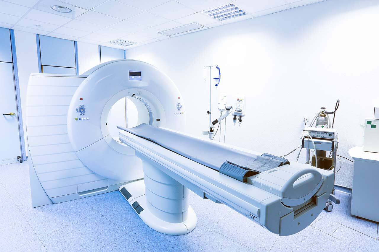
During the first visit, the doctor will conduct a clinical examination and go through the results of the available diagnostic tests. After that, you will undergo the necessary additional examination, such as the assessment of liver and kidney function, ultrasound scan, CT scan and MRI. This will allow the doctor to determine which vessels are feeding the tumor and its metastases, as well as determine how well you will tolerate the procedure.
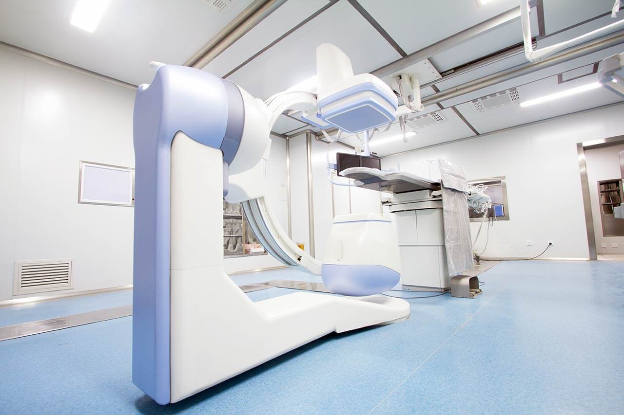
Chemoembolization begins with local anesthesia and catheterization of the femoral artery. The thin catheter is inserted through a few centimeters long incision of the blood vessel. The doctor gradually moves the catheter to the vessel feeding the primary tumor or its metastases. The procedure is carried out under visual control, an angiographic device is used for this. The vascular bed and the position of the catheter in it are displayed on the screen of the angiograph.
When the catheter reaches a suspected artery, a contrast agent is injected through it. Due to the introduction of the contrast agent, the doctor clearly sees the smallest vessels of the tumor and the surrounding healthy tissues on the screen of the angiograph. After that, he injects emboli into the tumor vessels through the same catheter.
Emboli are the spirals or the liquid microspheres. The type of embolus is selected individually, taking into account the diameter of the target vessel. When carrying out chemoembolization, a solution of a chemotherapy drug is additionally injected into the tumor vessel. Due to the subsequent closure of the vessel lumen with an embolus, the chemotherapy drug influences the tumor for a long time. In addition, the drug does not enter the systemic circulation, which allows doctors to use high doses of chemotherapeutic agents without the development of serious side effects. Chemoembolization leads to the destruction of the tumor or slowing down its progression.
After that, the catheter is removed from the artery. The doctor puts a vascular suture on the femoral artery and closes it with a sterile dressing. During chemoembolization, you will be awake. General anesthesia is not used, which significantly reduces the risks of the procedure and allows performing it on an outpatient basis, avoiding long hospital stay.
After the first procedure, you will stay under the supervision of an interventional oncologist and general practitioner. If necessary, you will receive symptomatic treatment. As a rule, a second chemoembolization procedure is performed in 3-5 days after the first one in order to consolidate the therapeutic effect. After that, you will receive recommendations for further follow-up and treatment.
Required documents
Medical records
MRI/CT scan (not older than 3 months)
Biopsy results (if available)
Service
Price from:
Type of program :
Price for 1 day:
Expected duration of the program:
The minimum duration of the program:
Select
You may also book:
Hospital directly:
Saving:
BookingHealth Price from:
About the department
The Department of Nuclear Medicine, Interventional Radiology and Neuroradiology at the University Hospital Greifswald provides the full range of medical services in the fields of its competence. The medical facility has advanced infrastructure and equipment for imaging tests and radioisotope diagnostics. The department's doctors also successfully perform many therapeutic procedures under imaging guidance and therapy with radionuclides. CT and MRI scans, including their modern modifications (for example, diffusion MRI or perfusion MRI), angiography, scintigraphy, PET-CT, SPECT-CT, and other tests are performed in the department's diagnostic rooms. Therapeutic procedures in the fields of interventional radiology and neuroradiology are performed under imaging guidance. These are stent implantation, vascular balloon dilatation, sympathicolysis, thrombolysis, chemoembolization, etc. The specialists in nuclear medicine treat patients with radiopharmaceuticals. Of particular interest to the department's doctors is the treatment of thyroid diseases, bone metastases, and liver tumors. The department's medical team has deep expert knowledge and high professional skills to provide patients with effective care. In addition, it is important for doctors to take into account the patient's individual needs and wishes, so they pay attention to personal communication and building trusting relationships.
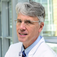
The Head Physician of the department is Prof. Dr. med. Norbert Hosten. Prof. Hosten is the author of a number of textbooks on radiology, including one on the X-ray diagnostics of eye tumors. He is the author of more than 250 scientific publications and has held numerous invited lectures in a broad range of subject fields, such as telemedicine, radiological diagnostics of the eye, MRI, etc.
The priority focus of the department's medical team is interventional radiology. Doctors most often perform angioplasty and stent implantation to eliminate obstructions and stenosis of the blood vessels and hollow internal organs, embolization and chemoembolization to treat malignant tumors and metastases, uterine artery embolization, and transjugular intrahepatic portosystemic shunting. All manipulations are performed under the guidance of X-ray, CT, or MRI scans, which ensure their highest accuracy. The specialists at the medical facility take a responsible approach to the choice of the treatment method, and therefore comprehensive diagnostics precede the prescription of an intervention, the results of which are assessed in cooperation with doctors from related disciplines. For example, interventional radiologists consider chemoembolization in patients with liver cancer during interdisciplinary tumor boards with oncologists, hepatologists, radiation therapists, and other specialists.
The medical facility also offers services in neuroradiology. The doctors in this specialty focus on patients with carotid artery stenosis, brain aneurysms, and dural arteriovenous fistulas. Despite the absence of severe symptoms, carotid stenosis significantly increases the risk of a stroke, so doctors strongly recommend treating the pathology in a timely manner. The department's neuroradiologists eliminate carotid artery stenosis with the help of stent implantation. The interventional procedure is performed under imaging guidance using local anesthesia. The pathological focus is approached through a puncture in the radial, brachial, or femoral artery, through which a stent (a special metal frame) is delivered to the carotid artery affected by atherosclerosis and implanted in its lumen. Doctors implant special platinum coils (coiling) to treat brain aneurysms. The implantation is performed using endovascular techniques under imaging guidance. Dural arteriovenous fistulas are treated with endovascular embolization procedures.
The department's range of services is complemented by the use of radiopharmaceuticals for treatment. Radioiodine therapy for treating benign and malignant thyroid pathologies is one of the most popular procedures in this area. Radioactive iodine therapy is indicated for hyperthyroidism and thyroid cancer (after surgery). Radioiodine therapy is the most sparing and safest for patients because radioactive iodine affects only the thyroid gland without harming other organs. A patient takes radioactive iodine in the form of a capsule or liquid. The therapeutic effect is achieved due to local radioactive irradiation, which leads to the gradual destruction of thyroid cells and metastases of thyroid tumors.
The specialists in nuclear medicine also successfully perform pain management with radiopharmaceuticals for bone metastases, for example, due to prostate cancer, breast cancer, or other cancers with osteoblastic metastases. This method improves the quality of life and relieves pain for up to 4 months. The advantages of this type of pain management include a low risk of side effects and a minor radiation exposure. In addition, the treatment is provided on an outpatient basis.
The department also offers selective internal radiation therapy (SIRT) with intra-arterial administration of Yttrium-90 to treat liver tumors. Another option for radionuclide therapy to treat liver cancer is iodine-131-lipiodol therapy. In this case, the radioactive agent accumulates only in the tumor and does not affect other organs. This ensures a higher radiation load on atypical cells, which contributes to their destruction.
The department's key clinical focuses include:
Nuclear medicine
Iodine-131 therapy for benign and malignant thyroid pathologies
Hyperthyroidism caused by nodular goiter, autoimmune process, etc.
Thyroid cancer after surgery (treatment of recurrences and metastases)
Pain management for bone metastases
Therapy for liver tumors
Selective internal radiation therapy (SIRT) using Yttrium-90
Internal radiation therapy with iodine-131-lipiodol
Antitumor therapy with radioactive antibodies
Interventional radiology
Angioplasty and stent implantation
Sympathicolysis
Embolization and chemoembolization for treating malignant tumors and metastases
Uterine artery embolization
Transjugular intrahepatic portosystemic shunting
Neuroradiology
Carotid artery stent implantation
Coiling for brain aneurysms
Endovascular embolization for dural arteriovenous fistulas
Interventional pain management for chronic back pain
Other medical services
Curriculum vitae
Higher Education and Professional Career
1980 - 1986 Human Medicine studies at the Free University of Berlin.
1986 Admission to medical practice.
1987 Thesis defense.
1987 - 1992 Internship as a Specialist in Radiology at the Westend Hospital and the University Hospital Charite Berlin.
1992 Board certification in Radiology.
1993 - 1994 Senior Physician, Charite University Hospital Berlin.
1994 - 2000 Head of the Department of Radiology.
1994 Habilitation and Venia Legendi in Radiology.
1999 Extraordinary Professorship at the Humboldt University of Berlin.
2001 Invitation to the Chair of Diagnostic and Interventional Radiology at the University of Greifswald, and Head Physician of the Department of Nuclear Medicine, Interventional Radiology and Neuroradiology at the University Hospital Greifswald.
Since 2008 Chairman of the Board of Directors "Telemedicine in the Euroregion Pomerania".
2013 - 2015 President of the German Radiological Society.
Prizes, Awards and Distinctions
October 2015 Pomerania Nostra Award for Merit in Telemedicine in Pomerania Euroregion, University of Greifswald and University of Szczecin.
Photo of the doctor: (c) Universitätsmedizin Greifswald
About hospital
According to the reputable Focus magazine, the University Hospital Greifswald is included in the ranking of the best medical complexes throughout Germany!
The hospital is one of the oldest healthcare facilities in Germany, with long traditions and an excellent reputation. The history of the hospital begins in 1456, when the Faculty of Medicine at the University of Greifswald was founded. During this time, the hospital has managed to earn recognition in the national medical arena and gain prestige abroad. The hospital has 19 institutes and 21 specialized departments. The key to successful clinical practice is the combination of state-of-the-art equipment and highly qualified medical personnel who are actively engaged in the development of effective medical techniques and implement them in everyday practice.
The medical team of the university hospital has more than 4,400 employees, including world-famous professors who regularly undergo advanced training in the leading European and American hospitals, where they share their experience with foreign specialists. The doctors at the medical facility annually treat about 180,000 patients. At the same time, the specialists often provide medical care to patients with complex clinical cases, even in those cases that doctors at other medical centers consider hopeless. The hospital has more than 1,000 beds for inpatients, and many highly-specialized outpatient clinics are available in the medical facility for counseling and outpatient medical care.
The hospital presents all the fields of modern medicine. According to Focus magazine, the medical complex is recognized as one of the best in Germany for treating bowel cancer, bladder cancer, malignant brain tumors, skin cancer, multiple sclerosis, Parkinson's disease, dementia, cardiovascular pathologies, knee and hip pathologies, as well as ophthalmic and proctologic diseases. Diagnostic and therapeutic procedures are carried out in strict accordance with national and international standards, which ensures top-class medical care.
The medical team at the hospital makes sure that each patient feels as comfortable as possible during the therapeutic process. Doctors and nursing staff show humanity and understanding, striving to support each patient in every possible way on the path to recovery.
Photo: (с) depositphotos
Accommodation in hospital
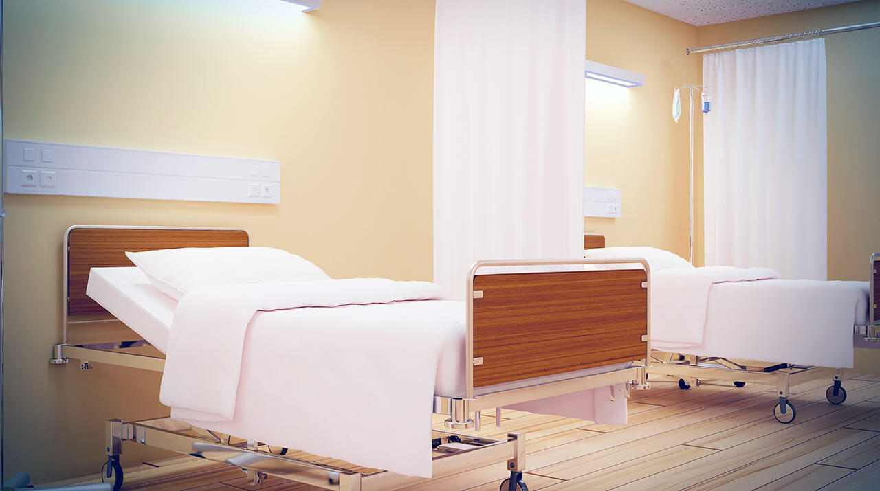
Patients rooms
The patients of the University Hospital Greifswald live in comfortable single and double rooms. The patient rooms are made in bright colors and are quite cozy. The furnishings of a standard patient room include an automatically adjustable bed, a bedside table, a TV, and a telephone. The patient room has a table and chairs for receiving visitors. There is also free Wi-Fi in the patient rooms.
If desired, patients can live in enhanced-comfort rooms. In such rooms, patients are additionally offered toiletries, a bathrobe, and a change of towels.
Meals and Menus
The patients of the hospital are offered three healthy and tasty meals a day: a buffet breakfast, a hearty lunch, and dinner. The hospital also houses a cafeteria where one can have a tasty snack, a cup of aromatic coffee, tea, or soft drinks.
Patients staying in enhanced-comfort rooms are offered a special menu with a wide range of main courses, appetizers, desserts, and drinks.
Further details
Standard rooms include:
Shower
Toilet
Wi-Fi
TV
Religion
The services of representatives of religions are available upon request.
Accompanying person
Your accompanying person may stay with you in your patient room or at the hotel of your choice during the inpatient program.
Hotel
You may stay at the hotel of your choice during the outpatient program. Our managers will support you for selecting the best option.
Select your currency:
Your guarantee
One year of support after the treatment
Insurance to cover unforeseen expenses arising from complications during and 48 months after treatment (coverage up to 200,000 €)
Reduced costs by 40-70% (contracts with Hospitals)
In addition, we are the only TÜV-certified company with an ISO 9001:2015 certificate
thank you for reaching out! One of our medical advisors is currently reviewing your details. You can expect a call from us within one business day to discuss your request.
Note: The incoming call will appear as a German number or your local area code. This call is entirely free of charge.
Booking Health guarantees:
Expert Placement
We use rigorous statistical analysis to ensure you are treated at the best-rated facility for your specific diagnosis.
Total Cost Protection
No hidden fees or unexpected bills. We provide a fixed price guarantee, backed by insurance that covers any additional complications.
12-Month Extended Care
Your recovery is our priority. Benefit from a full year of direct medical support following your procedure.
 BookingHealth Price from:
