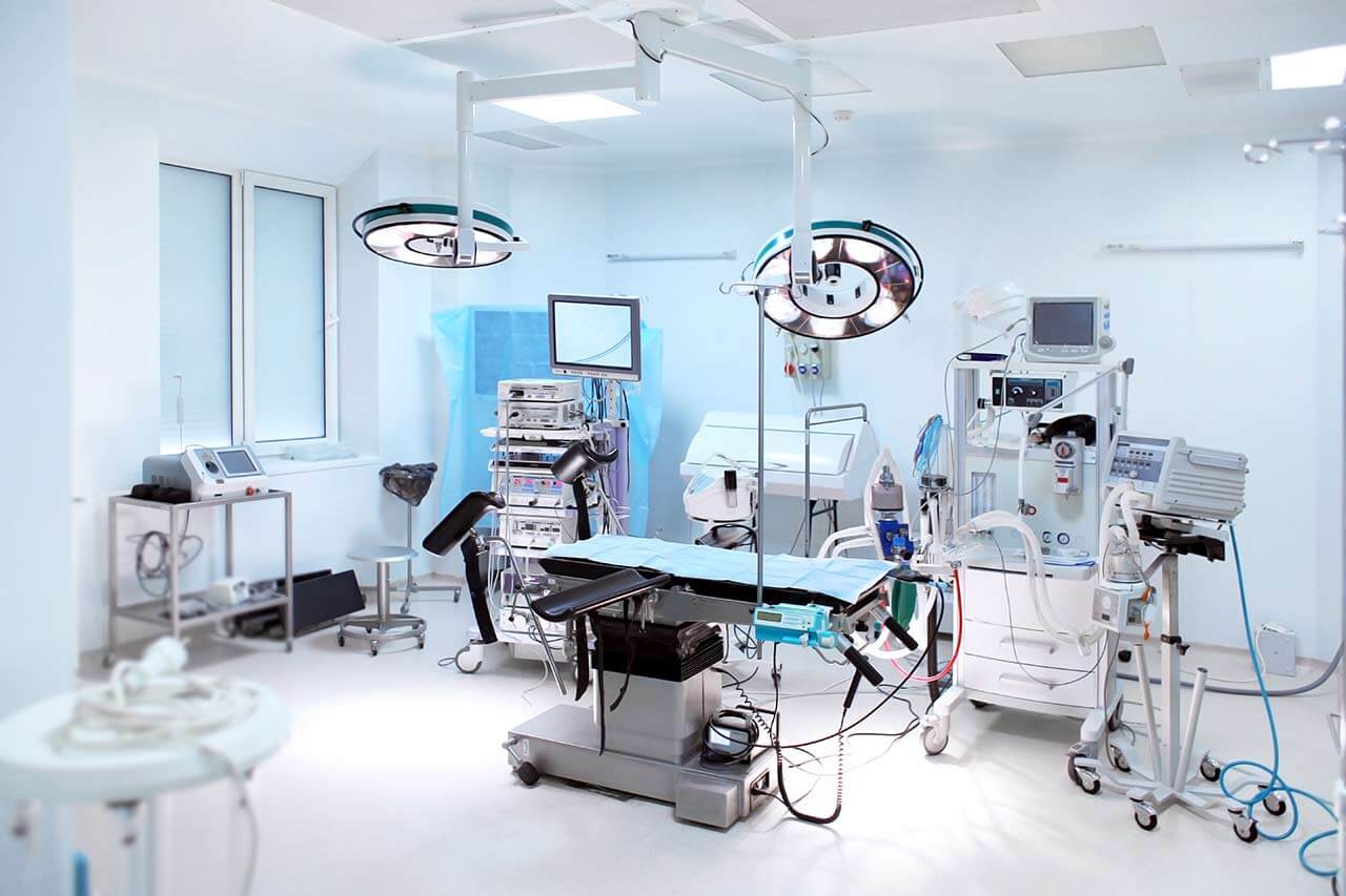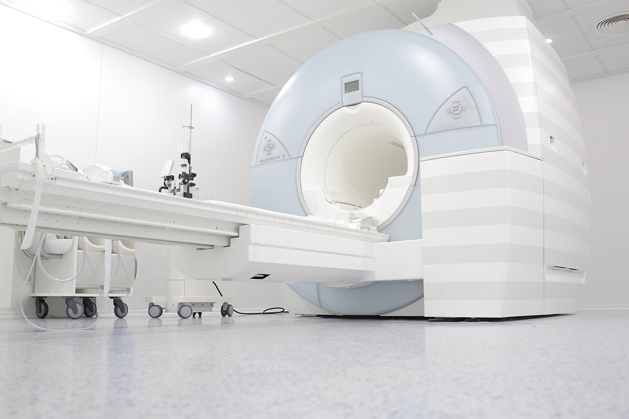
The program includes:
- Initial presentation in the hospital
- Clinical history taking
- Review of available medical records
- Physical examination
- Laboratory tests:
- Complete blood count
- General urine analysis
- Biochemical analysis of blood
- Tumor markers
- Inflammation indicators (CRP, ESR)
- Coagulogram
- Ultrasound scan
- CT scan / MRI
- Preoperative care
- Embolization or chemoembolization, 2 procedures
- Symptomatic treatment
- Cost of essential medicines
- Nursing services
- Elaboration of further recommendations
How program is carried out
During the first visit, the doctor will conduct a clinical examination and go through the results of the available diagnostic tests. After that, you will undergo the necessary additional examination, such as the assessment of liver and kidney function, ultrasound scan, CT scan and MRI. This will allow the doctor to determine which vessels are feeding the tumor and its metastases, as well as determine how well you will tolerate the procedure.
Chemoembolization begins with local anesthesia and catheterization of the femoral artery. The thin catheter is inserted through a few centimeters long incision of the blood vessel. The doctor gradually moves the catheter to the vessel feeding the primary tumor or its metastases. The procedure is carried out under visual control, an angiographic device is used for this. The vascular bed and the position of the catheter in it are displayed on the screen of the angiograph.
When the catheter reaches a suspected artery, a contrast agent is injected through it. Due to the introduction of the contrast agent, the doctor clearly sees the smallest vessels of the tumor and the surrounding healthy tissues on the screen of the angiograph. After that, he injects emboli into the tumor vessels through the same catheter.
Emboli are the spirals or the liquid microspheres. The type of embolus is selected individually, taking into account the diameter of the target vessel. When carrying out chemoembolization, a solution of a chemotherapy drug is additionally injected into the tumor vessel. Due to the subsequent closure of the vessel lumen with an embolus, the chemotherapy drug influences the tumor for a long time. In addition, the drug does not enter the systemic circulation, which allows doctors to use high doses of chemotherapeutic agents without the development of serious side effects. Chemoembolization leads to the destruction of the tumor or slowing down its progression.
After that, the catheter is removed from the artery. The doctor puts a vascular suture on the femoral artery and closes it with a sterile dressing. During chemoembolization, you will be awake. General anesthesia is not used, which significantly reduces the risks of the procedure and allows performing it on an outpatient basis, avoiding long hospital stay.
After the first procedure, you will stay under the supervision of an interventional oncologist and general practitioner. If necessary, you will receive symptomatic treatment. As a rule, a second chemoembolization procedure is performed in 3-5 days after the first one in order to consolidate the therapeutic effect. After that, you will receive recommendations for further follow-up and treatment.
Required documents
- Medical records
- MRI/CT scan (not older than 3 months)
- Biopsy results (if available)
Service
You may also book:
 BookingHealth Price from:
About the department
The Department of Interventional Radiology and Neuroradiology at the University Hospital Halle (Saale) offers a full range of advanced imaging diagnostics and minimally invasive treatments on both an inpatient and outpatient basis. The department has state-of-the-art medical equipment for imaging tests such as X-ray, computed tomography, magnetic resonance imaging, digital subtraction angiography, and mammography. The medical facility also performs many highly effective interventional therapeutic procedures under image guidance, which in many cases allow patients to avoid traumatic open surgery. For example, the department successfully performs local fibrinolysis, thrombectomy, percutaneous transluminal angioplasty, hemostasis, transarterial chemoembolization, uterine artery embolization, and other procedures. The department's neuroradiologists specialize in brain and spinal cord imaging and the treatment of central nervous system disorders. Interventional neuroradiology focuses on the treatment of carotid artery stenosis, brain aneurysms, arteriovenous malformations, dural fistulas, subdural hematomas, brain tumors, skull base and spinal tumors, and chronic back pain. The department's medical team has extensive clinical experience in their areas of expertise. The specialists are guided by the recommendations of the German Society for Interventional Radiology and Minimally Invasive Therapy (DeGIR) and the German Society for Neuroradiology (DGNR), which helps to achieve the best results. The Head Physician of the department is Prof. Dr. Dr. med. Walter Wohlgemuth.
The department's team of interventional radiologists specializes in performing the full range of diagnostic and therapeutic procedures available in this field. One of the most popular interventional therapeutic procedures is percutaneous transluminal angioplasty (PTA), which is indicated for patients with peripheral arterial occlusive disease and mesenteric and renal artery stenosis. Percutaneous transluminal angioplasty is performed under local anesthesia. At the first stage of the procedure, a radiopaque substance is administered intravenously and angiography is performed. At the second stage, a flexible catheter with a balloon attached to it is inserted through the femoral artery, advanced to the site of narrowing or occlusion of the blood vessel, and inflated to restore normal blood flow through the affected blood vessel. If clinically indicated, the PTA procedure may be supplemented with stenting. After dilation of the lumen of the blood vessel, a stent is placed in the stenosis site to prevent further narrowing of the artery.
The department's doctors also regularly admit patients with oncological diseases. The most common interventional treatments are for bleeding during tumor disintegration (hemostasis) and transarterial chemoembolization (TACE). The TACE procedure is performed for hepatocellular carcinoma, intrahepatic cholangiocarcinoma, and liver metastases. The essence of TACE treatment is the introduction of embolization particles with chemotherapy drugs into the artery supplying the tumor. The therapeutic effect is achieved by depriving the tumor of blood supply and the effect of the chemotherapy drug, resulting in the death of cancer cells (necrosis). It should be noted that the chemotherapy drug acts locally on the tumor without harming adjacent healthy tissues because it is delivered directly to the pathological focus without entering the systemic bloodstream. This fact is important because liver cancer is practically insensitive to systemic chemotherapy, and when cytostatics are administered into the hepatic artery, their effectiveness increases significantly.
An integral part of the clinical practice of the department's specialists is imaging diagnostics and interventional treatment of diseases of the central nervous system, the brain and spinal cord. During examinations, doctors perform magnetic resonance imaging on powerful MAGNETOM Sola and MAGNETOM Skyra devices, computed tomography on SOMATOM X cite and SOMATOM Force devices, and digital subtraction angiography on the latest ARTIS icono device. The department's team of neuroradiologists also performs many image-guided endovascular therapeutic procedures to treat carotid artery stenosis, brain aneurysms, arteriovenous malformations, dural fistulas, subdural hematomas, brain skull base, and spinal tumors, and chronic back pain. The department's neuroradiologists work closely with neurologists and neurosurgeons, so that in complex clinical cases, the decision to perform one type of treatment or another is made jointly by the specialists.
The range of medical services provided by the department includes the following:
- Interventional radiology
- Recanalization procedures
- Percutaneous transluminal angioplasty with and without subsequent stenting
- Local fibrinolysis
- Thrombectomy
- Occlusion procedures
- Transarterial chemoembolization (TACE) and radioembolization for the treatment of liver tumors and liver metastases
- Coiling for the removal of aneurysms, fistulas, and arteriovenous malformations
- Preoperative tumor embolization
- Hemostasis for internal bleeding, including tumor bleeding
- Uterine artery embolization
- Implantation of cava filters
- Percutaneous retrieval of foreign bodies (for example, catheter fragments or electrode remnants)
- Percutaneous transhepatic cholangiodrainage
- Transjugular intrahepatic portosystemic shunting (TIPS)
- Interventional pain management
- Sciatic nerve block
- Recanalization procedures
- Interventional neuroradiology
- Percutaneous transluminal angioplasty with and without subsequent stenting for cerebral artery stenosis
- Endovascular procedures for the treatment of brain aneurysms
- Coiling
- Implantation of Contour Device, pCANvas, and other systems
- Implantation of stents (pCONus, Enterprise, Accero, Leo Baby, and others)
- Endovascular procedures for the closure of cranial and spinal dural fistulas
- Endovascular procedures for the closure of arteriovenous malformations of the brain and spinal cord
- Endovascular procedures for the treatment of hemangiomas
- Endovascular procedures for the treatment of idiopathic intracranial hypertension
- Endovascular procedures for the treatment of chronic subdural hematomas
- Interventional pain management
- Sympathetic ganglion block
- Periradicular therapy and facet joint block
- Other therapeutic options
Curriculum vitae
Prof. Walter Wohlgemuth studied medicine at the University of Regensburg, the Technical University of Munich, and the Ludwig Maximilian University of Munich, and healthcare economics at the University of Bayreuth. He received his license to practice medicine in 1994 and his doctorate in the same year from the Technical University of Munich. After receiving his doctorate, he worked as an Assistant Physician in the Department of Diagnostic Radiology and Neuroradiology, as well as in the Department of Neurology and Clinical Neurophysiology at the Augsburg Hospital and in the Department of Radiology at the Ingolstadt Hospital.
Medical Practice
- Since 2001 Senior Resident with the specialization "angiography", and since 2003 ‒ Senior Physician and Head Physician of the Department of Interventional Radiology.
- Since 2002 Research Assistant at the Institute of Medical Management and Health Sciences at the University of Bayreuth.
- 2005 Habilitation and Venia legendi in "Medical Management and Health Sciences", Faculty of Economics, University of Bayreuth.
- 2006 Board certification in Neuroradiology.
- 2009 - 2011 Head of the Center for Vascular Malformations, Augsburg Hospital.
- Since October 2011 Professor and Head Physician of the Department of Interventional Radiology at the University Hospital Regensburg.
- 2012 - 2017 Head of the interdisciplinary Center for Vascular Anomalies at the University Hospital Regensburg.
- 2014 Research Fellowship at Boston Children's Hospital, Harvard Medical School, and the Children's Hospital of Philadelphia.
- 2015 - 2017 Founder and Director of the first interdisciplinary Center for Pediatric Interventional Radiology in Germany.
- Since January 2017 President of the German Interdisciplinary Society for Vascular Anomalies.
- Since June 2017 Head Physician, Department of Interventional Radiology and Neuroradiology, University Hospital Halle (Saale), Professor, Martin Luther University Halle-Wittenberg.
Memberships in Professional Societies
- German Interdisciplinary Society for Vascular Anomalies (GISVA).
- German Radiological Society (DRG).
- German Society for Interventional Radiology and Minimally Invasive Therapy (DeGIR).
- German Society for Angiology (DGA).
- German Society for Neuroradiology (DGNR).
- European Society of Radiology (ESR).
- Cardiovascular and Interventional Radiological Society of Europe (CIRSE).
- European Board of Interventional Radiology (EBIR).
Photo of the doctor: (c) Universitätsklinikum Halle (Saale)
About hospital
According to the prestigious Focus magazine, the University Hospital Halle (Saale) is one of the best medical institutions in Germany!
The history of the hospital goes back more than 300 years, and during this time it has managed to gain an excellent reputation not only in Germany, but also throughout the world. The hospital positions itself as a specialized healthcare facility for the treatment of severe and rare diseases and injuries. The hospital provides medical care to patients of all ages in compliance with the latest scientific achievements. The hospital is distinguished by successful research activities, especially in the field of cardiovascular diseases and oncopathologies – the specialists in these areas have made significant contributions to the development of the very latest diagnostic methods and therapeutic approaches.
The University Hospital Halle (Saale) has 30 specialized departments representing almost all areas of modern medicine, as well as 17 narrowly focused institutes. About 35,000 patients receive qualified medical care of European standards in the hospital every year, and more than 212,000 patients are served on an outpatient basis. This number of patients is evidence of the high efficiency of medical services and the excellent image of the hospital in the international medical arena; patients from all over the world regularly seek medical attention here.
Some of the hospital's structural units deserve special attention. For example, the Central Emergency Department (the largest in Saxony-Anhalt), modern dental clinics, the Perinatal Center, and the Transplant Center, which has a history of more than 40 years. The Transplant Center performs more than 40 kidney transplants annually, most of them from living donors.
Thanks to the use of the latest medical technologies and the availability of state-of-the-art equipment, many previously high-risk surgeries and procedures can now be performed in the hospital using sparing techniques. In this context, hybrid cardiac surgery and robotic surgery using the innovative da Vinci Si® system in urology are worthy of mention.
An integral part of the successful clinical practice of the University Hospital Halle (Saale) is the availability of experienced and competent medical staff. The total number of employees at the hospital is more than 4,450. Many physicians are known far beyond the borders of Germany: they regularly conduct important research that enables the development of modern medicine. In addition, the hospital specializes in training medical students, so qualified doctors and professors are willing to pass on their experience to the younger generation.
The hospital has many quality certificates such as DIN EN ISO 9001:2015 certificate, German Cancer Society (DKG) certificate, JACIE certificate, EndoCert certificate, ClarCert certificate, German Spine Society (DWG) certificate, German Trauma Society (DGU) certificate, CERT iQ certificate, LGA InterCetert certificate, and others.
Photo: (с) depositphotos
Accommodation in hospital
Patients rooms
The patients of the University Hospital Halle (Saale) stay in comfortable single, double, and triple rooms with a modern design. All patient rooms have an ensuite bathroom with a toilet and a shower. The standard patient room includes a comfortable automatically adjustable bed, a bedside table, a wardrobe, a table and chairs for receiving visitors, a TV, a radio, and a telephone. The patient rooms have access to Wi-Fi. For safety reasons, the use of laptops and cell phones is prohibited in some areas, including the intensive care units. The hospital also offers enhanced-comfort patient rooms.
Meals and Menus
The hospital offers delicious and well-balanced meals three times a day: breakfast, lunch, and dinner. Patients and their companions can choose from three daily menus, which always include dietary dishes. If necessary, an individual menu can be prepared for the patient. Children are offered a special menu with healthy and tasty dishes, rich in nutrients necessary for a growing body.
Further details
Standard rooms include:
![]() Toilet
Toilet
![]() Shower
Shower
![]() Wi-Fi
Wi-Fi
![]() TV
TV
Religion
Religious services are available upon request.
Accompanying person
Your accompanying person may stay with you in your patient room or at the hotel of your choice during the inpatient program.
Hotel
Your accompanying person may stay with you in your patient room or at the hotel of your choice during the inpatient program.


