Treatment of bladder cancer with embolization or chemoembolization in University Hospital Hamburg-Eppendorf
University Hospital Hamburg-Eppendorf
Hamburg, Germany
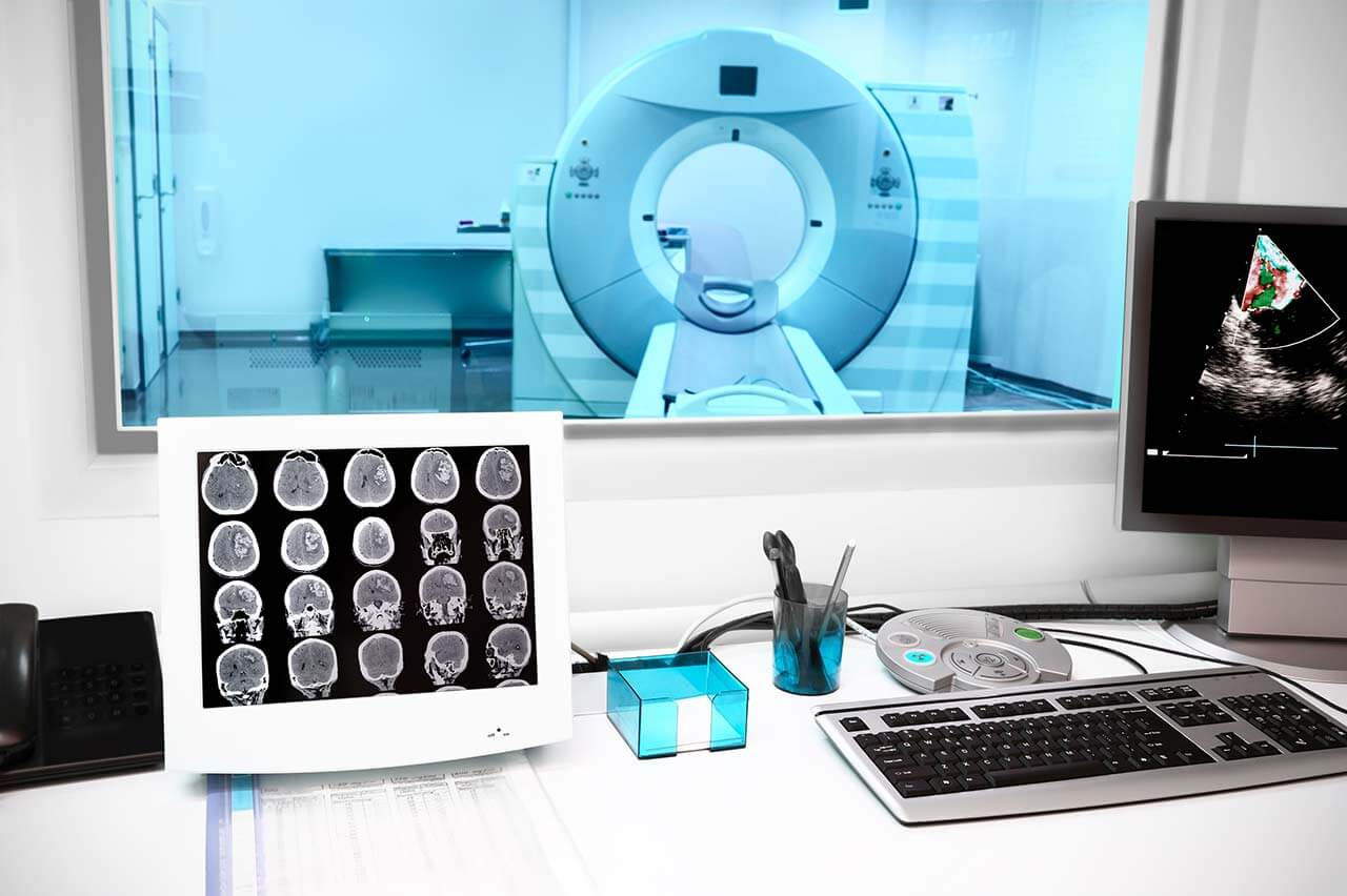
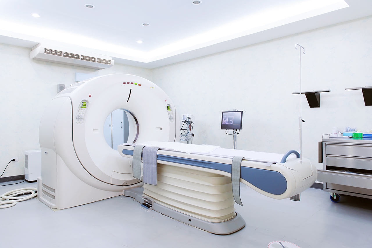


During the first visit, the doctor will conduct a clinical examination and go through the results of the available diagnostic tests. After that, you will undergo the necessary additional examination, such as the assessment of liver and kidney function, ultrasound scan, CT scan and MRI. This will allow the doctor to determine which vessels are feeding the tumor and its metastases, as well as determine how well you will tolerate the procedure.
Chemoembolization begins with local anesthesia and catheterization of the femoral artery. The thin catheter is inserted through a few centimeters long incision of the blood vessel. The doctor gradually moves the catheter to the vessel feeding the primary tumor or its metastases. The procedure is carried out under visual control, an angiographic device is used for this. The vascular bed and the position of the catheter in it are displayed on the screen of the angiograph.
When the catheter reaches a suspected artery, a contrast agent is injected through it. Due to the introduction of the contrast agent, the doctor clearly sees the smallest vessels of the tumor and the surrounding healthy tissues on the screen of the angiograph. After that, he injects emboli into the tumor vessels through the same catheter.
Emboli are the spirals or the liquid microspheres. The type of embolus is selected individually, taking into account the diameter of the target vessel. When carrying out chemoembolization, a solution of a chemotherapy drug is additionally injected into the tumor vessel. Due to the subsequent closure of the vessel lumen with an embolus, the chemotherapy drug influences the tumor for a long time. In addition, the drug does not enter the systemic circulation, which allows doctors to use high doses of chemotherapeutic agents without the development of serious side effects. Chemoembolization leads to the destruction of the tumor or slowing down its progression.
After that, the catheter is removed from the artery. The doctor puts a vascular suture on the femoral artery and closes it with a sterile dressing. During chemoembolization, you will be awake. General anesthesia is not used, which significantly reduces the risks of the procedure and allows performing it on an outpatient basis, avoiding long hospital stay.
After the first procedure, you will stay under the supervision of an interventional oncologist and general practitioner. If necessary, you will receive symptomatic treatment. As a rule, a second chemoembolization procedure is performed in 3-5 days after the first one in order to consolidate the therapeutic effect. After that, you will receive recommendations for further follow-up and treatment.
 BookingHealth Price from:
The Department of Interventional Radiology at the University Hospital Hamburg-Eppendorf provides a full range of medical services in its area of expertise. It performs highly accurate imaging tests of various organs and anatomical systems, including X-ray, computed tomography, magnetic resonance imaging, mammography, and angiography. The department also offers advanced image-guided therapeutic procedures, the most popular of which are liver tumor ablation, radiofrequency ablation (RFA) for osteoid osteomas, CT-guided drainage, transjugular intrahepatic portosystemic shunting (TIPS), and transarterial chemoembolization (TACE). Diagnostic and therapeutic procedures are performed with state-of-the-art equipment: the department has 3 Philips and Siemens 3 Tesla MRI machines, 3 Philips and Siemens CT scanners, two Philips Azurion angiography machines, a Philips Vereos PET/CT system, and a magnetic particle imaging (MPI) system. Most diagnostic tests and treatments are performed on an outpatient basis. The department's medical team has extensive clinical experience. The specialists work in strict accordance with the standards of the German Society of Interventional Radiology and Minimally Invasive Therapy (DeGIR) and the European Society of Radiology (ESR), which contributes to the high quality of medical services. The department is headed by Prof. Dr. med. Gerhard Adam.
The priority focus of the daily work of the department's specialists is the treatment of benign and malignant tumors using interventional procedures under the guidance of computed tomography. One of the most popular interventional procedures in clinical practice is radiofrequency ablation for the treatment of primary and secondary malignancies. It is most often used as an adjunct to surgery, chemotherapy, and radiation therapy. In the case of inoperable tumors, radiofrequency ablation may be the only therapeutic manipulation. An important advantage of this method is that it can be used repeatedly if new tumor foci are detected in the liver, and it has a high safety profile. The essence of radiofrequency ablation is as follows: electrodes are inserted into the tumor site through a puncture in the skin and soft tissues, with the help of which the malignant neoplasm is locally heated to high temperatures (60-100°C), which leads to its death. Radiofrequency ablation is performed in the department under the guidance of computed tomography. It is a minimally traumatic, highly effective method of treatment, which at the same time practically does not affect healthy tissues in the vicinity of the cancer focus.
The department's team of interventional radiologists has excellent qualifications in the field of transjugular intrahepatic portosystemic shunting (TIPS). This treatment is indicated for patients with portal hypertension, a complication of cirrhosis. Portal hypertension carries many serious risks: for example, the patient may develop ascites (fluid accumulation in the abdominal cavity), internal bleeding from varicose veins in the esophagus and stomach, and other complications. The TIPS procedure is performed using catheter-based techniques with an approach through the internal jugular vein. Under ultrasound guidance, the central branch of the portal vein is punctured and a stent is inserted through a thin guidewire. After assessing the pressure in the portal vein, a thin balloon is used to dilate the affected area and place a shunt to allow normal blood flow from the liver and other gastrointestinal organs to the right atrium. Transjugular intrahepatic portosystemic shunting allows for a significant reduction in portal vein pressure and eliminates the risk of complications due to portal hypertension. The TIPS procedure is performed under local anesthesia (sometimes sedation can be used). The duration of the therapeutic manipulation is about 60-90 minutes. The specialists of the department perform more than 100 transjugular intrahepatic portosystemic shunting procedures annually, which has earned the medical institution the reputation of being one of the most competent in this field in Germany. After completion of the treatment, the patient is recommended to undergo regular examinations and follow-up duplex sonography.
The department also offers transarterial chemoembolization (TACE) for the treatment of hepatocellular carcinoma (liver cancer). The essence of the treatment is to block the lumen of the arteries feeding the tumor with emboli containing chemotherapeutic agents. TACE treatment is performed under angiographic guidance using catheter-based technologies and injection of a contrast agent to visualize hepatocellular carcinoma foci. After transarterial chemoembolization, the department's specialists manage to achieve local control of the tumor – the local progression of the disease is significantly slowed down or completely stopped. The TACE procedure is performed in a medical facility under local anesthesia, in the hospital (hospital stay is 1-2 days). If necessary, TACE can be repeated.
The range of medical services provided by the department includes the following:
Higher Education and Postgraduate Training
Professional Career
Editorial and Scientific Activities
Other Research Focuses
Prizes, Awards, and Honorable Memberships
Scientific Leadership Positions
Memberships in Professional Societies
Photo of the doctor: (c) Universitätsklinikum Hamburg-Eppendorf (UKE)
According to the Focus magazine, the University Hospital Hamburg-Eppendorf is one of the top ten hospitals in Germany!
Since its foundation in 1889, the hospital has taken a leading position in the European medical arena, which it still holds today. A highly competent medical team of more than 15,300 employees takes care of the health of patients. Approximately 2,900 of them are physicians and researchers, and more than 3,400 work as nurses and therapists. The hospital has 1,738 beds for inpatient treatment, and many diagnostic and therapeutic services are provided on an outpatient basis. A solid foundation for successful clinical practice in the medical complex is formed by a combination of research achievements with state-of-the-art equipment and the highest professionalism of doctors. In addition, the hospital has a modern and extremely comfortable infrastructure. The most important value for every employee of the University Hospital Hamburg-Eppendorf is the health and well-being of every patient.
The medical facility was the first university hospital in Europe to implement an electronic system for storing patient medical reports. As a result, all diagnostic and treatment protocols are stored electronically. In 2011, the hospital was certified as the first fully digital hospital in Europe.
The hospital represents all areas of modern medicine. The doctors of the healthcare facility have a wealth of theoretical knowledge and vast clinical experience, which allows them to easily cope with the treatment of both common and extremely rare, complex clinical cases. About 550,000 patients are treated here each year, over 450,000 of whom receive outpatient medical care.
An important part of the work of the University Hospital Hamburg-Eppendorf is research activities aimed at developing innovative diagnostic and treatment methods. The main areas of research of the hospital include neurobiology, oncology, cardiovascular research, and research on infectious and inflammatory diseases. Special attention is also given to research in molecular imaging and skeletal biology.
The hospital is distinguished by its first-class level of medical care, which is confirmed by numerous quality certificates of European and international standards: DIN EN IS0 9001 certificate, certificates of the German Cancer Society (DKG) in the treatment of breast cancer, colon cancer, gynecological cancer, prostate cancer, and other oncological diseases, certificate of the German Cardiac Society (DGK) in the treatment of acute coronary syndrome, certificate of the German Spine Society (DWG), and others.
Photo: (с) depositphotos
The patients of the University Hospital Hamburg-Eppendorf stay in comfortable single and double bright rooms with a modern design. Each patient room has an ensuite bathroom with a shower and a toilet. The standard patient room furnishings include an automatically adjustable bed with an orthopedic mattress, a bedside table, a wardrobe, a table and chairs for receiving visitors, a telephone, a radio, and a TV. Wi-Fi access is available in patient rooms and throughout the hospital.
If desired, patients can stay in single enhanced-comfort rooms. These rooms are more spacious and are equipped with upholstered furniture, a safe, and a mini-fridge.
The hospital offers three meals a day: breakfast, lunch, and dinner. Breakfast and dinner are served in the form of buffets, and for lunch you can choose from several set menus – in total, more than 20 dishes are served for lunch, including vegetarian ones.
If, for some reason, you cannot eat all of the foods, you will be offered an individual menu. Please inform the medical staff about your dietary preferences prior to the treatment.
Standard rooms include:
![]() Toilet
Toilet
![]() Shower
Shower
![]() Wi-Fi
Wi-Fi
![]() TV
TV
Religious services are available upon request.
During the inpatient program, an accompanying person may stay with you in a patient room or hotel of your choice.
During the outpatient program, you may stay in a hotel at the hospital.
During the outpatient program, you may stay in a hotel of your choice. Managers will help you choose the most suitable options.
The hospital offers a full range of laboratory tests (general, hormonal, tests for infections, antibodies, tumor markers, etc.), genetic tests, various modifications of ultrasound scans, CT scans, MRI and PET / CT, angiography, myelography, biopsy and other examinations. Treatment with medications, endoscopic and robotic operations, stereotaxic interventions is carried out here, modern types of radiation therapy are also used. The hospital offers patients all the necessary therapeutic techniques.
These are arteriovenous malformations and angiomas, vascular aneurysms, pathologies of the mammary glands, pelvic organ prolapse, urinary incontinence, malignant tumors of various localizations (area of special attention is treatment of intestinal cancer), pathologies of liver and pancreas, cataracts and rare ophthalmic pathologies (aphakia, aniridia ), infertility and other diseases.
Over 2,900 highly qualified physicians and researchers work at the hospital.
