The program includes:
- Initial presentation in the clinic
- clinical history taking
- review of medical records
- physical examination
- laboratory tests:
- complete blood count
- general urine analysis
- biochemical analysis of blood
- inflammation of indicators (CRP, ESR)
- indicators of blood coagulation
- gynecological examination
- ultrasound scan: pelvis, abdomen
- preoperative care
- da Vinci treatment
- symptomatic treatment
- control examinations
- the cost of essential medicines and materials
- nursing services
- full hospital accommodation
- explanation of future recommendations
How program is carried out
During the first visit, the physician will conduct a clinical examination and go through the results of the available diagnostic tests. After that, you will undergo the necessary additional examination, such as the assessment of liver and kidney function, ultrasound scan of the abdominal and pelvic organs. Based on the results of the examination, the physician will choose the surgical technique and the type of anesthesia.
Surgery with the da Vinci robot starts with general anesthesia. After anesthesia, the surgeon makes small incisions through which the manipulators of the da Vinci robot and a video camera are inserted into the pelvic cavity. With the help of manipulators, the surgeon fixes the prolapsed pelvic organs in the physiological position, and the video camera continuously transmits a three-dimensional image of the operating field in 12-fold magnification to the monitor.
To fix the pelvic organs, the surgeon can use own muscles and ligaments of the vagina, uterus, rectum and other organs, or mesh prostheses. The use of mesh implants improves the functional outcomes of the operation and reduces the risk of recurrence.
After the completion of the operation, you will be transferred back to the ward, under the supervision of the attending physician and nursing staff. Due to the minimal invasiveness of the operation and the short duration of general anesthesia, you will not need to stay in the intensive care unit for a long time.
Finally, the attending physician will evaluate the results of control examinations, schedule the date of discharge from the hospital and give you detailed recommendations for further follow-up and treatment.
Required documents
- Medical records
- Pelvic ultrasoud (if available)
- Pelvic MRI/CT scan (if available)
Service
You may also book:
 BookingHealth Price from:
About the department
According to Focus, the Department of Adult and Pediatric Gynecology, Mammology, Reproductive Medicine at the University Hospital Jena ranks among the top German medical facilities specializing in breast cancer treatment!
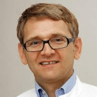
The department offers the full range of surgical and conservative treatment of diseases of the female reproductive system. One of the priorities is breast cancer treatment. Other important focuses include the treatment of gynecologic cancers, endometriosis, uterine myomas, dysplasia, pelvic prolapse, urinary incontinence. In addition, the competence covers the use of all modern assisted reproductive technologies. The department is headed by Prof. Dr. med. Ingo Bernard Runnebaum.
In the field of gynecology, of particular interest is the diagnostics and treatment of oncological diseases of the female genital sphere, for example, uterine, cervical, ovarian cancers, etc. The department's surgeons annually perform more than 250 interventions for the treatment of such pathologies. Thanks to the excellent technical equipment, many operations are carried out using sparing minimally invasive techniques. The decision on the most effective treatment concept is made at interdisciplinary tumor boards with the participation of the experienced gynecologists, radiation therapists, radiologists, oncologists, as well as representatives of clinical trials.
Another key focus of the department’s work is breast cancer therapy. The department offers all progressive diagnostic methods for the early detection of pathology. In addition, the department implements a special breast cancer screening program, which allows to detect the disease as soon as possible and to ensure the proper treatment. In many cases, the doctors manage to deal with breast cancer using organ-sparing surgical techniques, but some clinical cases still require breast resection. Nevertheless, the department's range is complemented by modern reconstructive operations, which allow the doctors to guarantee a good aesthetic result.
In addition, the competence includes the diagnostics and treatment of infertility in women, hormonal disorders in patients of all age groups, including girls, medical care in puberty disorders, as well as during menopause. In the case of problems with conception, the doctors conduct in vitro fertilization, intracytoplasmic sperm injection. In some cases, it is enough to monitor the menstrual cycle and ovulation, which allows for the promotion of natural conception without additional manipulations.
The service range of the department includes:
- Diagnostics and treatment of gynecologic cancers
- All gynecological interventions
- Special operations (laparoscopic radical surgery with the preservation of nerves and/or organs)
- Multivisceral surgery at the advanced stages of oncopathology
- Cryopreservation of eggs for fertility preservation
- Reconstructive surgery on the reproductive organs
- Radiation therapy
- Chemotherapy
- Antibody therapy
- Immunotherapy
- Hormone therapy
- Treatment within the clinical trials
- Psycho-oncological support
- Second opinion
- Naturopathic methods and nutrition counseling
- Diagnostics and treatment of breast cancer
- Diagnostics
- Breast cancer screening
- Clinical examination
- Sonography and mammography
- Biopsy
- Magnetic resonance imaging
- Therapy
- Surgical treatment
- Chemotherapy
- Radiation therapy
- Follow up care
- Treatment within the clinical trials
- Naturopathic methods
- Diagnostics
- Diagnostics and treatment of urinary and fecal incontinence
- Diagnostics
- Ultrasound diagnostics for the assessment of bladder function and the degree of uterine prolapse
- Cystoscopy and endoscopic examination of the rectum in the case of hemorrhage, incontinence or infection
- Urodynamic diagnostics with the evaluation of urine loss and differentiation of stress and imperative urinary incontinence
- Anal sphincter manometry in functional impairment after childbirth
- Defecography in fecal incontinence or emptying disorders due to pelvic prolapse
- Therapy
- Correction of pelvic prolapse (with and without organ preservation)
- Anterior and posterior colporrhaphy
- Apical fixation
- Amreich-Richter fixation
- Laparoscopic sacropexy using a special mesh
- Vaginal sacropexy (colporectosacropexy)
- Imposition of a special mesh through vaginal access in prolapse recurrence
- Treatment of urinary incontinence
- TVT procedure
- Colposuspension after Burch procedure (open or laparoscopic)
- Bulkamid™ paraurethral injection
- Botox injections
- Special treatment methods
- Anal sphincter reconstruction after birth injuries
- Incisional gynecological hernia repair
- Surgery to treat hemorrhoid due to pregnancy
- Correction of pelvic prolapse (with and without organ preservation)
- Diagnostics
- Diagnostics and treatment of endometriosis
- Diagnostics
- Study of medical history and gynecological examination
- Imaging diagnostics (MRI)
- Diagnostic procedures of the urological range (for example, pyelography)
- Diagnostic procedures of the gastroenterological range (proctoscopy, rectal endosonography, irrigoscopy)
- Cystoscopy
- Gynecological laparoscopy with biopsy and subsequent histological examination
- Therapy
- Drug therapy
- Surgical interventions (mainly endoscopic minimally invasive surgical techniques)
- Follow-up consultations
- Interdisciplinary medical care in extragenital infection
- Diagnostics
- Diagnostics and treatment of uterine fibroids
- Diagnostics
- Ultrasound diagnostics
- MRI
- Hysteroscopy
- Laparoscopy
- Therapy
- Therapy with the uterus preservation
- Uterine fibroid enucleation
- Uterine fibroid embolization
- Radiofrequency ablation
- HOLA laser procedure
- Drug therapy
- Diagnostics
- Diagnostics and treatment of cervical dysplasia
- Diagnostics and treatment of pathological changes of the cervix, vagina and vulva
- Diagnostics and treatment of HPV infection
- Recommendations for HPV vaccination
- Treatment of genital warts
- Treatment of vulvar pain syndromes
- Plastic, reconstructive and aesthetic surgery
- Reconstructive breast surgery
- Breast reconstruction using the patient’s own tissues
- Breast reconstruction using implants
- Nipple reconstruction
- Breast asymmetry correction
- Preventive breast removal with the subsequent reconstruction
- Aesthetic surgery
- Breast surgery
- Breast augmentation
- Breast reduction
- Inverted nipple correction
- Gynecomastia surgery
- Abdominoplasty
- Thigh lift
- Arm lift
- Liposuction
- Breast surgery
- Other operations
- Hyperhidrosis correction
- Scar revision
- Aesthetic surgery of the external genitalia
- Labia correction
- Labia augmentation
- Pubic correction
- Reconstructive breast surgery
- Diagnostics and treatment of infertility
- Stimulation of natural conception
- In vitro fertilization (IVF)
- Intracytoplasmic sperm injection (ICSI)
- Cryopreservation of eggs
- Insemination
- Testicular sperm extraction
- Assisted hatching
- Polarization microscopy
- Preservation of fertility in women and men
- Diagnostics and treatment of hormonal disorders in women and girls
- Menopause, including early menopause
- Endometriosis
- Menstrual disorders
- Contraception
- Hirsutism and hair loss due to the hormonal disorders
- Decreased libido
- Puberty disorders
- Other medical services
Curriculum vitae
Since 1980 till 1982 Dr. Runnebaum studied Philosophy and Chemistry in Heidelberg, whereas since 1982 till 1988 he studied Human Medicine at the Universities of Mainz, Berlin and Munich. Since 1989 till 1992, he was a Postdoctoral Fellow at the Salk Institute for Biological Studies in San Diego, where he became a Member of the Working Group on p53 gene in breast cancer, which was headed by the Nobel Prize winner Renato Dulbecco jointly with Saraswati Sukumar. In 1990, he received his Doctorate at the Ludwig Maximilian University of Munich in the Department of Dermatology and Allergology under the guidance of Otto Braun-Falco. In 1997, he had his board certification by the Medical Association of Baden-Württemberg and habilitation at the University of Ulm under the supervision of his curator Rolf Kreienberg, which gave him Venia Legendi in Gynecology and Obstetrics. Until 1999, Runnebaum worked in the Department of Gynecology at the University Hospital Ulm, after which he accepted an offer to hold the position of Professor at the University of Freiburg (he also worked here as Leading Senior Physician). This was followed by the position of Deputy Head of the department with a focus on Gynecological Surgery at the University Hospital of Ludwig Maximilian. In addition, in 2005, he received his Master's Degree in Business Administration in the New Ulm High School. In the same year, he held the position of W3 Professor at the Friedrich Schiller University Jena and Head of the Department of Adult and Pediatric Gynecology, Mammology, Reproductive Medicine (till nowadays). In addition to the successful clinical and surgical activities, Prof. Runnebaum is known for publishing more than 200 scientific works, giving over 500 lectures in Germany and worldwide, teaching and university training of doctors (ESGO congresses, etc.), as well as participation in television programs ( Sat 1, RTL, MDR Erfurt und Leipzig).
Specialization
Prof. Runnebaum is an expert in the treatment of breast cancer and gynecological tumors. He specializes in organ-sparing surgeries on the mammary gland, oncoplastic and reconstructive operations, as well as in minimally invasive surgeries for the removal of sentinel lymph nodes and pelvic organ tumors (laparoscopy in uterine myomas, deep infiltrating endometriosis, nerve‐sparing radical hysterectomy, lymphadenectomy and complex surgical interventions in cancer of the reproductive organs in combination with multivisceral surgery). In addition, he specializes in reproductive medicine, perinatal medicine and special obstetrics. His research projects focus on ovarian, cervical and breast cancer.
Scientific Сontribution
Prof. Runnebaum demonstrated the role of p53 gene (TP53) as a tumor suppressor gene in breast and ovarian cancer. In the field of clinical trials, he became the first researcher in Germany, who studied adenoviral gene therapy in patients with ovarian cancer. He led the first randomized HIPEC research using heated chemotherapy agents in ovarian cancer in Germany. After the participation in creation of the University Cancer Center Jena (2008), he founded the Endometriosis Center (2009), the University Center for Pelvic Floor Diseases and the Network for Pelvic Floor Diseases in Thuringia. In 2010, the doctor also founded the Gynecological Cancer Center Jena, and then the Thuringian Interdisciplinary Center for Uterine Myoma.
Honors
For his researches of p53 gene as a tumor suppressor and cell cycle regulator in breast cancer cells, Prof. Runnebaum won the Walter Hohlweg Prize (1994). Since 2007, he has been regularly ranked among the top German gynecologists (Focus magazine and Gute Rat rating).
Membership in Scientific Societies
- International societies
- American Association for Cancer Research (AACR), USA.
- American Society of Clinical Oncology (ASCO), USA.
- European Society for Gynecological Endoscopy (ESGE).
- European Society of Gynecological Oncology (ESGO).
- European Society of Breast Cancer Specialists (EUSOMA).
- European Group for Breast Cancer Screening.
- National Societies
- Association of Gynecologic Oncology (AGO).
- Working Group on Immunology in Gynecology and Obstetrics (AIGM)
- Working Group on Molecular Biology in Gynecology (AMF).
- Advisory Board of the International Prevention Organization (IPO).
- Professional Association of Gynecologists.
- German Working Group on Gene Therapy (DAGGT).
- German Society of Gynecology and Obstetrics (DGGG).
- German Society for Senology (DGS).
- German Cancer Society (DKG).
- German Cancer Society, Department of Experimental Cancer Research (AEK).
- Commission on Ovarian Diseases, Working Group on Gynecological Oncology.
- Commission on the Treatment of Uterine Diseases, Working Group on Gynecological Oncology.
- Commission on the Development of Therapeutic Guidelines: S3 ovarian carcinoma, S2E hysterectomy, S3 endometrial carcinoma.
- Board of the Runnebaum Foundation, Heidelberg.
Photo of the doctor: (c) Universitätsklinikum Jena
About hospital
According to the prestigious Focus magazine, the University Hospital Jena regularly ranks among the top German medical facilities!
The hospital has positioned itself as a multidisciplinary medical facility with a long history of more than 200 years. Since its foundation, the hospital has been constantly developing and modernizing, thanks to which nowadays it offers patients the highest level of treatment in Germany based on the use of innovative technologies and the very latest therapeutic techniques. The hospital consists of 26 specialized departments and 25 research institutes. It treats more than 53,600 inpatients and about 274,000 outpatients every year. The staff of the hospital includes more than 5,600 competent doctors.
The extensive resources of the university hospital, high treatment standards, and the introduction of new research developments provide first-class treatment in Germany meeting the stringent international standards. The hospital has an excellent reputation not only in Germany, but also far beyond its borders, due to which it accepts a large number of foreign patients for the diagnostics and treatment.
Despite the technical progress and the availability of accurate computerized systems, the patient’s physical health and emotional state is the main value of each employee of the hospital, since some diagnoses cause emotional distress in patients. The doctors of the hospital believe that the key to a successful result is a comprehensive and individual approach, so they spend a lot of time talking with patients, listen carefully to all their wishes and support at all stages of the therapeutic process. All this in combination with high-precision diagnostic techniques and the very latest types of therapy forms a solid basis for the achievement of an optimal treatment result.
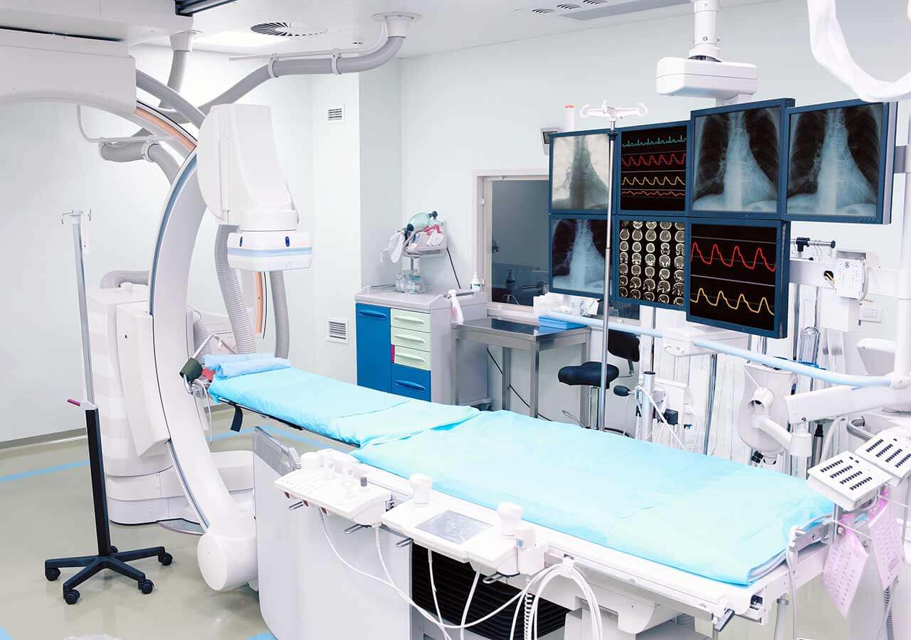
Photo: (c) depositphotos
Accommodation in hospital
Patients rooms
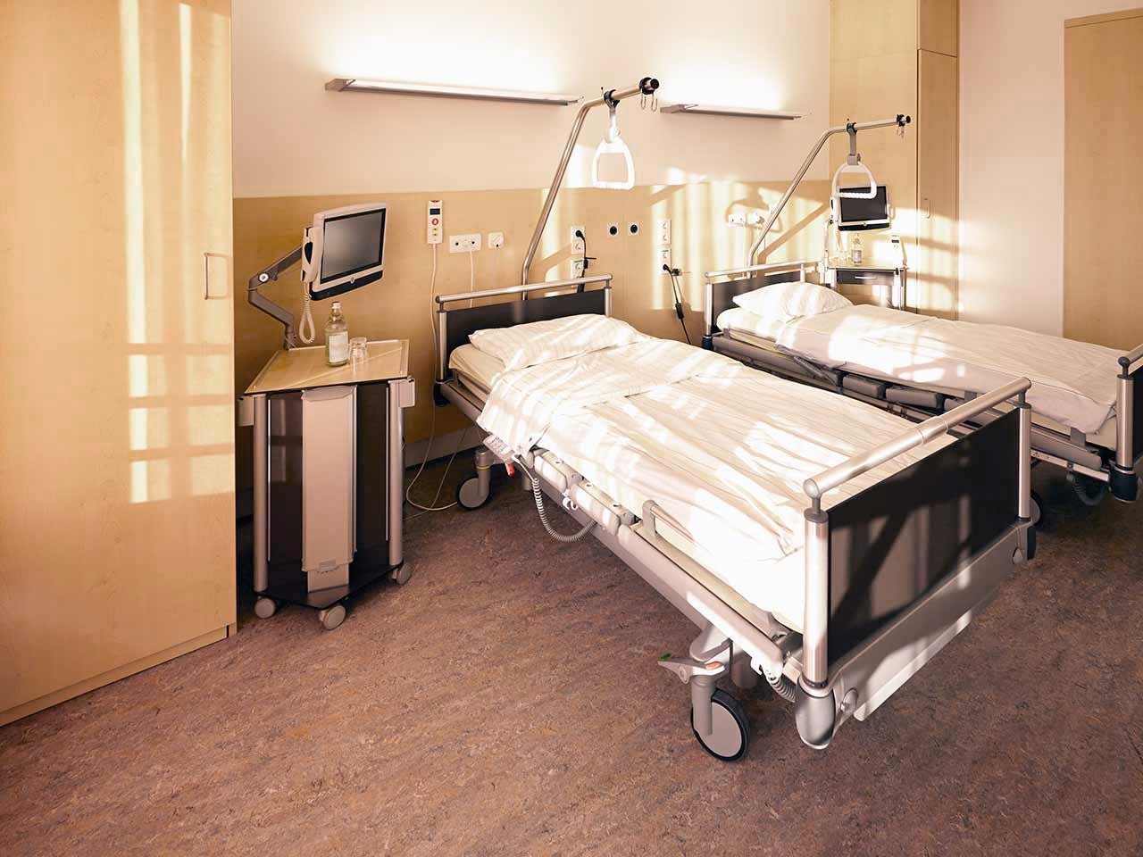
The patients of the University Hospital Jena live in comfortable single and double rooms made in a modern design. Each patient room is equipped with an ensuite bathroom with shower and toilet. The room has enough space to store personal belongings, as well as a table and chairs for receiving visitors. A bedside table can be converted into a table so that patients can eat right in their bed. Each room has a TV, and there is also access to the Internet. In addition, the hospital offers enhanced-comfort rooms.
Meals and Menus
The patient and his accompanying person have a daily choice of three menus. If for some reason the patient does not eat all the foods, he will be offered an individual menu. Please inform the medical staff about your dietary preferences prior to the treatment.
Further details
Standard rooms include:
Religion
Religious services are available upon request.
Accompanying person
During the inpatient program, an accompanying person may stay with you in a room or at the hotel of your choice.
Hotel
During the outpatient program, you can live at a hotel of your choice. Managers will help you to choose the most suitable options.


