Treatment of laryngeal cancer with embolization or chemoembolization in University Hospital Oldenburg
University Hospital Oldenburg
Oldenburg, Germany
During the first visit, the doctor will conduct a clinical examination and go through the results of the available diagnostic tests. After that, you will undergo the necessary additional examination, such as the assessment of liver and kidney function, ultrasound scan, CT scan and MRI. This will allow the doctor to determine which vessels are feeding the tumor and its metastases, as well as determine how well you will tolerate the procedure.
Chemoembolization begins with local anesthesia and catheterization of the femoral artery. The thin catheter is inserted through a few centimeters long incision of the blood vessel. The doctor gradually moves the catheter to the vessel feeding the primary tumor or its metastases. The procedure is carried out under visual control, an angiographic device is used for this. The vascular bed and the position of the catheter in it are displayed on the screen of the angiograph.
When the catheter reaches a suspected artery, a contrast agent is injected through it. Due to the introduction of the contrast agent, the doctor clearly sees the smallest vessels of the tumor and the surrounding healthy tissues on the screen of the angiograph. After that, he injects emboli into the tumor vessels through the same catheter.
Emboli are the spirals or the liquid microspheres. The type of embolus is selected individually, taking into account the diameter of the target vessel. When carrying out chemoembolization, a solution of a chemotherapy drug is additionally injected into the tumor vessel. Due to the subsequent closure of the vessel lumen with an embolus, the chemotherapy drug influences the tumor for a long time. In addition, the drug does not enter the systemic circulation, which allows doctors to use high doses of chemotherapeutic agents without the development of serious side effects. Chemoembolization leads to the destruction of the tumor or slowing down its progression.
After that, the catheter is removed from the artery. The doctor puts a vascular suture on the femoral artery and closes it with a sterile dressing. During chemoembolization, you will be awake. General anesthesia is not used, which significantly reduces the risks of the procedure and allows performing it on an outpatient basis, avoiding long hospital stay.
After the first procedure, you will stay under the supervision of an interventional oncologist and general practitioner. If necessary, you will receive symptomatic treatment. As a rule, a second chemoembolization procedure is performed in 3-5 days after the first one in order to consolidate the therapeutic effect. After that, you will receive recommendations for further follow-up and treatment.
 BookingHealth Price from:
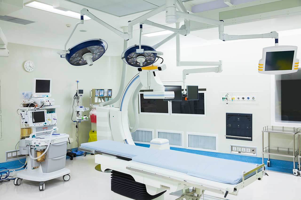
The Department of Interventional Radiology at the University Hospital Oldenburg offers a full range of modern imaging tests and image-guided therapeutic procedures. The department performs imaging examinations for patients from all departments of the University Hospital Oldenburg: X-ray, computed tomography, magnetic resonance imaging, sonography, mammography, and angiography; diagnostic examinations are performed on an outpatient basis. To ensure accurate diagnosis and effective treatment, the department has state-of-the-art medical equipment, including a 64-slice spiral CT scanner, two high-performance 1.5 Tesla MRI scanners, advanced equipment for digital subtraction angiography, pulsed fluoroscopy, and many other devices. The department's team of physicians also specializes in image-guided therapeutic interventional procedures – the treatment of vascular stenoses and occlusions, malignant tumors, and chronic back pain. Therapeutic manipulations are often performed on an outpatient basis. The department adheres strictly to radiation safety standards, so patients can expect highly effective and safe treatment. The Head Physician of the department is Prof. Dr. med. Martin Maurer.
One of the most popular interventional procedures in the department is balloon angioplasty (percutaneous transluminal angioplasty), which is indicated for patients with severe symptoms of critical lower limb ischemia or with the formation of trophic ulcers and non-healing wounds. The essence of balloon angioplasty is as follows: a specialist inserts a special catheter with a tiny balloon attached to it through the femoral artery (sometimes through the radial artery) to the site of occlusion or narrowing of the blood vessel under imaging guidance, after which the balloon is inflated, thus normalizing blood flow in the pathological area of the blood vessel. In many cases, percutaneous transluminal angioplasty is performed at the same time as stenting. A stent is implanted to prevent repeated occlusion of the artery. The duration of the therapeutic manipulation is about 2 hours. The procedure is performed under local anesthesia.
The department's doctors also specialize in transarterial chemoembolization (TACE) for the treatment of liver tumors. The therapeutic effect is achieved by depriving the tumor of blood supply through embolization, the introduction of microparticles into the lumen of the blood supply vessel. Embolization is performed in combination with the administration of a high dose of chemotherapy drugs through a selective catheter directly into the tumor focus, which makes it possible to significantly shrink the tumor, and, in some cases, destroy it completely. As a rule, the TACE procedure is prescribed for advanced stages of hepatocellular carcinoma and cholangiocarcinoma; an important condition for its performance is the absence of metastases outside the liver. Within 24-48 hours after transarterial chemoembolization, a follow-up MRI or CT scan is done to assess the results of the manipulation. In many cases, the procedure is repeated 2-3 times with an interval of four weeks.
The department's therapeutic options are complemented by infiltration therapy for the treatment of chronic back pain. This treatment method is considered when oral medications have failed. Before infiltration therapy is prescribed, computerized tomography and/or magnetic resonance imaging is performed to plan the upcoming treatment. Infiltration therapy involves the targeted injection of a potent painkiller directly into the pathological focus near the nerve root under CT guidance. The procedure is highly effective and can be repeated. It is also painless for the patient and poses virtually no health risks.
The department's clinical focuses include the following:
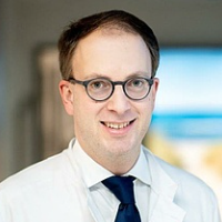
Since October 1, 2022, Prof. Dr. med. Martin Maurer has been the Head Physician of the Department of Interventional Radiology at the University Hospital Oldenburg. Previously, he was a Senior Physician in the Department of Interventional Radiology at the University Hospital Bern (Switzerland). The specialist began his clinical career at the Charité University Hospital Berlin, where he worked in the Department of Radiology for 7 years.
Prof. Maurer studied medicine in Muenster, Berlin, Dublin, and Paris. He received his medical degree from the Charité Medical University of Berlin in 2007. Dr. Martin Maurer received his Master of Business Administration in Healthcare from the University of Erlangen-Nuremberg and his Master of Science in Healthcare Economics, Policy and Management from London, England. In 2019, he was appointed Extraordinary Professor at the Charité Medical University of Berlin.
Prof. Maurer has a special clinical and research interest in image-guided interventional procedures for the treatment of abdominal pathologies. In 2015, he started a successful clinical program for minimally invasive image-guided treatment of malignant liver diseases using computer navigation, which he is now also implementing at the University Hospital Oldenburg.
Prof. Maurer serves as a reviewer for renowned radiology scientific journals and is a member of numerous professional societies, including the German Roentgen Society (DRG) and the European Society of Radiology (ESR).
Photo of the doctor: (c) Klinikum Oldenburg AöR
The University Hospital Oldenburg is a multidisciplinary medical complex offering top-notch services of the European standard. The hospital has 20 specialized departments, 15 highly specialized centers, and more than 10 institutes. The hospital provides services in almost all areas of modern medicine: general and abdominal surgery, cardiac surgery, orthopedics, traumatology, cardiology, oncology, dermatology, gastroenterology, gynecology, mammology, urology, and pediatric and adolescent medicine.
The hospital has 832 beds, and many diagnostic and therapeutic procedures are performed on an outpatient basis. More than 150,000 patients are treated here annually, 37,000 of them receive medical care in a hospital setting. The medical complex has a highly professional medical team of more than 2,900 employees and most departments are headed by professors with extensive clinical experience.
The hospital is home to one of the best comprehensive cancer centers in Germany (Nordwestdeutsches Tumorzentrum), certified according to the requirements of the German Cancer Society (DKG). The center successfully treats breast cancer, prostate cancer, colorectal cancer, pancreatic cancer, esophageal cancer, head and neck tumors, skin tumors, and other oncological diseases. The center uses modern cancer treatment methods with proven effectiveness and also offers allogeneic and autologous bone marrow transplantation for the treatment of severe hematologic diseases (European certification JACIE).
The University Hospital Oldenburg is an expert medical facility in robotic surgery. The hospital has a specialized da Vinci Surgery Center, where gastrointestinal, urological, and gynecological diseases are treated with excellence. Patients are operated on by highly qualified professors with in-depth expertise and extensive experience in robotic surgery.
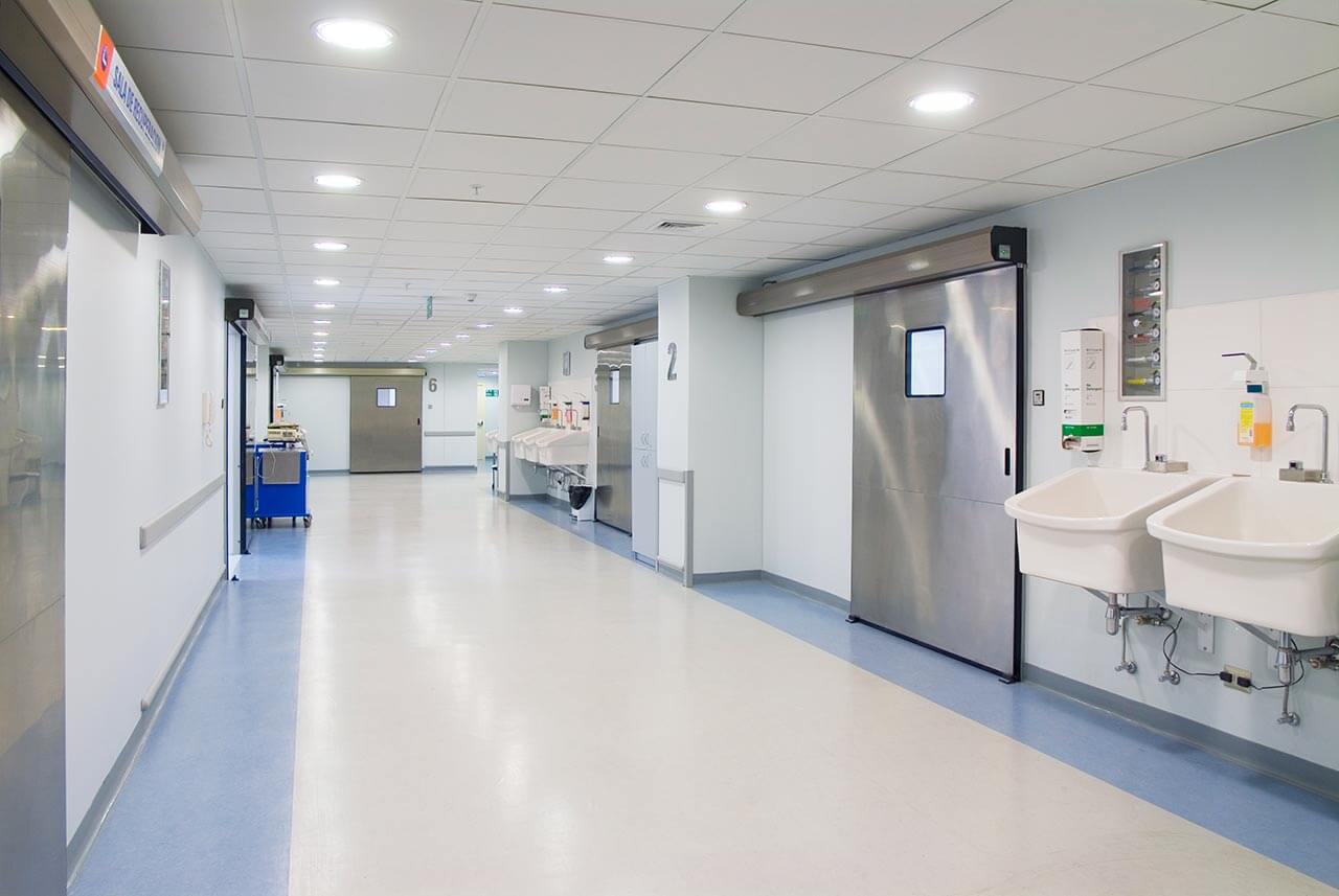
The University Hospital Oldenburg is one of the leading medical institutions in Germany because it offers excellent medical and technical resources, qualified personnel, and a comfortable infrastructure designed with the needs of patients in mind. The hospital's physicians treat patients with complex clinical cases and achieve incredible results.
The basis of medical care in the hospital is humane attitude to the patient, empathy and understanding of his needs. During the therapeutic process, the patient is surrounded by care and, if necessary, receives professional psychological support.
Photo: (с) depositphotos
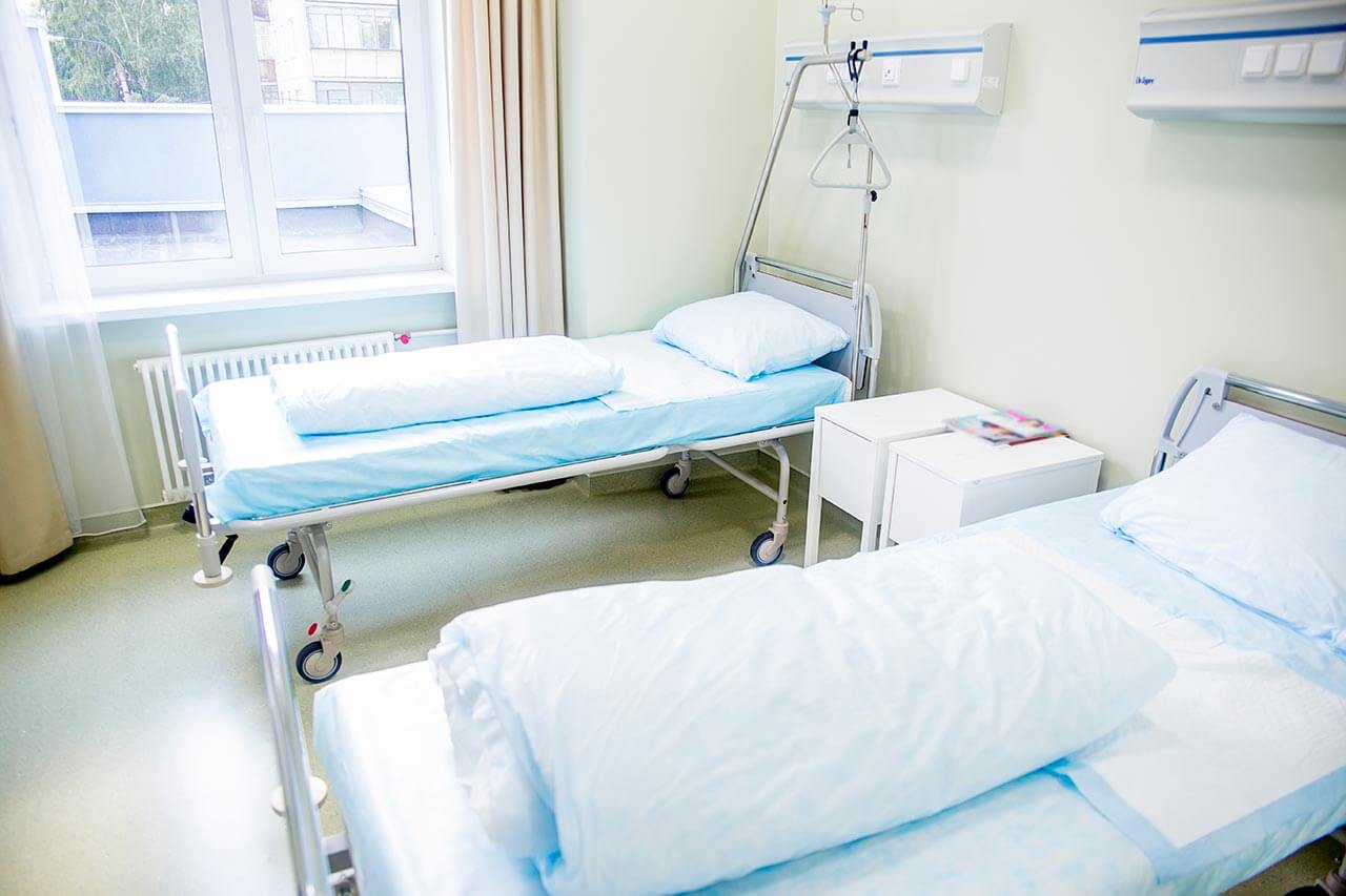
The patients at the University Hospital Oldenburg stay in comfortable single, double, and triple rooms. Each patient room has an ensuite bathroom with a toilet and a shower. The patient rooms have a TV, a telephone, and Wi-Fi. The patient rooms in the pediatric departments are designed in an appropriate interior to make the young patients feel as comfortable as possible. A child can stay in the same room with one of the parents.
The hospital has an excellent infrastructure: there is a pharmacy, a hairdresser, an ATM, and a library with audio books, DVD movies, and board games.
The hospital offers delicious and balanced meals three times a day: breakfast, lunch, and dinner. Since 2022, a new menu "Vitalessen" has been introduced here, which excludes the addition of preservatives, dyes, and flavor enhancers to food. The menu takes into account all preferences and wishes of the patients.
There is also a cozy cafe in the hospital, where you can enjoy a tasty snack or a cup of aromatic coffee or hot tea with dessert.
Standard rooms include:
![]() Toilet
Toilet
![]() Shower
Shower
![]() Wi-Fi
Wi-Fi
![]() TV
TV
There is a chapel on the ground floor of the hospital where Catholic and Evangelical services are held regularly. Services by representatives of other religions are available upon request.
Your accompanying person may stay with you in your patient room or at the hotel of your choice during the inpatient program.
You may stay at the hotel of your choice during the outpatient program. Our managers will support you for selecting the best option.
