Astrocytoma treatment with MRI-guided laser ablation in University Hospital Regensburg
University Hospital Regensburg
Regensburg, Germany
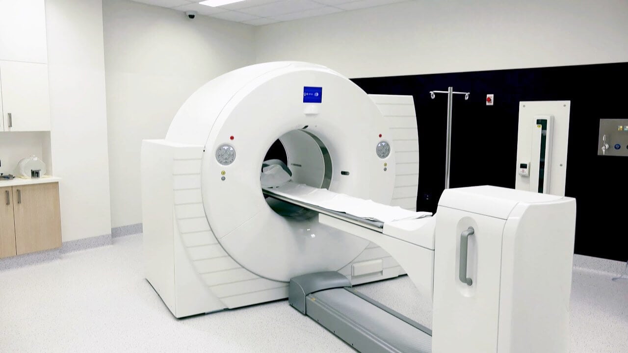


During the first visit, the physician will conduct a clinical examination and neurological examination, and go through the results of the available diagnostic tests. After that, you will undergo the necessary additional examination, such as the laboratory blood and urine tests, MRI scan with obtaining 3D images and creating volumetric images of the whole brain. Based on the results of an additional examination, the physician will clarify the localization of brain metastases. After that, the preparation according to the preoperative standard will begin.
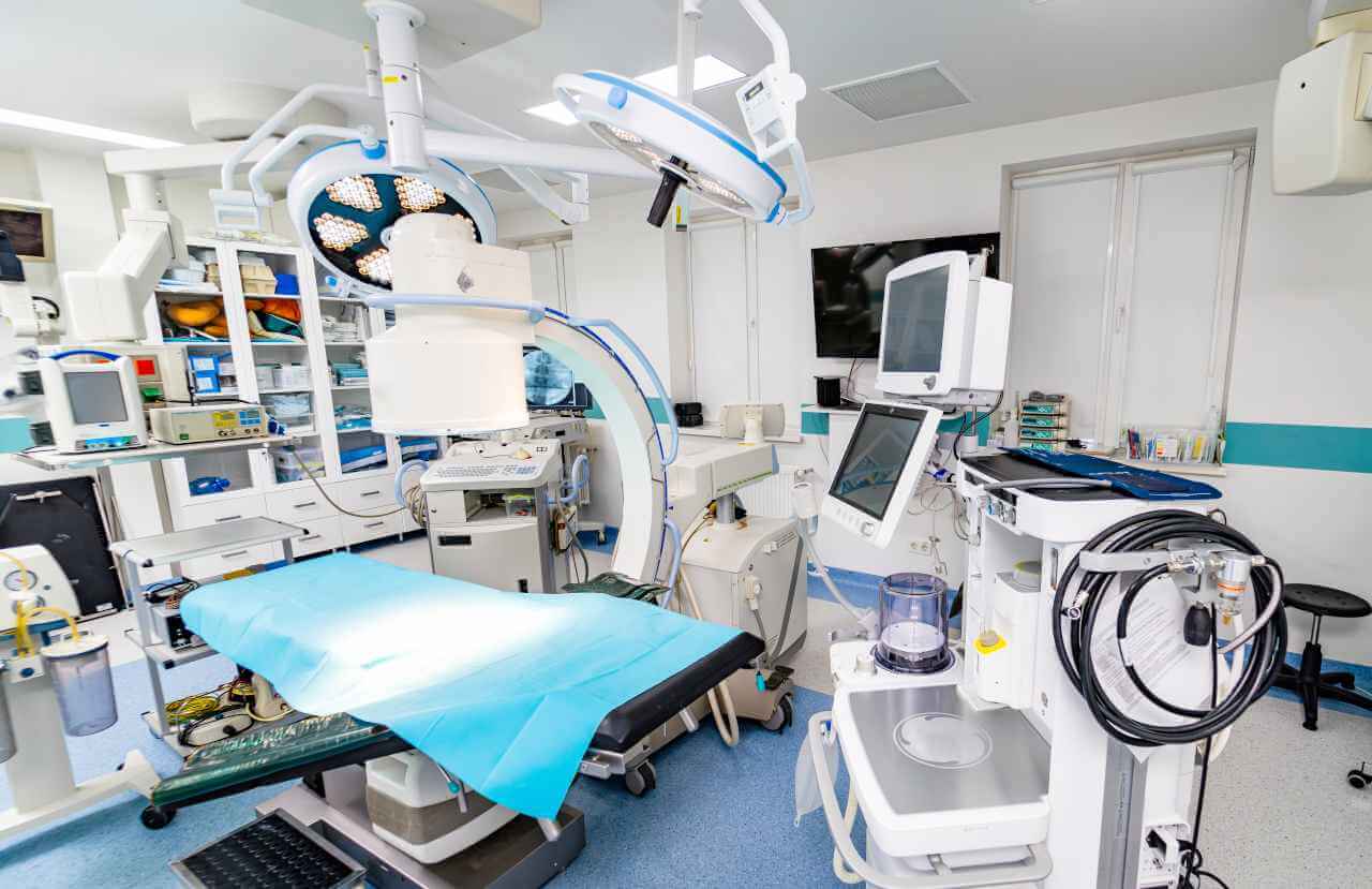
Laser ablation of metastases begins with the formation of access to the brain. When using a catheter with a laser fiber, a hole with a diameter of 3.2 mm is sufficient for this. The miniature size and flexibility of the catheter help preserve healthy brain tissue.
When the catheter reaches metastasis, energy is delivered through the laser fiber. Under the influence of high temperature, metastases are destroyed. The procedure is guided by real-time MRI. Surgeons also assess the temperature of healthy brain tissue using thermographic images to avoid overheating.
After the completion of the operation, you will be transferred to the ward, under the round-the-clock supervision of doctors and nurses. In several days, the attending physician will evaluate the results of the postoperative MRI scans, schedule the date of discharge from the hospital and give you detailed recommendations for further follow-up and treatment.
 BookingHealth Price from:
The Department of Adult and Pediatric Neurosurgery, Spinal Surgery at the University Hospital Regensburg provides the full range of modern diagnostics and treatment of diseases of the central and peripheral nervous systems for patients of all ages, ranging from newborns to the elderly. The department's neurosurgeons focus on patients with brain and spinal cord tumors, cerebrovascular diseases, movement disorders, peripheral nerve compression syndromes, and spinal pathologies. In the field of pediatric neurosurgery, particular interest is in the surgical treatment of brain tumors, congenital malformations of the nervous system, and traumatic brain injuries. The department carries out comprehensive diagnostic examinations of the nervous system, including laboratory tests, neurophysiological studies, computed tomography, magnetic resonance imaging, including functional MRI and diffusion-weighted MRI, and positron emission tomography. The department's team of surgeons carefully plans each surgical procedure, as it is important to the specialists that the patient receives the most effective and safest treatment. For this purpose, the NeuroVis 3D planning computer program is used here. Brain and spinal cord surgery is performed using advanced neuronavigation systems, intraoperative monitoring, and fluorescent navigation with 5-aminolevulinic acid, which eliminates the risk of damaging vital anatomical structures of the brain and spinal cord.
The department is headed by Prof. Dr. med. Nils Ole Schmidt. The neurosurgeon has more than 20 years of successful clinical experience. He has performed thousands of operations on the brain and spinal cord, including the most complex ones. He keeps abreast of the latest innovations in neurosurgery and uses modern sparing techniques in his practice. Prof. Schmidt is also actively involved in research, publishing scientific papers, and sharing his experience with colleagues at national and international conferences.
The department has earned a reputation as one of the best in Germany for the surgical treatment of brain tumors. Patient care is provided in a specialized center certified according to the standards of the German Cancer Society (DKG). The center has an interdisciplinary team of highly skilled neurosurgeons, neurologists, neuroradiologists, oncologists, radiation therapists, and nuclear medicine specialists. The experts study each clinical case at regular tumor boards and develop the most effective treatment regimen for the patient. The department's neurosurgeons most commonly operate on patients with glioblastomas, oligodendrogliomas, astrocytomas, ependymomas, brain metastases, and rare brain neoplasms (such as hemangioblastomas and epidermoids). The department also successfully performs surgery for benign brain neoplasms, particularly meningiomas and neuromas. Surgery for both benign and malignant brain tumors is performed in the department using minimally invasive, microsurgical, and endoscopic techniques. For brain tumor resection surgery, the department's neurosurgeons use fluorescent navigation with 5-aminolevulinic acid, intraoperative monitoring, and neuromonitoring. When indicated, the department's specialists perform brain tumor removal surgery in the awake state under local anesthesia. Such surgical procedures are characterized by high safety and avoid postoperative speech and motor disorders.
The medical facility also excels in functional neurosurgery. This area includes surgical treatment of movement disorders (Parkinson's disease, essential tremor, and dystonia), epilepsy, spasticity, chronic back pain, pain due to trigeminal neuralgia, and pain after peripheral nerve traumatic injuries. To deal with such pathologies, the department's neurosurgeons perform deep brain stimulation, vagus nerve stimulation, spinal cord stimulation, peripheral nerve stimulation, facet joint blocks, nerve root blocks, and percutaneous thermocoagulation. The optimal treatment option for each patient is determined on an individual basis after a comprehensive neurological examination. Depending on individual indications, neurosurgeons perform functional procedures under local or general anesthesia.
In cooperation with neuroradiologists, the department's neurosurgeons treat neurovascular diseases, the most common of which are brain aneurysms, arteriovenous malformations, cavernomas, and neurovascular compression syndromes. Surgery for these pathologies is performed with endovascular techniques, if possible, and a surgical approach is provided through small blood vessel incisions or punctures under imaging guidance. The department's neurosurgeons also specialize in minimally invasive and open microsurgical interventions. For example, microsurgical clipping is successfully performed for the treatment of brain aneurysms, during which a special titanium clip is placed on the aneurysm, effectively excluding it from blood circulation and eliminating the risk to the patient's life.
The department's team of neurosurgeons also admits young patients with diseases of the nervous system. Particular attention is paid to the surgical treatment of brain tumors, congenital malformations, traumatic brain injuries, and spinal cord injuries. Medical care for children with diseases of the nervous system is provided in close cooperation with pediatric oncologists, pediatric surgeons, pediatric neurologists, radiation therapists, otolaryngologists, oral and maxillofacial surgeons, neuroradiologists, and other specialists from related medical fields. When performing neurosurgical operations on children, the department's specialists use surgical microscopes with fluorescent navigation technology with 5-aminolevulinic acid, neuronavigation systems, intraoperative neuromonitoring devices, intraoperative ultrasound, etc. All these technological devices allow the doctors to carry out highly effective and safe treatment without the risk of developing neurological deficits.
An integral part of the department's clinical practice is spinal surgery. The department's team of spinal surgeons has vast experience in the field of its competence, brilliantly handling clinical cases of any severity. Diagnostics and treatment of patients with suspected spinal pathology take place in a highly specialized center, where a highly qualified team consisting of neurosurgeons, neuroradiologists, traumatologists, radiologists, physiotherapists, and other specialists is in charge. Of particular interest to the department's neurosurgeons are the operations for herniated discs, spinal canal stenosis, and spondylolisthesis (slipped vertebrae). Surgical procedures for herniated discs and spinal canal stenosis are performed using minimally traumatic microsurgical techniques, so there is no need for a prolonged hospital stay. For the treatment of spondylolisthesis, the surgeons of the department implant screw systems that ensure the stabilization of the spine and eliminate pain syndromes caused by the pathology. It is worth mentioning that the medical facility is one of the few in Germany where the innovative Mazor robotic guidance system is used during surgeries to implant screw systems for spinal stabilization. This device ensures the safe and millimeter-accurate placement of the screw system in the spine.
The department specializes in the diagnostics and treatment of the following diseases:
The department's range of surgical options includes the following:
Higher Education and Postgraduate Training
Clinical Interests
Photo of the doctor: (c) Universitätsklinikum Regensburg
The University Hospital Regensburg is known as one of the best and largest medical complexes in Germany. The hospital was opened in 1984 and has been providing effective medical care in accordance with the highest standards of modern university medicine ever since. The hospital has more than 30 highly specialized departments, centers, and facilities. Practically all fields of modern medicine are represented here, including ophthalmology, general and abdominal surgery, vascular surgery, neurosurgery, cardiothoracic surgery, dermatology, cardiology, pulmonology, gastroenterology, nephrology, neurology, oncology, hematology, orthopedics, traumatology, nuclear medicine, radiation therapy, and others.
The hospital employs an experienced and highly qualified medical team of more than 5,000 people. Specialists make every effort to provide each patient with a comprehensive treatment that meets their individual needs and wishes. More than 35,500 inpatients and about 154,000 outpatients receive medical care at the hospital annually. The hospital has 839 beds to accommodate patients.
As one of the largest and most progressive hospitals in Germany, the University Hospital Regensburg offers its patients advanced and minimally invasive treatment. The medical facility excels in laser, endoscopic, arthroscopic, thoracoscopic, minimally invasive, microsurgical, and robotic surgery. Such surgical procedures are characterized by low trauma rates and efficiency comparable to the results of classical open surgery. The hospital houses a Transplant Center where experts perform heart, kidney, liver, and bone marrow transplants. In addition, this center is the only one in the state of Bavaria to offer liver transplantation for children.
The successful clinical practice of the medical complex has been recognized with numerous prestigious quality certificates, including the ISO 9001 TÜV SÜD certificate, the Aktion Saubere Hände certificate, and certificates from the German Cancer Society (DKG) in the treatment of brain cancer, skin cancer, head and neck tumors, gastrointestinal cancer, liver cancer, and pancreatic cancer.
Despite all the achievements and progressiveness of the hospital, a compassionate approach to patients and their life situations is of great importance. The specialists of the hospital show understanding and empathy to the patients and give them moral support on their way to recovery.
Photo: (с) depositphotos
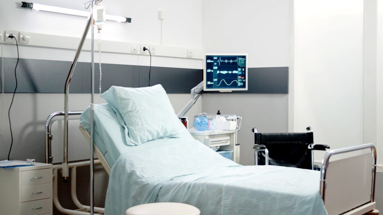
Patients of the University Hospital Regensburg stay in comfortable single and double rooms. The furnishings of a standard room include an automatically adjustable bed, a bedside table with a pull-out tray, a table and chairs, a wardrobe, a TV, and a telephone. Wi-Fi is also available in the patient rooms.
Each room has an ensuite bathroom with a shower and toilet. The bathroom has toiletries, changeable towels, a bathrobe, and a hairdryer.
The patient and his accompanying person are offered three delicious and balanced meals a day: breakfast, lunch, and dinner. If, for some reason, you do not eat all the foods, you will be offered an individual menu. Please inform the medical staff about your dietary preferences prior to treatment.
Standard rooms include:
![]() Shower
Shower
![]() Toilet
Toilet
![]() Wi-Fi
Wi-Fi
![]() TV
TV
Your accompanying person may stay with you in your patient room or at the hotel of your choice during the inpatient program.
You may stay at the hotel of your choice during the outpatient program. Our managers will support you for selecting the best option.
