google_counter
Treatment of bladder cancer with embolization or chemoembolization (688847) | University Hospital Saarland Homburg - BookingHealth
{"translation_price":"50","translation_doc_price":"40","child_coefficient":"1.1","transfer_price":"2.00","transfer_price_vip":"5.00","constant_transfer_price_vip":350,"constant_transfer_price":150,"constant_transfer_distanse":60,"type":"treatment","program_full_story":"<ul>\n\t<li>Initial presentation in the hospital<\/li>\n\t<li>Clinical history taking<\/li>\n\t<li>Review of available medical records<\/li>\n\t<li>Physical examination<\/li>\n\t<li>Laboratory tests:\n\t<ul>\n\t\t<li>Complete blood count<\/li>\n\t\t<li>General urine analysis<\/li>\n\t\t<li>Biochemical analysis of blood<\/li>\n\t\t<li>Tumor markers<\/li>\n\t\t<li>Inflammation indicators (CRP, ESR)<\/li>\n\t\t<li>Coagulogram<\/li>\n\t<\/ul>\n\t<\/li>\n\t<li>Ultrasound\u200b scan<\/li>\n\t<li>CT scan \/ MRI<\/li>\n\t<li>Preoperative care<\/li>\n\t<li>Embolization or chemoembolization, 2 procedures<\/li>\n\t<li>Symptomatic treatment<\/li>\n\t<li>Cost of essential medicines<\/li>\n\t<li>Nursing services<\/li>\n\t<li>Elaboration of further recommendations<\/li>\n<\/ul>\n<div class=\"program_how_program_going mt-4\"><h4>How program is carried out<\/h4><p style=\"text-align:justify\"><strong>During the first visit<\/strong>, the doctor will conduct a clinical examination and go through the results of the available diagnostic tests. After that, you will undergo the necessary additional examination, such as the assessment of liver and kidney function, ultrasound scan, CT scan and MRI. This will allow the doctor to determine which vessels are feeding the tumor and its metastases, as well as determine how well you will tolerate the procedure.<\/p>\n\n<p style=\"text-align:justify\"><strong>Chemoembolization <\/strong>begins with local anesthesia and catheterization of the femoral artery. The thin catheter is inserted through a few centimeters long incision of the blood vessel. The doctor gradually moves the catheter to the vessel feeding the primary tumor or its metastases. The procedure is carried out under visual control, an angiographic device is used for this. The vascular bed and the position of the catheter in it are displayed on the screen of the angiograph.<\/p>\n\n<p style=\"text-align:justify\">When the catheter reaches a suspected artery, a contrast agent is injected through it. Due to the introduction of the contrast agent, the doctor clearly sees the smallest vessels of the tumor and the surrounding healthy tissues on the screen of the angiograph. After that, he injects emboli into the tumor vessels through the same catheter.<\/p>\n\n<p style=\"text-align:justify\">Emboli are the spirals or the liquid microspheres. The type of embolus is selected individually, taking into account the diameter of the target vessel. When carrying out chemoembolization, a solution of a chemotherapy drug is additionally injected into the tumor vessel. Due to the subsequent closure of the vessel lumen with an embolus, the chemotherapy drug influences the tumor for a long time. In addition, the drug does not enter the systemic circulation, which allows doctors to use high doses of chemotherapeutic agents without the development of serious side effects. Chemoembolization leads to the destruction of the tumor or slowing down its progression.<\/p>\n\n<p style=\"text-align:justify\">After that, the catheter is removed from the artery. The doctor puts a vascular suture on the femoral artery and closes it with a sterile dressing. During chemoembolization, you will be awake. General anesthesia is not used, which significantly reduces the risks of the procedure and allows performing it on an outpatient basis, avoiding long hospital stay.<\/p>\n\n<p style=\"text-align:justify\"><strong>After the first procedure<\/strong>, you will stay under the supervision of an interventional oncologist and general practitioner. If necessary, you will receive symptomatic treatment. As a rule, a second chemoembolization procedure is performed in 3-5 days after the first one in order to consolidate the therapeutic effect. After that, you will receive recommendations for further follow-up and treatment.<\/p>\n<\/div><div class=\"program_required_documents mt-4\"><h4>Required documents<\/h4><ul>\n\t<li style=\"text-align: justify;\">Medical records<\/li>\n\t<li style=\"text-align: justify;\">MRI\/CT scan (not older than 3 months)<\/li>\n\t<li style=\"text-align: justify;\">Biopsy results (if available)<\/li>\n<\/ul>\n<\/div>","program_full_story_crm":"<ul>\n\t<li>Initial presentation in the hospital<\/li>\n\t<li>Clinical history taking<\/li>\n\t<li>Review of available medical records<\/li>\n\t<li>Physical examination<\/li>\n\t<li>Laboratory tests:\n\t<ul>\n\t\t<li>Complete blood count<\/li>\n\t\t<li>General urine analysis<\/li>\n\t\t<li>Biochemical analysis of blood<\/li>\n\t\t<li>Tumor markers<\/li>\n\t\t<li>Inflammation indicators (CRP, ESR)<\/li>\n\t\t<li>Coagulogram<\/li>\n\t<\/ul>\n\t<\/li>\n\t<li>Ultrasound\u200b scan<\/li>\n\t<li>CT scan \/ MRI<\/li>\n\t<li>Preoperative care<\/li>\n\t<li>Embolization or chemoembolization, 2 procedures<\/li>\n\t<li>Symptomatic treatment<\/li>\n\t<li>Cost of essential medicines<\/li>\n\t<li>Nursing services<\/li>\n\t<li>Elaboration of further recommendations<\/li>\n<\/ul>\n<div class=\"program_how_program_going mt-4\"><h4>How program is carried out<\/h4><p style=\"text-align:justify\"><strong>During the first visit<\/strong>, the doctor will conduct a clinical examination and go through the results of the available diagnostic tests. After that, you will undergo the necessary additional examination, such as the assessment of liver and kidney function, ultrasound scan, CT scan and MRI. This will allow the doctor to determine which vessels are feeding the tumor and its metastases, as well as determine how well you will tolerate the procedure.<\/p>\n\n<p style=\"text-align:justify\"><strong>Chemoembolization <\/strong>begins with local anesthesia and catheterization of the femoral artery. The thin catheter is inserted through a few centimeters long incision of the blood vessel. The doctor gradually moves the catheter to the vessel feeding the primary tumor or its metastases. The procedure is carried out under visual control, an angiographic device is used for this. The vascular bed and the position of the catheter in it are displayed on the screen of the angiograph.<\/p>\n\n<p style=\"text-align:justify\">When the catheter reaches a suspected artery, a contrast agent is injected through it. Due to the introduction of the contrast agent, the doctor clearly sees the smallest vessels of the tumor and the surrounding healthy tissues on the screen of the angiograph. After that, he injects emboli into the tumor vessels through the same catheter.<\/p>\n\n<p style=\"text-align:justify\">Emboli are the spirals or the liquid microspheres. The type of embolus is selected individually, taking into account the diameter of the target vessel. When carrying out chemoembolization, a solution of a chemotherapy drug is additionally injected into the tumor vessel. Due to the subsequent closure of the vessel lumen with an embolus, the chemotherapy drug influences the tumor for a long time. In addition, the drug does not enter the systemic circulation, which allows doctors to use high doses of chemotherapeutic agents without the development of serious side effects. Chemoembolization leads to the destruction of the tumor or slowing down its progression.<\/p>\n\n<p style=\"text-align:justify\">After that, the catheter is removed from the artery. The doctor puts a vascular suture on the femoral artery and closes it with a sterile dressing. During chemoembolization, you will be awake. General anesthesia is not used, which significantly reduces the risks of the procedure and allows performing it on an outpatient basis, avoiding long hospital stay.<\/p>\n\n<p style=\"text-align:justify\"><strong>After the first procedure<\/strong>, you will stay under the supervision of an interventional oncologist and general practitioner. If necessary, you will receive symptomatic treatment. As a rule, a second chemoembolization procedure is performed in 3-5 days after the first one in order to consolidate the therapeutic effect. After that, you will receive recommendations for further follow-up and treatment.<\/p>\n<\/div>","is_ambulant":"1","bh_fee":"0","only_for_children":"0","no_service":"0","with_prepayment":"1","show_calculator":"1","paket_type":"1","btn_type":"0","clinic_icon":"5ffc47990b13a.jpg","city":"Homburg","clinic_site":"http:\/\/www.uniklinikum-saarland.de\/de\/","department_recommend":"1","country":"Germany","country_id":"1","clinic_name":"University Hospital Saarland Homburg","cinic_name":"University Hospital Saarland Homburg","department_id":"2326","duration":"7","direction":"Interventional radiology","min_duration":0,"clinic_id":"1702","paketPrice":7800,"paket":"<ul>\n <li>Interpreter up to 17 hours<\/li>\n <li>Translation up to 10 pages<\/li>\n <li>Visa support<\/li>\n\n \n<\/ul>","title":"Treatment of bladder cancer with embolization or chemoembolization","price":{"val":26139.95,"type":"val"},"price_surcharge":0,"price_surcharge_clear":0,"extra_service_clinic":[],"extra_service":[],"translation_hours":"0","translation_doc_count":null,"roads":[{"id":"6","distance":"159","airport_title":"Frankfurt a.M."},{"id":"19","distance":"193","airport_title":"Stuttgart"},{"id":"7","distance":"265","airport_title":"Cologne-Bonn"}],"pakets":[],"lang":{"day":"Day","days":"days","ambulatory":"Outpatient","stationaryProgram":"Inpatient"}}

Treatment of bladder cancer with embolization or chemoembolization

University Hospital Saarland Homburg

Homburg, Germany
Program id # 688847
Doctor photo
Prof. Dr. med. Michael Stöckle
Department of Adult and Pediatric Urology, Andrology
Specialized in: adult and pediatric urology, andrology

The program includes:

  • Initial presentation in the hospital
  • Clinical history taking
  • Review of available medical records
  • Physical examination
  • Laboratory tests:
    • Complete blood count
    • General urine analysis
    • Biochemical analysis of blood
    • Tumor markers
    • Inflammation indicators (CRP, ESR)
    • Coagulogram
  • Ultrasound​ scan
  • CT scan / MRI
  • Preoperative care
  • Embolization or chemoembolization, 2 procedures
  • Symptomatic treatment
  • Cost of essential medicines
  • Nursing services
  • Elaboration of further recommendations

How program is carried out

During the first visit, the doctor will conduct a clinical examination and go through the results of the available diagnostic tests. After that, you will undergo the necessary additional examination, such as the assessment of liver and kidney function, ultrasound scan, CT scan and MRI. This will allow the doctor to determine which vessels are feeding the tumor and its metastases, as well as determine how well you will tolerate the procedure.

Chemoembolization begins with local anesthesia and catheterization of the femoral artery. The thin catheter is inserted through a few centimeters long incision of the blood vessel. The doctor gradually moves the catheter to the vessel feeding the primary tumor or its metastases. The procedure is carried out under visual control, an angiographic device is used for this. The vascular bed and the position of the catheter in it are displayed on the screen of the angiograph.

When the catheter reaches a suspected artery, a contrast agent is injected through it. Due to the introduction of the contrast agent, the doctor clearly sees the smallest vessels of the tumor and the surrounding healthy tissues on the screen of the angiograph. After that, he injects emboli into the tumor vessels through the same catheter.

Emboli are the spirals or the liquid microspheres. The type of embolus is selected individually, taking into account the diameter of the target vessel. When carrying out chemoembolization, a solution of a chemotherapy drug is additionally injected into the tumor vessel. Due to the subsequent closure of the vessel lumen with an embolus, the chemotherapy drug influences the tumor for a long time. In addition, the drug does not enter the systemic circulation, which allows doctors to use high doses of chemotherapeutic agents without the development of serious side effects. Chemoembolization leads to the destruction of the tumor or slowing down its progression.

After that, the catheter is removed from the artery. The doctor puts a vascular suture on the femoral artery and closes it with a sterile dressing. During chemoembolization, you will be awake. General anesthesia is not used, which significantly reduces the risks of the procedure and allows performing it on an outpatient basis, avoiding long hospital stay.

After the first procedure, you will stay under the supervision of an interventional oncologist and general practitioner. If necessary, you will receive symptomatic treatment. As a rule, a second chemoembolization procedure is performed in 3-5 days after the first one in order to consolidate the therapeutic effect. After that, you will receive recommendations for further follow-up and treatment.

Required documents

  • Medical records
  • MRI/CT scan (not older than 3 months)
  • Biopsy results (if available)

Service

Price:
Type of program :
Price for 1 day:
Expected duration of the program:
The minimum duration of the program:
Select

You may also book:

Guarantee Price:

About the department

The Department of Adult and Pediatric Urology, Andrology at the University Hospital Saarland Homburg provides the full range of services in the fields of its competence, meeting the high standards of modern university medicine. The department specializes in the diagnostics and treatment of diseases of the kidneys, adrenal glands, ureters, bladder, and urethra. The medical facility also successfully treats diseases of the prostate gland, testicles, penis, male infertility, and erectile dysfunction. In addition, the department's doctors admit children with congenital and acquired diseases of the urinary system, as well as boys with congenital malformations of the reproductive system. A unique feature of the department is an advanced Center for da Vinci Robotic Surgery, which performs many effective minimally invasive interventions for prostate gland, bladder, and kidney cancer. Since 2006, when the center first opened its doors to its patients, the specialists have performed a total of more than 7,000 robot-assisted surgical procedures, including more than 5,000 prostatectomies, about 260 cystectomies, and more than 1,500 nephrectomies. Since 2016, the center's surgeons have also performed 23 kidney transplants with the da Vinci surgical system. The department is headed by Prof. Dr. med. Michael Stöckle.

Prostate cancer is one of the most common oncological diseases in men, so the department's doctors often treat this pathology and have the widest opportunities to deal with it. The PSA test, a digital rectal exam, a transrectal ultrasound scan, and a biopsy are used to diagnose prostate cancer. Imaging tests help the department's urologists determine the stage of the oncological process. A treatment regimen is developed individually for each patient based on his diagnostic data, cancer stage, age, and general health condition. In most cases, the therapy is based on tumor resection surgery along with the prostate gland (prostatectomy), which is performed in the department with the innovative da Vinci surgical system. Robot-assisted interventions are the most sparing of all the surgical treatment options available. In addition, da Vinci robot-assisted surgery has been practiced in the department since 2006, and the specialists have tremendous experience in this medical field. Modern chemotherapy regimens are also often used. Many patients with prostate cancer are also prescribed brachytherapy (internal radiation therapy). The essence of this type of treatment is the placement of radioactive seeds directly into the tumor. In this case, the radiation source has the maximum effect on the tumor but practically does not cause any damage to the adjacent healthy tissues.

The department also successfully treats bladder, kidney, adrenal, ureteral, and urethral cancers. The results of the patient's diagnostic examinations are considered at an interdisciplinary tumor board, during which doctors cooperatively develop a treatment regimen for a particular clinical case. The main treatment method for urinary system cancers is surgery. As an advanced medical facility, the department offers its patients the most sparing type of surgery, namely da Vinci robot-assisted surgery. The intervention is complemented by conservative treatment methods, such as chemotherapy, radiation therapy, targeted therapy, immunotherapy, and others.

The department's pride is the Center for da Vinci Robotic Surgery, which became one of the first medical facilities of this kind in Germany. The specialists initially practiced only robot-assisted surgery for prostate cancer, but today they also successfully perform kidney and bladder tumor removal surgery, partial and total kidney and bladder removal surgery, as well as kidney transplants. The Center for da Vinci Robotic Surgery has two state-of-the-art robotic surgeons with which more than 600 interventions of varying complexity are performed annually. The department is one of the most competent medical facilities in the field of da Vinci robot-assisted surgery, which is increasingly replacing classical surgery. This is not surprising, since robot-assisted surgery has many advantages. The main benefits of using the da Vinci surgical system include minimal pain, minimal blood loss, and a short hospital stay. It is worth noting that the surgical system does not work independently but is controlled by the operating doctor, thanks to which the robot acts as an "assistant" that implements the surgeon's decisions absolutely accurately and without the slightest deviation.

The department's urologists treat patients with benign pathologies, including benign prostatic hyperplasia, prostatitis, urinary incontinence in women and men, urethral strictures, kidney stone disease, urinary tract infections, and sexually transmitted diseases. When treating benign diseases, the specialists mainly use conservative treatment methods. For example, antibiotic therapy is prescribed for infectious diseases, and kidney stone disease can be treated with extracorporeal shock wave lithotripsy.

The department has excellent resources to provide medical care to young patients with urologic diseases. Pediatric urologists most often treat phimosis, undescended testicles, hydrocele, hypospadias, enuresis (nocturnal and daytime), and vesicoureteral reflux. A child needs comprehensive diagnostics, which may include blood and urine tests, X-ray scans, ultrasound scans, urodynamic tests, uroflowmetry, and other diagnostic procedures, to provide optimal treatment. Therapeutic options in pediatric urology include both conservative and surgical methods. Depending on the complexity of the clinical case, the department's specialists use drug therapy, injection therapy, biofeedback, and surgical procedures. Surgery is often performed for congenital malformations of the reproductive system in boys. Doctors perform operations on an outpatient basis, which makes hospitalization unnecessary.

The work of the department's andrologists is focused on treating male infertility and erectile dysfunction. In the first case, it is often enough to normalize the man's hormonal levels or treat urologic pathologies (for example, testicular diseases, ejaculatory duct obstruction, or ejaculatory disorders) that prevent him from conceiving a child. Men with complex forms of male infertility may require interventional procedures such as testicular sperm aspiration (TESA) or microsurgical epididymal sperm aspiration (MESA). As for erectile dysfunction, the department's doctors often use only modern medications. The department's doctors also perform microsurgical procedures to repair the penile curvature in men.

The department provides the diagnostics and treatment of the following diseases:

  • Adult urology
    • Prostate diseases
      • Prostate cancer
      • Benign prostatic hyperplasia
      • Prostatitis
    • Testicular and penile diseases 
      • Testicular cancer
      • Penile cancer
      • Penile curvature
      • Inflammatory processes in the testicles and epididymis
    • Bladder diseases
      • Bladder cancer
      • Bladder stones
      • Urinary incontinence in men and women
      • Inflammatory processes in the bladder
    • Kidney, adrenal and ureteral diseases
      • Kidney cancer
      • Adrenal cancer
      • Renal pelvis and ureteral cancers
      • Kidney and ureteral stones
      • Renal pelvis outflow stenosis
      • Inflammatory processes in the renal pelvis
    • Urethral diseases
      • Urethral stricture
      • Urethral cancer
  • Pediatric urology
    • Undescended testicles
    • Testicular torsion
    • Phimosis (foreskin narrowing)
    • Hydrocele
    • Hypospadias
    • Vesicoureteral reflux
    • Megaureter
    • Neurogenic bladder
    • Urethral valves
    • Duplex kidney
    • Enuresis (urinary incontinence in children)
    • Renal pelvis outflow stenosis
    • Tumors (Wilms' tumor, testicular tumors, etc.)
  • Andrology
    • Male infertility
    • Erectile dysfunction
    • Peyronie's disease
  • Other diseases

The department's range of therapeutic options includes:

  • Conservative treatment
    • Drug therapy with pills
    • Injection therapy
    • Extracorporeal shock wave lithotripsy
    • Ultrasound therapy
    • Biofeedback therapy 
  • Surgical treatment
    • Da Vinci robot-assisted surgery
    • Endoscopic interventions
    • Laser interventions (ND:YAG neodymium laser)
    • Brachytherapy (internal irradiation)
  • Other treatment methods

Curriculum vitae

Higher Education and Professional Career

  • 1976 - 1983 Medicine studies at the Universities of Hanover, Bonn and Mainz.
  • 1983 German and American state examinations in Medicine.
  • 1983 Doctor of Medicine, Institute of Medical Microbiology, University of Mainz.
  • 1983 -1985 Training in Surgery, Nordwest Clinic, Frankfurt am Main.
  • 1985 - 1989 Training in Urology: University Hospital Mainz, University Hospital Leiden (the Netherlands), Cantonal Hospital St. Gallen (Switzerland).
  • 1986 Scholarship from the German Research Foundation, Institute of Cytochemistry and Cytometry, University of Leiden, the Netherlands.
  • 1989 Board certification in Urology, Medical Association of Rheinhessen.
  • 1990 Habilitation, University of Mainz.
  • 1989 - 1994 Senior Physician in the Department of Adult and Pediatric Urology, University Hospital Mainz.
  • 1994 -1995 Managing Senior Physician and Deputy Head of the Department of Adult and Pediatric Urology at the University Hospital Mainz.
  • 1996 - 2000 Head of the Department of Urology at the University Hospital Kiel.
  • Since 01.04.2000 Head of the Department of Adult and Pediatric Urology, Andrology at the University Hospital Saarland Homburg.

Memberships in Professional Societies

  • German Society of Urology.
  • Southwestern German Society of Urology.
  • Association of Urologists of Northern Germany.
  • European Urological Association.
  • American Urological Association.
  • International Society of Urology.
  • German Cancer Society.
  • Consortium of German Prostate Cancer Organizations.

Photo of the doctor: (c) Universitätsklinikum des Saarlandes


About hospital

The University Hospital Saarland Homburg is the largest hospital in the city of Homburg and the most important medical facility in the region. The hospital, which currently has 30 specialized departments and 20 institutes, was founded in 1947 and operates on the basis of Saarland University. The hospital plays a leading role in medical education, research, and medical care both in the state of Saarland and throughout Germany. With vast experience in serving foreign patients, the medical facility is also widely known in the international medical arena.

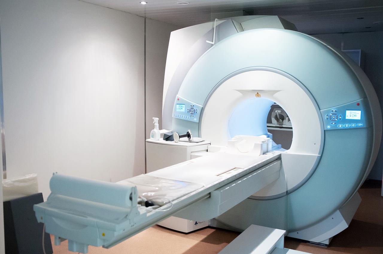
The pride of the hospital is state-of-the-art equipment that allows the doctors to perform high-precision comprehensive diagnostics and the most sparing treatment even if a patient has a severe pathology. Patients are offered innovative medicine based on the very latest scientific achievements. At the same time, the hospital offers many therapeutic methods that are used only in leading medical centers in Europe, including da Vinci robot-assisted surgery, CAR T-cell therapy, TAVI and MitraClip catheter-based procedures, innovative laser procedures, etc. Great importance is paid to ethical and social competence. The hospital is constantly improving the work of its medical personnel and infrastructure to provide medical services that meet the highest standards.

As a multidisciplinary medical complex, the hospital offers highly effective treatment of the full range of common diseases as well as rare and severe pathologies. The efforts of the medical staff, which includes over 600 doctors and 2,000 nurses, are focused on the patient and their unique needs and desires. The doctors always devote enough time to personal communication with their patients, provide them with moral support, and are sympathetic to every life situation.

Photo: (с) depositphotos

Accommodation in hospital

Patients rooms

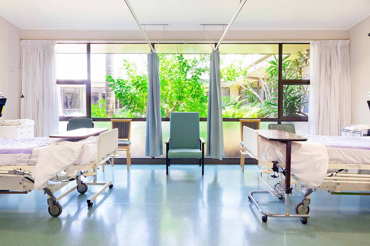
The patients of the University Hospital Saarland Homburg live in comfortable single, double, and triple patient rooms with a modern design. Each room is equipped with an ensuite bathroom with a shower and a toilet, as well as everything else necessary: a comfortable bed, a bedside table, a TV, and a telephone. In addition, enhanced-comfort rooms and specially equipped rooms for people with disabilities are available for the patients.

Meals and Menus

The hospital offers healthy and delicious meals three times a day: buffet breakfast, dinner with a wide choice of dishes for every taste, and a light supper. The menu features dietary and vegetarian dishes. There is also a cafeteria on the territory where one can taste delicious dishes and have a cup of coffee, tea, or some refreshing drinks

Further details

The standard patient rooms include:

Shower
Toilet
Wi-Fi
TV

Religion

The hospital regularly hosts catholic and evangelical devine services. The services of representatives of other religions are available upon request.

Accompanying person

During an inpatient program, an accompanying person can stay with you in the patient room or in a hotel of your choice.

Hotel

During an outpatient program, you can stay in a hotel of your choice. The managers will help you choose the most suitable options.