The program includes:
- Initial presentation in the clinic
- clinical history taking
- review of medical records
- physical examination
- laboratory tests:
- complete blood count
- general urine analysis
- biochemical analysis of blood
- inflammation indicators (CRP, ESR)
- indicators of blood coagulation
- neurological examination
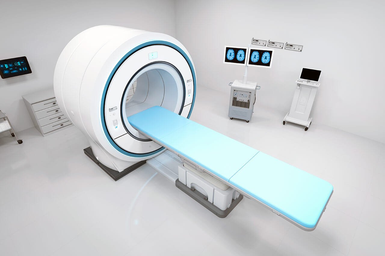
- CT/MRI scan
- neuropsychological tests (if indicated):
- ENMG (electroneuromyography)
- EEG (electroencephalography)
- SEPs (somatosensory evoked potentials)
- VEPs (visually evoked potentials)
- BAEP tests (brainstem auditory evoked potentials)
- preoperative care
- MRI-guided laser ablation of brain metastases
- postoperative MRI control
- symptomatic treatment
- control examinations
- the cost of essential medicines and materials
- nursing services
- full hospital accommodation
- developing of further guidance by specialists
in oncology and radiotherapy
How program is carried out
During the first visit, the physician will conduct a clinical examination and neurological examination, and go through the results of the available diagnostic tests. After that, you will undergo the necessary additional examination, such as the laboratory blood and urine tests, MRI scan with obtaining 3D images and creating volumetric images of the whole brain. Based on the results of an additional examination, the physician will clarify the localization of brain metastases. After that, the preparation according to the preoperative standard will begin.
Laser ablation of metastases begins with the formation of access to the brain. When using a catheter with a laser fiber, a hole with a diameter of 3.2 mm is sufficient for this. The miniature size and flexibility of the catheter help preserve healthy brain tissue.
When the catheter reaches metastasis, energy is delivered through the laser fiber. Under the influence of high temperature, metastases are destroyed. The procedure is guided by real-time MRI. Surgeons also assess the temperature of healthy brain tissue using thermographic images to avoid overheating.
After the completion of the operation, you will be transferred to the ward, under the round-the-clock supervision of doctors and nurses. In several days, the attending physician will evaluate the results of the postoperative MRI scans, schedule the date of discharge from the hospital and give you detailed recommendations for further follow-up and treatment.
Required documents
- Medical records
- MRI/CT scan (not older than 3 months)
- Biopsy results (if available)
Service
You may also book:
 BookingHealth Price from:
About the department
The Department of Neurosurgery and Spinal Surgery at the University Hospital Ulm provides top-class medical care for patients with diseases of the central and peripheral nervous systems. The medical facility performs surgery of any complexity on the brain, spinal cord, spine, and peripheral nerves. The department is the largest healthcare facility of this kind in Germany and has a team of highly professional neurosurgeons with long clinical experience. The department's specialists focus on patients with brain tumors (gliomas, meningiomas, and brain metastases), neurovascular diseases (aneurysms, arteriovenous malformations, and dural arteriovenous fistulas), and traumatic brain injuries. Spinal surgeons operate on patients with herniated discs, spinal canal stenosis, spondylolisthesis, spondylodiscitis, spinal fractures, and spinal tumors. The department extensively uses minimally invasive surgical techniques that allow for good therapeutic results with high safety in surgical manipulations. The medical facility has two hybrid operating rooms with state-of-the-art equipment, including the Zeego® intraoperative robotic angiography system and the intraoperative Brainsuite® MRI. The Head Physician of the department is Prof. Dr. med. Christian Rainer Wirtz.
The department's neurosurgeons often admit patients with brain tumors, such as gliomas, meningiomas, and brain metastases. The main treatment method is physical tumor removal. At the same time, neurosurgeons are faced with the task of preserving vital brain structures and preventing the development of neurological deficits. During brain tumor resection surgery, the department's specialists use advanced intraoperative imaging, intraoperative neuromonitoring devices, and neuronavigation systems. In most cases, craniotomy is indicated for patients with brain tumors, during which the doctor removes a part of the cranium and approaches the brain. The neoplasm is resected using special surgical instruments under imaging guidance, after which the surgical wound is sutured. Depending on the complexity of the clinical case, the duration of the operation varies from 2 to 5 hours. The surgical intervention is usually performed under general anesthesia.
An important focus for the department's neurosurgeons is the treatment of neurovascular diseases. They regularly operate on patients with brain aneurysms. The pathology is characterized by protrusion of the brain artery wall. The essence of aneurysm treatment is to "exclude" it from the bloodstream in order to prevent its rupture, which may potentially lead to a fatal outcome. The department's specialists perform endovascular treatment of brain aneurysms using coiling and also perform clipping using classical open surgical techniques. During the coiling procedure, surgeons insert a platinum coil through a puncture in the femoral artery under angiography guidance. It is directed to the pathological focus along the vascular channel and fills the cavity of the aneurysm. As a result, the aneurysm is excluded from the blood flow, and the risk of its rupture disappears. For brain aneurysm clipping, a neurosurgeon forms a hole in the skull, through which they isolate a section of artery with an aneurysm and clamp the neck of the aneurysm with a titanium clip. After applying the clip, the specialist opens the aneurysm and, if necessary, partially removes its walls. The clip remains on the aneurysm permanently. The choice of a particular type of surgical intervention depends on various factors, including the size and shape of the aneurysm, its localization, and the general health condition of the patient.
Spinal surgery is well-developed in the department. The specialists brilliantly perform minimally invasive surgery for herniated discs, spinal canal stenosis, spinal instability, spinal fractures, spinal inflammatory processes, and malignant tumors. Herniated discs are a widespread spinal pathology. The hernia most often develops in the lumbar spine. This disease causes severe back pain. In the early stages of the pathology, conservative treatment is usually sufficient, including physiotherapy, oral painkillers, and injections of analgesic drugs into the pathological focus. If these measures do not give the desired result, surgery may be indicated. The department's spinal surgeons operate on patients with herniated discs using minimally invasive and endoscopic techniques. Neuronavigation and intraoperative neuromonitoring are used during these interventions to ensure the maximum safety of the procedure, as damage to vital anatomical structures in the spine may lead to significant mobility restrictions.
The department's range of surgical services includes the following options:
- Neurosurgery
- Surgical treatment of brain tumors
- Surgery for glioma removal
- Surgery for meningioma removal
- Surgery for brain metastasis removal
- Surgical treatment of neurovascular diseases
- Surgery for intracerebral hemorrhages: placement of a drainage system into the brain ventricle and microsurgical operations
- Surgery for brain aneurysms: coiling and clipping
- Surgery for arteriovenous malformations
- Surgery for arteriovenous fistulas: endovascular and classical open surgical interventions
- Surgical treatment of peripheral nerve diseases
- Surgery for peripheral nerve compression syndromes (for example, carpal tunnel syndrome)
- Peripheral nerve reconstructive plastic surgery (for example, after injuries)
- Surgery for peripheral nerve tumor resection
- Surgical treatment of traumatic brain injuries
- Surgical treatment of brain tumors
- Spinal surgery
- Surgery for herniated discs
- Surgery for spinal canal stenosis
- Surgery for spondylolisthesis
- Surgery for spondylodiscitis
- Surgery for spinal fractures
- Surgery for sacroiliac joint arthritis
- Surgery for spinal tumors
- Other medical services
Curriculum vitae
Higher Education and Postgraduate Training
- 1980 - 1986 Medical studies, RWTH Aachen University.
- 1986 Admission to medical practice.
- 1992 Thesis defense and doctoral degree, RWTH Aachen University.
- 2001 Habilitation. Subject: "Surgical interventions using imaging systems in neurosurgery. Neuronavigation and intraoperative magnetic resonance imaging".
Professional Career
- 1986 - 1987 One-year practice at the John Radcliffe Hospital, Oxford, and at the University Hospital Aachen.
- 1989 Assistant Physician and Research Fellow, Department of Neurosurgery, University Hospital Heidelberg.
- 1997 - 2005 Senior physician, Department of Neurosurgery, University Hospital Heidelberg.
- 2005 Managing Senior Physician, Department of Neurosurgery, University Hospital Heidelberg.
- 2006 Extraordinary Professorship at Heidelberg University.
- Since 2009 Head Physician, Department of Neurosurgery and Spinal Surgery, University Hospital Ulm.
Awards and Honors
- 2001 Educational Prize from the Faculty of Medicine at Heidelberg University.
Memberships in Professional Societies
- 2001 Founding Member of the German Society for Computer- and Robot-Assisted Surgery (CURAC).
- 2004 Deputy Head of the Neurosurgical Academy for Training, Advanced and Continuing Education of the German Society of Neurosurgery (DGNC).
Photo of the doctor: (c) Universitätsklinikum Ulm
About hospital
The University Hospital Ulm is an advanced medical complex that provides patients with high-class medical care using the very latest scientific achievements. The medical facility has been performing successful clinical activities for more than 40 years and has long earned an excellent reputation throughout Europe. The hospital regularly demonstrates high treatment success rates, takes an active part in the training of medical students, and works tirelessly on promising research projects.
The university hospital consists of 29 specialized departments and 16 scientific institutes, where more than 7,000 highly qualified employees work for the benefit of their patients. More than 55,000 inpatients and about 300,000 outpatients are treated here every year. The hospital has 1,274 beds. The medical team of the hospital is focused on providing personalized medical services using the most modern and sparing diagnostic and treatment methods.
The University Hospital Ulm is the largest medical complex in the region, and practically all areas of modern medicine are represented here. Transplantology and oncology are among the priority areas of clinical activity in the medical facility. The hospital holds leading positions in the world in bone marrow transplantation. In addition, the hospital has advanced experience in cancer treatment. The Comprehensive Cancer Center is recognized as the leading facility of this kind in the country, and it is certified by the German Cancer Society (DKG). It provides effective treatment for various types of cancer. The center also offers innovative CAR T-cell therapy. In addition, the Cancer Center is actively engaged in research activities to improve available treatment methods and develop innovative therapeutic techniques to fight cancer.
Along with the use of advanced technologies, doctors show respect, understanding, and a humane attitude toward the patient. The medical team includes competent psychologists, who are always ready to provide assistance and support to the patients and their families during the therapeutic process.
Photo: (с) depositphotos
Accommodation in hospital
Patients rooms
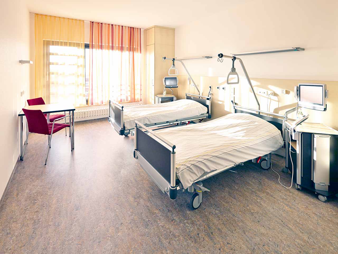
The patients of the University Hospital Ulm live in comfortable single and double rooms with a modern design and light colors. All patient rooms have an ensuite bathroom with a toilet and a shower. The patient room furnishings include a comfortable automatically adjustable bed, a bedside table, a wardrobe, a table and chairs, a telephone, a radio, and a TV. Wi-Fi access is also available in patient rooms.
The hospital also offers enhanced-comfort rooms, which additionally have a safe, a refrigerator, and upholstered furniture. The bathroom in the enhanced-comfort room has changeable towels, a cosmetic mirror, a hairdryer, and toiletries.
Meals and Menus
Patients and their accompanying person are offered three meals a day: breakfast, lunch, and dinner. The patient and accompanying person have a choice of three menus every day, including a vegetarian menu. Patients staying in the enhanced-comfort rooms are also offered light snacks, fruits, desserts, and hot and cold drinks in the comfortable lounge area.
If, for some reason, you do not eat all the foods, you will be offered an individual menu. Please inform the medical staff about your dietary preferences prior to treatment.
Further details
Standard rooms include:
![]() Shower
Shower
![]() Toilet
Toilet
![]() Wi-Fi
Wi-Fi
![]() TV
TV
Religion
The hospital has a chapel where Catholic and Protestant services are held weekly. The services are also broadcast on the internal television channel of the hospital. The chapel is open 24 hours a day for visits and prayers.
The services of other religious representatives are available upon request.
Accompanying person
Your accompanying person may stay with you in your patient room or at the hotel of your choice during the inpatient program.
Hotel
You may stay at the hotel of your choice during the outpatient program. Our managers will support you for selecting the best option.


