{"translation_price":"50","translation_doc_price":"40","child_coefficient":"1.1","transfer_price":"2.00","transfer_price_vip":"5.00","constant_transfer_price_vip":350,"constant_transfer_price":150,"constant_transfer_distanse":60,"type":"treatment","program_full_story":"<ul>\n\t<li>Initial presentation in the hospital<\/li>\n\t<li>Clinical history taking<\/li>\n\t<li>Review of available medical records<\/li>\n\t<li>Physical examination<\/li>\n\t<li>Laboratory tests:\n\t<ul>\n\t\t<li>Complete blood count<\/li>\n\t\t<li>General urine analysis<\/li>\n\t\t<li>Biochemical analysis of blood<\/li>\n\t\t<li>Tumor markers<\/li>\n\t\t<li>Inflammation indicators (CRP, ESR)<\/li>\n\t\t<li>Coagulogram<\/li>\n\t<\/ul>\n\t<\/li>\n\t<li>Ultrasound\u200b scan<\/li>\n\t<li>CT scan \/ MRI<\/li>\n\t<li>Preoperative care<\/li>\n\t<li>Embolization or chemoembolization, 2 procedures<\/li>\n\t<li>Symptomatic treatment<\/li>\n\t<li>Cost of essential medicines<\/li>\n\t<li>Nursing services<\/li>\n\t<li>Elaboration of further recommendations<\/li>\n<\/ul>\n<div class=\"program_how_program_going mt-4\"><h4>How program is carried out<\/h4><p style=\"text-align: justify;\"><strong>During the first visit<\/strong>, the doctor will conduct a clinical examination and go through the results of the available diagnostic tests. After that, you will undergo the necessary additional examination, such as the assessment of liver and kidney function, ultrasound scan, CT scan and MRI. This will allow the doctor to determine which vessels are feeding the tumor and its metastases, as well as determine how well you will tolerate the procedure.<\/p>\n\n<p style=\"text-align: justify;\"><strong>Chemoembolization <\/strong>begins with local anesthesia and catheterization of the femoral artery. The thin catheter is inserted through a few centimeters long incision of the blood vessel. The doctor gradually moves the catheter to the vessel feeding the primary tumor or its metastases. The procedure is carried out under visual control, an angiographic device is used for this. The vascular bed and the position of the catheter in it are displayed on the screen of the angiograph.<\/p>\n\n<p style=\"text-align: justify;\">When the catheter reaches a suspected artery, a contrast agent is injected through it. Due to the introduction of the contrast agent, the doctor clearly sees the smallest vessels of the tumor and the surrounding healthy tissues on the screen of the angiograph. After that, he injects emboli into the tumor vessels through the same catheter.<\/p>\n\n<p style=\"text-align: justify;\">Emboli are the spirals or the liquid microspheres. The type of embolus is selected individually, taking into account the diameter of the target vessel. When carrying out chemoembolization, a solution of a chemotherapy drug is additionally injected into the tumor vessel. Due to the subsequent closure of the vessel lumen with an embolus, the chemotherapy drug influences the tumor for a long time. In addition, the drug does not enter the systemic circulation, which allows doctors to use high doses of chemotherapeutic agents without the development of serious side effects. Chemoembolization leads to the destruction of the tumor or slowing down its progression.<\/p>\n\n<p style=\"text-align: justify;\">After that, the catheter is removed from the artery. The doctor puts a vascular suture on the femoral artery and closes it with a sterile dressing. During chemoembolization, you will be awake. General anesthesia is not used, which significantly reduces the risks of the procedure and allows performing it on an outpatient basis, avoiding long hospital stay.<\/p>\n\n<p style=\"text-align: justify;\"><strong>After the first procedure<\/strong>, you will stay under the supervision of an interventional oncologist and general practitioner. If necessary, you will receive symptomatic treatment. As a rule, a second chemoembolization procedure is performed in 3-5 days after the first one in order to consolidate the therapeutic effect. After that, you will receive recommendations for further follow-up and treatment.<\/p>\n<\/div><div class=\"program_required_documents mt-4\"><h4>Required documents<\/h4><ul>\n\t<li style=\"text-align: justify;\">Medical records<\/li>\n\t<li style=\"text-align: justify;\">MRI\/CT scan (not older than 3 months)<\/li>\n\t<li style=\"text-align: justify;\">Biopsy results (if available)<\/li>\n<\/ul>\n<\/div>","program_full_story_crm":"<ul>\n\t<li>Initial presentation in the hospital<\/li>\n\t<li>Clinical history taking<\/li>\n\t<li>Review of available medical records<\/li>\n\t<li>Physical examination<\/li>\n\t<li>Laboratory tests:\n\t<ul>\n\t\t<li>Complete blood count<\/li>\n\t\t<li>General urine analysis<\/li>\n\t\t<li>Biochemical analysis of blood<\/li>\n\t\t<li>Tumor markers<\/li>\n\t\t<li>Inflammation indicators (CRP, ESR)<\/li>\n\t\t<li>Coagulogram<\/li>\n\t<\/ul>\n\t<\/li>\n\t<li>Ultrasound\u200b scan<\/li>\n\t<li>CT scan \/ MRI<\/li>\n\t<li>Preoperative care<\/li>\n\t<li>Embolization or chemoembolization, 2 procedures<\/li>\n\t<li>Symptomatic treatment<\/li>\n\t<li>Cost of essential medicines<\/li>\n\t<li>Nursing services<\/li>\n\t<li>Elaboration of further recommendations<\/li>\n<\/ul>\n<div class=\"program_how_program_going mt-4\"><h4>How program is carried out<\/h4><p style=\"text-align: justify;\"><strong>During the first visit<\/strong>, the doctor will conduct a clinical examination and go through the results of the available diagnostic tests. After that, you will undergo the necessary additional examination, such as the assessment of liver and kidney function, ultrasound scan, CT scan and MRI. This will allow the doctor to determine which vessels are feeding the tumor and its metastases, as well as determine how well you will tolerate the procedure.<\/p>\n\n<p style=\"text-align: justify;\"><strong>Chemoembolization <\/strong>begins with local anesthesia and catheterization of the femoral artery. The thin catheter is inserted through a few centimeters long incision of the blood vessel. The doctor gradually moves the catheter to the vessel feeding the primary tumor or its metastases. The procedure is carried out under visual control, an angiographic device is used for this. The vascular bed and the position of the catheter in it are displayed on the screen of the angiograph.<\/p>\n\n<p style=\"text-align: justify;\">When the catheter reaches a suspected artery, a contrast agent is injected through it. Due to the introduction of the contrast agent, the doctor clearly sees the smallest vessels of the tumor and the surrounding healthy tissues on the screen of the angiograph. After that, he injects emboli into the tumor vessels through the same catheter.<\/p>\n\n<p style=\"text-align: justify;\">Emboli are the spirals or the liquid microspheres. The type of embolus is selected individually, taking into account the diameter of the target vessel. When carrying out chemoembolization, a solution of a chemotherapy drug is additionally injected into the tumor vessel. Due to the subsequent closure of the vessel lumen with an embolus, the chemotherapy drug influences the tumor for a long time. In addition, the drug does not enter the systemic circulation, which allows doctors to use high doses of chemotherapeutic agents without the development of serious side effects. Chemoembolization leads to the destruction of the tumor or slowing down its progression.<\/p>\n\n<p style=\"text-align: justify;\">After that, the catheter is removed from the artery. The doctor puts a vascular suture on the femoral artery and closes it with a sterile dressing. During chemoembolization, you will be awake. General anesthesia is not used, which significantly reduces the risks of the procedure and allows performing it on an outpatient basis, avoiding long hospital stay.<\/p>\n\n<p style=\"text-align: justify;\"><strong>After the first procedure<\/strong>, you will stay under the supervision of an interventional oncologist and general practitioner. If necessary, you will receive symptomatic treatment. As a rule, a second chemoembolization procedure is performed in 3-5 days after the first one in order to consolidate the therapeutic effect. After that, you will receive recommendations for further follow-up and treatment.<\/p>\n<\/div>","is_ambulant":"1","bh_fee":"0","only_for_children":"0","no_service":"0","with_prepayment":"1","show_calculator":"1","paket_type":"1","btn_type":"0","clinic_icon":"5ff71ab6e4686.jpg","city":"Ulm","clinic_site":"http:\/\/www.uniklinik-ulm.de\/","department_recommend":"0","country":"Germany","country_id":"1","clinic_name":"University Hospital Ulm","cinic_name":"University Hospital Ulm","department_id":"2807","duration":"7","direction":"Interventional radiology","min_duration":0,"clinic_id":"835","paketPrice":7800,"paket":"<ul>\n <li>Interpreter up to 17 hours<\/li>\n <li>Translation up to 10 pages<\/li>\n <li>Visa support<\/li>\n\n \n<\/ul>","title":"Treatment of thyroid cancer with embolization or chemoembolization","price":{"val":29261.51,"type":"val"},"price_surcharge":0,"price_surcharge_clear":0,"extra_service_clinic":[],"extra_service":[],"translation_hours":"0","translation_doc_count":null,"roads":[{"id":"19","distance":"79","airport_title":"Stuttgart"},{"id":"2","distance":"163","airport_title":"Munich"},{"id":"6","distance":"277","airport_title":"Frankfurt a.M."}],"pakets":[],"lang":{"day":"Day","days":"days","ambulatory":"Outpatient","stationaryProgram":"Inpatient"}}
Treatment of thyroid cancer with embolization or chemoembolization in University Hospital Ulm
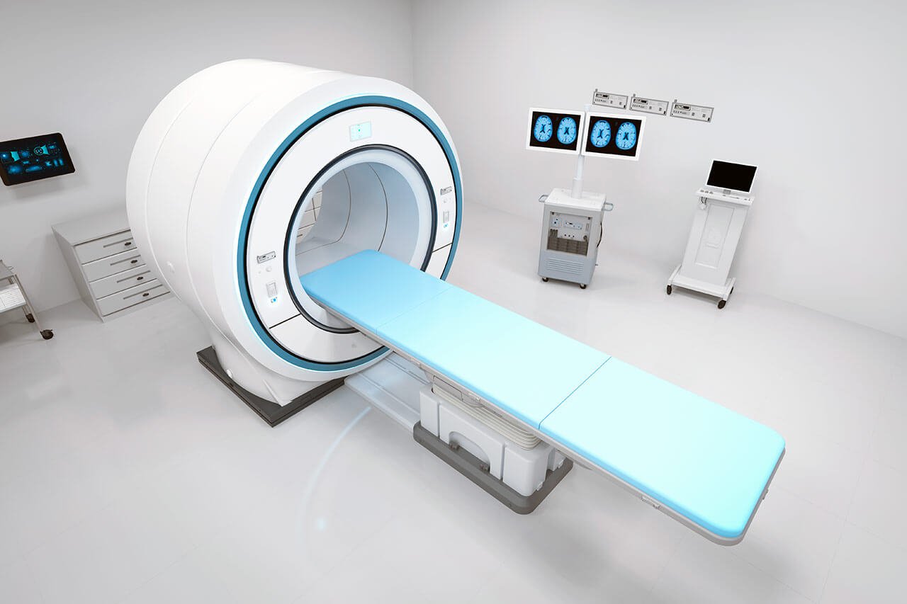
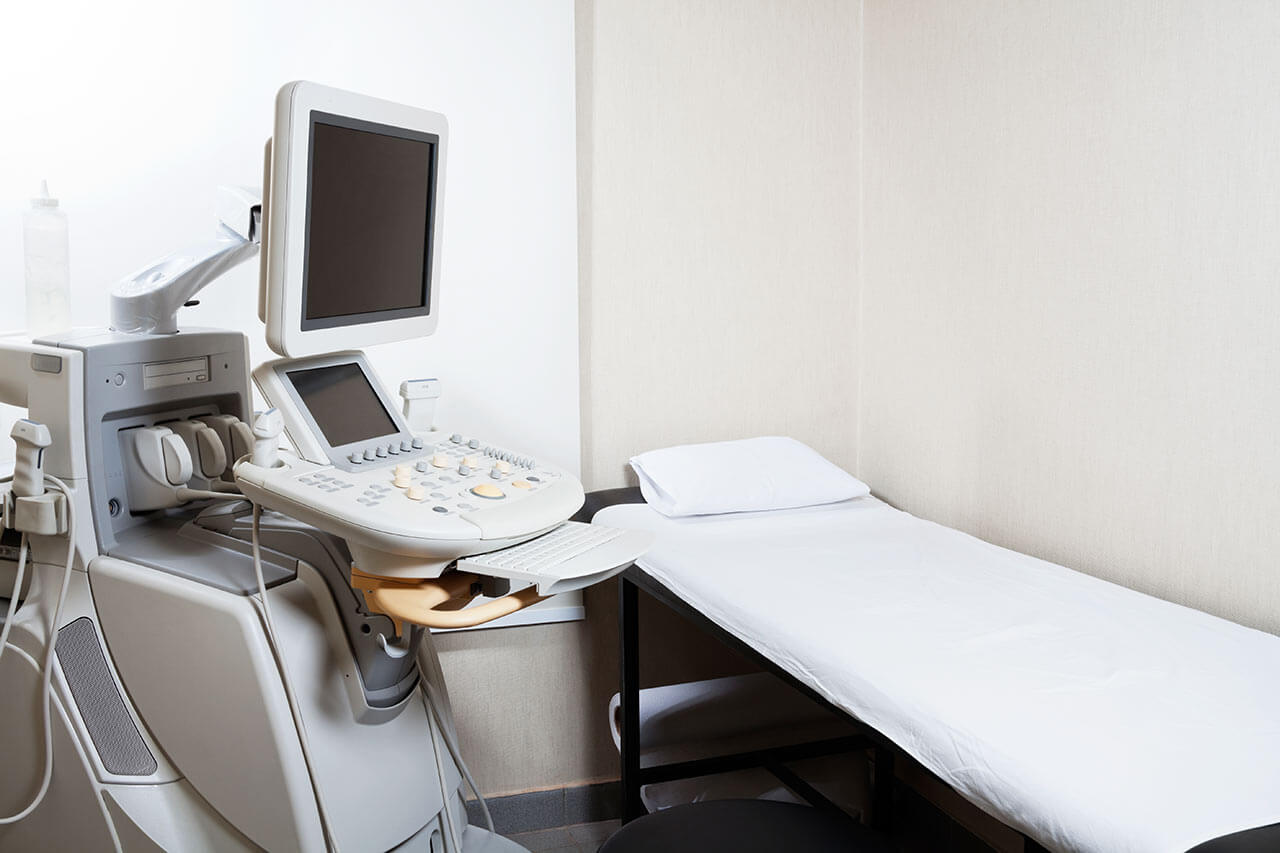
During the first visit, the doctor will conduct a clinical examination and go through the results of the available diagnostic tests. After that, you will undergo the necessary additional examination, such as the assessment of liver and kidney function, ultrasound scan, CT scan and MRI. This will allow the doctor to determine which vessels are feeding the tumor and its metastases, as well as determine how well you will tolerate the procedure.
Chemoembolization begins with local anesthesia and catheterization of the femoral artery. The thin catheter is inserted through a few centimeters long incision of the blood vessel. The doctor gradually moves the catheter to the vessel feeding the primary tumor or its metastases. The procedure is carried out under visual control, an angiographic device is used for this. The vascular bed and the position of the catheter in it are displayed on the screen of the angiograph.
When the catheter reaches a suspected artery, a contrast agent is injected through it. Due to the introduction of the contrast agent, the doctor clearly sees the smallest vessels of the tumor and the surrounding healthy tissues on the screen of the angiograph. After that, he injects emboli into the tumor vessels through the same catheter.
Emboli are the spirals or the liquid microspheres. The type of embolus is selected individually, taking into account the diameter of the target vessel. When carrying out chemoembolization, a solution of a chemotherapy drug is additionally injected into the tumor vessel. Due to the subsequent closure of the vessel lumen with an embolus, the chemotherapy drug influences the tumor for a long time. In addition, the drug does not enter the systemic circulation, which allows doctors to use high doses of chemotherapeutic agents without the development of serious side effects. Chemoembolization leads to the destruction of the tumor or slowing down its progression.
After that, the catheter is removed from the artery. The doctor puts a vascular suture on the femoral artery and closes it with a sterile dressing. During chemoembolization, you will be awake. General anesthesia is not used, which significantly reduces the risks of the procedure and allows performing it on an outpatient basis, avoiding long hospital stay.
After the first procedure, you will stay under the supervision of an interventional oncologist and general practitioner. If necessary, you will receive symptomatic treatment. As a rule, a second chemoembolization procedure is performed in 3-5 days after the first one in order to consolidate the therapeutic effect. After that, you will receive recommendations for further follow-up and treatment.
Required documents
Medical records
MRI/CT scan (not older than 3 months)
Biopsy results (if available)
Service
Price from:
Type of program :
Price for 1 day:
Expected duration of the program:
The minimum duration of the program:
Select
You may also book:
Hospital directly:
Saving:
BookingHealth Price from:
About the department
The Department of Adult and Pediatric Interventional Radiology, Neuroradiology at the University Hospital Ulm specializes in modern imaging examinations and minimally traumatic image-guided interventional procedures. Of particular interest to the department's experienced medical team is individualized minimally invasive therapy for cancer and the treatment of pain syndromes and vascular pathologies. The medical facility has state-of-the-art equipment for X-ray scanning, angiography, ultrasound scanning, CT, MRI, and PET/MRI. The diagnostic devices are adapted for children, including newborns. During examinations and treatment, the department's specialists strictly meet radiation protection standards. As for treatment, special attention in clinical practice is paid to interventional procedures for vascular diseases and malignant tumors. These procedures are performed using catheter-based techniques under CT, MRI, or angiography guidance. Despite the low trauma rate of interventional manipulations, their effectiveness is comparable to full-fledged surgery. The medical facility cooperates closely with other departments at the hospital, thus providing comprehensive medical care at the highest level of university medicine. The department's successful clinical practice has been recognized by prestigious quality certificates, including the ClarZert certificate and certificates from the German Cancer Society (DKG) and the German Radiological Society (DRG). The Head Physician of the department is Prof. Dr. med. Meinrad Beer.
The department often performs catheter-based treatment of malignant tumors using transarterial chemoembolization (TACE) and selective internal radiation therapy (SIRT). During the TACE procedure, emboli (microscopic spheres) containing a chemotherapeutic agent are injected into the blood vessels supplying the tumor using a catheter. After transarterial chemoembolization, the tumor is deprived of blood supply and locally exposed to chemotherapeutic agents, which leads to the death of the neoplasm. The essence of selective internal radiation therapy is the administration of tiny radioactive particles into the blood vessels supplying the tumor, which locally irradiate the tumor while practically not damaging the adjacent healthy tissues. The SIRT procedure is also performed using catheter-based techniques through the inguinal artery under imaging guidance. TACE and SIRT techniques are used to treat primary and secondary liver tumors.
The department's specialists also successfully perform radiofrequency ablation, microwave ablation, and cryoablation for the treatment of tumors of various localizations. The above-mentioned procedures are performed under CT guidance, which ensures their high accuracy and safety. The tumor focus is approached using a special probe, after which the tumor tissue is destroyed by extremely high or low temperatures. The ablation procedure is indicated for patients with tumors no larger than 5 cm.
The department's interventional radiologists regularly admit patients with vascular stenosis and obstructions, abdominal and thoracic aortic aneurysms, and vascular malformations. Balloon dilatation, followed by stent implantation, is performed to normalize blood flow in cases of stenosis. During this interventional procedure, specialists direct a balloon dilator, a long catheter with a deflated balloon attached to its distal end, to the narrowed area along the lumen of the blood vessel under angiography guidance. With the help of a special instrument, a liquid is injected into the balloon, creating a certain pressure, due to which it increases to the required diameter and restores the patency of the affected blood vessel. To prevent recurrent stenosis in the narrowed blood vessel, a stent is placed to maintain it in an open state.
The department also offers neuroradiology services. Doctors specializing in this field carry out imaging diagnostics and interventional catheter-based treatment of diseases of the brain and spinal cord. They perform carotid artery stenting for stenosis, intra-arterial lysis therapy for stroke, coiling for brain aneurysms, and endovascular interventions for arteriovenous fistulas in the department's operating rooms. The department also has vast experience in the treatment of chronic back pain using facet joint infiltration and periradicular therapy. The above-mentioned therapeutic procedures are performed under CT or MRI guidance under local anesthesia.
A special offer in the department is imaging diagnostics for children of all age groups, ranging from premature babies and infants to adolescents up to 18 years old. The department has state-of-the-art equipment to perform accurate examinations of children. All devices work in such a way that the child receives the minimum radiation dose and the growing organism is not harmed. Moreover, the rich experience and high professionalism of the department's doctors specializing in diagnostic imaging in children play a crucial role in successful diagnosis.
The department's range of therapeutic services includes the following options:
Interventional radiology
Interventional treatment of malignant tumors
Transarterial chemoembolization (TACE)
Selective internal radiation therapy (SIRT)
Radiofrequency ablation, microwave ablation, and cryoablation
Interventional treatment of vascular stenoses and obstructions
Balloon dilatation followed by stent implantation
Coiling for abdominal and thoracic aortic aneurysms and vascular malformations
Percutaneous transhepatic cholangiography followed by drainage and/or stent implantation
CT-guided puncture and drainage
Neuroradiology
Interventional treatment of cerebrovascular diseases
Carotid artery stenting
Intra-arterial lysis therapy
Coiling
Interventional treatment of chronic back pain
Facet joint block
Sacroiliac joint block
Periradicular therapy
Other medical services
Curriculum vitae
Higher Education and Professional Career
1988 - 1995 Study of Human Medicine and Philosophy, University of Regensburg and Julius Maximilian University of Wuerzburg.
1995 - 2003 Research Fellow and Assistant Physician, Institute of X-ray Diagnostics, University Hospital Wuerzburg.
2003 - 2007 Senior Physician (specialization: Cardiovascular Imaging, Cancer Imaging, and Pediatric Radiology), Institute of X-ray Diagnostics, University Hospital Wuerzburg.
2007 - 2012 Managing Senior Physician and Head of Pediatric Radiology, Institute of X-ray Diagnostics, University Hospital Wuerzburg.
2009 Invitation to the position of W3 Professor for Radiology, University Hospital Goettingen (rejection).
2009 - 2012 Professor for Radiology, Julius Maximilian University of Wuerzburg.
2012 Invitation to the position of Professor for Pediatric Radiology, Medical University of Graz.
2012 - 2013 Head Physician, Department of Pediatric Radiology, University Hospital Graz.
2013 Invitation to the position of W3 Professor for Radiology, University of Ulm.
Since 2013 Head Physician, Department of Adult and Pediatric Interventional Radiology, Neuroradiology, University Hospital Ulm.
Qualifications
Board certification in Diagnostic Radiology.
Specialization in Pediatric Radiology.
Permission for advanced training in Diagnostic Radiology.
Permission for advanced training in Pediatric Radiology.
Instructor of the German Society of Ultrasound in Medicine (DEGUM), level II.
Memberships in Professional Societies
German Radiological Society (DRG).
European Society of Cardiovascular Radiology (ESCR).
European Society of Radiology (ESR).
Society of Pediatric Radiology (GPR).
Radiological Society of North America (RSNA).
Photo of the doctor: (c) Universitätsklinikum Ulm
About hospital
The University Hospital Ulm is an advanced medical complex that provides patients with high-class medical care using the very latest scientific achievements. The medical facility has been performing successful clinical activities for more than 40 years and has long earned an excellent reputation throughout Europe. The hospital regularly demonstrates high treatment success rates, takes an active part in the training of medical students, and works tirelessly on promising research projects.
The university hospital consists of 29 specialized departments and 16 scientific institutes, where more than 7,000 highly qualified employees work for the benefit of their patients. More than 55,000 inpatients and about 300,000 outpatients are treated here every year. The hospital has 1,274 beds. The medical team of the hospital is focused on providing personalized medical services using the most modern and sparing diagnostic and treatment methods.
The University Hospital Ulm is the largest medical complex in the region, and practically all areas of modern medicine are represented here. Transplantology and oncology are among the priority areas of clinical activity in the medical facility. The hospital holds leading positions in the world in bone marrow transplantation. In addition, the hospital has advanced experience in cancer treatment. The Comprehensive Cancer Center is recognized as the leading facility of this kind in the country, and it is certified by the German Cancer Society (DKG). It provides effective treatment for various types of cancer. The center also offers innovative CAR T-cell therapy. In addition, the Cancer Center is actively engaged in research activities to improve available treatment methods and develop innovative therapeutic techniques to fight cancer.
Along with the use of advanced technologies, doctors show respect, understanding, and a humane attitude toward the patient. The medical team includes competent psychologists, who are always ready to provide assistance and support to the patients and their families during the therapeutic process.
Photo: (с) depositphotos
Accommodation in hospital
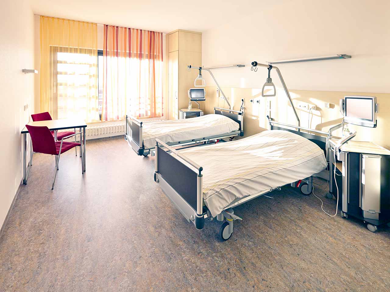
Patients rooms
The patients of the University Hospital Ulm live in comfortable single and double rooms with a modern design and light colors. All patient rooms have an ensuite bathroom with a toilet and a shower. The patient room furnishings include a comfortable automatically adjustable bed, a bedside table, a wardrobe, a table and chairs, a telephone, a radio, and a TV. Wi-Fi access is also available in patient rooms.
The hospital also offers enhanced-comfort rooms, which additionally have a safe, a refrigerator, and upholstered furniture. The bathroom in the enhanced-comfort room has changeable towels, a cosmetic mirror, a hairdryer, and toiletries.
Meals and Menus
Patients and their accompanying person are offered three meals a day: breakfast, lunch, and dinner. The patient and accompanying person have a choice of three menus every day, including a vegetarian menu. Patients staying in the enhanced-comfort rooms are also offered light snacks, fruits, desserts, and hot and cold drinks in the comfortable lounge area.
If, for some reason, you do not eat all the foods, you will be offered an individual menu. Please inform the medical staff about your dietary preferences prior to treatment.
Further details
Standard rooms include:
Shower
Toilet
Wi-Fi
TV
Religion
The hospital has a chapel where Catholic and Protestant services are held weekly. The services are also broadcast on the internal television channel of the hospital. The chapel is open 24 hours a day for visits and prayers.
The services of other religious representatives are available upon request.
Accompanying person
Your accompanying person may stay with you in your patient room or at the hotel of your choice during the inpatient program.
Hotel
You may stay at the hotel of your choice during the outpatient program. Our managers will support you for selecting the best option.
Select your currency:
Your guarantee
We are the only TÜV-certified company with an ISO 9001:2015 certificate
thank you for reaching out! One of our medical advisors is currently reviewing your details. You can expect a call from us within one business day to discuss your request.
Note: The incoming call will appear as a German number or your local area code. This call is entirely free of charge.
Booking Health guarantees:
Expert Placement
We use rigorous statistical analysis to ensure you are treated at the best-rated facility for your specific diagnosis.
Total Cost Protection
No hidden fees or unexpected bills. We provide a fixed price guarantee, backed by insurance that covers any additional complications.
12-Month Extended Care
Your recovery is our priority. Benefit from a full year of direct medical support following your procedure.


 BookingHealth Price from:
