The program includes:
- Initial presentation in the hospital
- Clinical history taking
- Review of available medical records
- Physical examination
- Laboratory tests:
- Complete blood count
- General urine analysis
- Biochemical analysis of blood
- Tumor markers
- Inflammation indicators (CRP, ESR)
- Coagulogram
- Ultrasound scan
- CT scan / MRI
- Preoperative care
- Embolization or chemoembolization, 2 procedures
- Symptomatic treatment
- Cost of essential medicines
- Nursing services
- Elaboration of further recommendations
How program is carried out
During the first visit, the doctor will conduct a clinical examination and go through the results of the available diagnostic tests. After that, you will undergo the necessary additional examination, such as the assessment of liver and kidney function, ultrasound scan, CT scan and MRI. This will allow the doctor to determine which vessels are feeding the tumor and its metastases, as well as determine how well you will tolerate the procedure.
Chemoembolization begins with local anesthesia and catheterization of the femoral artery. The thin catheter is inserted through a few centimeters long incision of the blood vessel. The doctor gradually moves the catheter to the vessel feeding the primary tumor or its metastases. The procedure is carried out under visual control, an angiographic device is used for this. The vascular bed and the position of the catheter in it are displayed on the screen of the angiograph.
When the catheter reaches a suspected artery, a contrast agent is injected through it. Due to the introduction of the contrast agent, the doctor clearly sees the smallest vessels of the tumor and the surrounding healthy tissues on the screen of the angiograph. After that, he injects emboli into the tumor vessels through the same catheter.
Emboli are the spirals or the liquid microspheres. The type of embolus is selected individually, taking into account the diameter of the target vessel. When carrying out chemoembolization, a solution of a chemotherapy drug is additionally injected into the tumor vessel. Due to the subsequent closure of the vessel lumen with an embolus, the chemotherapy drug influences the tumor for a long time. In addition, the drug does not enter the systemic circulation, which allows doctors to use high doses of chemotherapeutic agents without the development of serious side effects. Chemoembolization leads to the destruction of the tumor or slowing down its progression.
After that, the catheter is removed from the artery. The doctor puts a vascular suture on the femoral artery and closes it with a sterile dressing. During chemoembolization, you will be awake. General anesthesia is not used, which significantly reduces the risks of the procedure and allows performing it on an outpatient basis, avoiding long hospital stay.
After the first procedure, you will stay under the supervision of an interventional oncologist and general practitioner. If necessary, you will receive symptomatic treatment. As a rule, a second chemoembolization procedure is performed in 3-5 days after the first one in order to consolidate the therapeutic effect. After that, you will receive recommendations for further follow-up and treatment.
Required documents
- Medical records
- MRI/CT scan (not older than 3 months)
- Biopsy results (if available)
Service
You may also book:
 BookingHealth Price from:
About the department
The Department of Adult and Pediatric Diagnostic, Interventional Radiology at the University Hospital Würzburg offers the full range of modern diagnostic and therapeutic procedures in this field. For this purpose, it has advanced medical equipment: magnetic resonance imaging (MRI), multispiral computed tomography (CT), duplex sonography and digital X-ray, fluoroscopy, etc. In the field of interventional radiology, the focus is on the treatment of vascular diseases, for example, the doctors of the department conduct special measures for dilating the vessels in peripheral arterial occlusive disease and closing the vessels in complicated bleedings. Also, the range of services in this field is complemented by the installation of drainage and port systems, punctures, and special percutaneous methods for tumor treatment. The Chief Physician of the department is Prof. Dr. med. Thorsten Bley, who has many years of clinical experience and regularly demonstrates high rates of successful treatment results.
A feature of the department is the Section of Experimental Radiology, which serves to conduct research on innovative procedures to improve diagnostic and therapeutic methods. Thanks to this, the department is able to offer patients unique techniques that are not available in other clinics.
The department also specializes in the modern imaging diagnostics in children and adolescents. All studies are conducted by specially trained doctors, on medical equipment adapted to the children's body and working with a minimal radiation dose. There are performed about 3,000 X-ray examinations, 9,000 ultrasound examinations, 2,000 MRI and 250 CT scans every year. All diagnostic procedures are performed in strict accordance with international standards of pediatric radiological diagnostics.
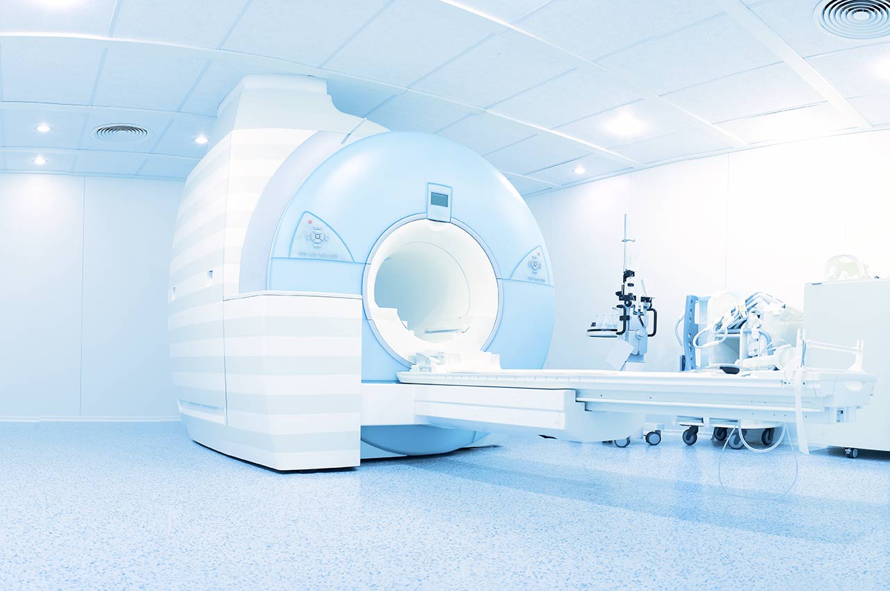
In October 2012, the department installed a new, modern "large-scale" MRI scanner, which, in addition to the full range of standard images, allows making full-body images, cardiographic studies, spectroscopy and diffusion images. MRI scans of the skull and spine are performed in collaboration with experienced neuroradiologists.
The goal of the medical team is to obtain all the necessary information about the child’s state of health using methods with minimal irradiation or without it. For example, the department applies the very latest ultrasound systems, harmonic imaging, contrast agents (for example, to diagnose reflux disease), ultrasound elastography. The advanced X-ray machines provide excellent image quality and minimal use of X-rays.
The diagnostic options of the department include:
- All types of ultrasound examinations, including high-resolution ultrasound, contrast-enhanced ultrasound
- Ultrasound diagnosis of the abdominal organs, intestines, kidneys and blood vessels
- Contrast-enhanced ultrasound of the liver and other organs
- Ultrasound-guided puncture (for example, puncture of the liver, kidneys, spleen, pancreas, lymph nodes, etc.)
- Preoperative lymph node labeling
- Computed tomography (CT), including contrast-enhanced dynamic CT
- Magnetic resonance imaging (MRI), also contrast-enhanced one
- Classic X-ray studies
- Fluoroscopy
- Mammography (breast diagnosis)
- Pediatric imaging diagnostics
- Ultrasound examinations of various organs
- Classic X-ray studies
- Computed tomography (CT)
- Magnetic resonance imaging (MRI)
- Other diagnostic services
The department offers the following methods of interventional radiology:
- Catheter procedures for the treatment of vascular stenosis and occlusion in the pelvis, lower limbs, kidneys – balloon dilation of the vessels or stenting (for example, percutaneous transluminal angioplasty)
- Treatment of acute ischemia of the lower extremities using local intra-arterial lysis at the stage of compensated ischemia (stage I and IIa) and surgical recanalization at the stage of uncompensated ischemia (stage IIb and III), removal of a blood clot by catheter aspiration
- Arterial embolization in acute arterial bleedings
- Minimally invasive treatment (stenting) of aortic ruptures, aortic aneurysms and aortic dissection
- Radiofrequency ablation for the treatment of liver cancer and liver metastases (usually ultrasound- or CT-guided)
- Transarterial chemoembolization (TACE) for liver cancer treatment
- Selective internal radiation therapy (SIRT) for the treatment of liver cancer, liver metastases in colorectal cancer, breast cancer and neuroendocrine tumors
- Transjugular intrahepatic portosystemic shunting (TIPS) for the treatment of portal hypertension
- Other therapeutic services
Photo of the doctor: (c) Universitätsklinikum Würzburg
About hospital
According to the FOCUS magazine in 2019, the University Hospital Würzburg ranks among the top national German hospitals!
The hospital is one of the oldest medical facilities in Germany. The centuries-old traditions of first-class treatment are combined with the very latest achievements of modern evidence-based medicine and advanced expert experience. The hospital is the maximum care medical center and covers all fields of modern medicine. The hospital has an impeccable international reputation and treats a large number of patients from other countries every year.
A distinctive peculiarity of the hospital is active interdisciplinary cooperation. A large number of diseases are diagnosed and treated within the specialized centers, which medical teams rely on the unique experience of treatment of a wide variety of clinical cases. For example, such centers include the General Cancer Center, the Stem Cell Therapy Center, the Breast Health Center, the Gastrointestinal Center, the Thoracic Surgery Center, etc. In total, the hospital has more than 40 centers of this kind. Therefore, the patients of the hospital are confident that they will be offered the most relevant treatment in accordance with the very latest medical recommendations.
In addition to the most advanced achievements and methods of modern medicine, the medical team of the hospital makes every effort to create a comfortable, friendly atmosphere, which promotes patient well-being and recovery.
Photo: (с) depositphotos
Accommodation in hospital
Patients rooms
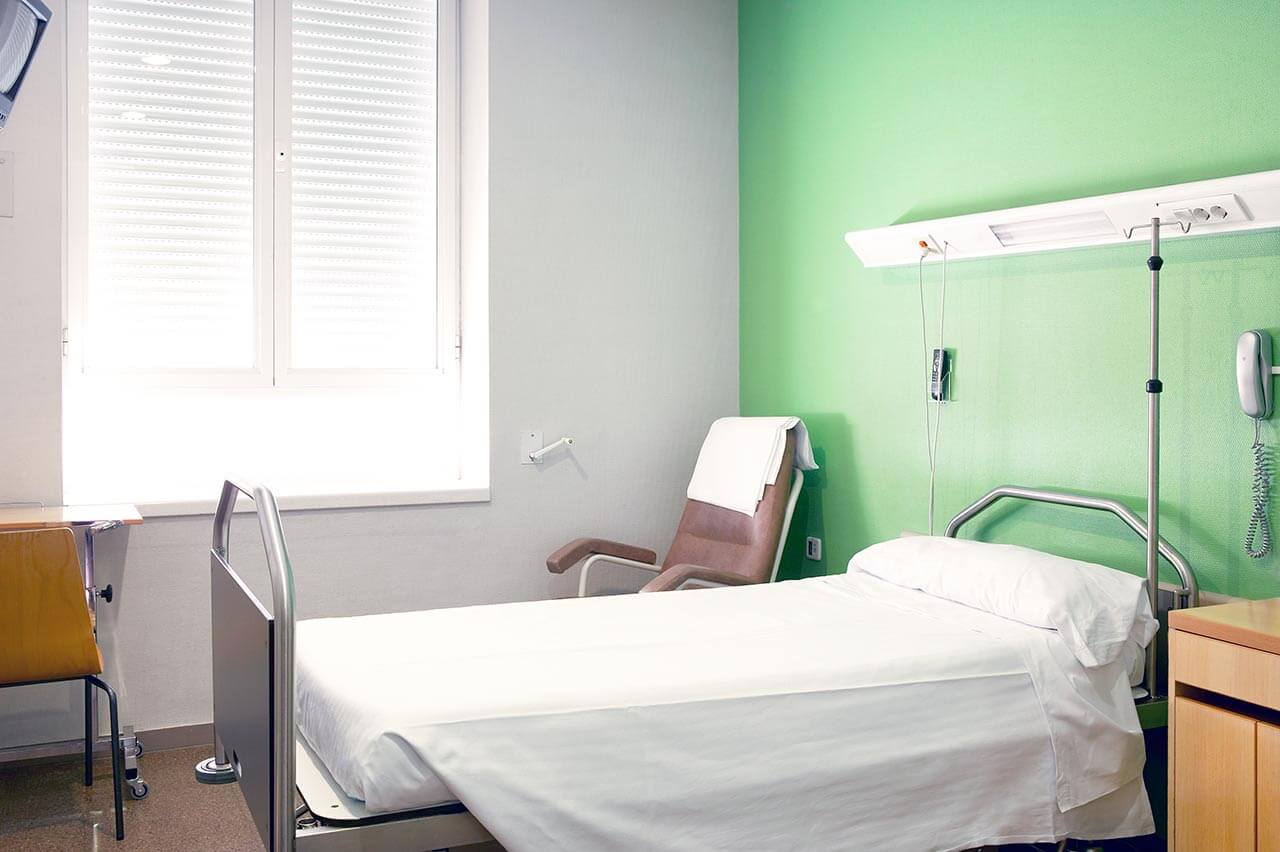
The patients of the University Hospital Würzburg live in comfortable rooms made in a modern design and bright colors. The patient room includes a bed with a remote control, a bedside table with a sliding table, a wardrobe, a TV. The patient rooms have the possibility of Internet connection. The enhanced-comfort rooms are equipped with a safe, a fridge and upholstered furniture.
Meals and Menus
The patients of the hospital are offered a varied and tasty three meals a day. There is a daily choice of several menus, including a dietary one. In case of intolerance to any food, please inform the medical staff about your food preferences in advance. After that, you will be offered an individual menu. Also, the hospital houses the cafes and cafeterias with a wide assortment of drinks, fresh pastries, fresh salads, sweets and other dishes.
Further details
Standard rooms include:
Religion
Christian priests are available for the patients at any time. Representatives of other religions may be requested at any time.
Accompanying person
Your companion may stay with you in your room or at a hotel of your choice during the fixed program.
Hotel
You may stay at the clinic hotel or a hotel of your choice during the outpatient program. Our manager will help you choose the best option.


