{"translation_price":"50","translation_doc_price":"40","child_coefficient":"1.1","transfer_price":"2.00","transfer_price_vip":"5.00","constant_transfer_price_vip":350,"constant_transfer_price":150,"constant_transfer_distanse":60,"type":"treatment","program_full_story":"<ul>\n\t<li>Initial presentation in the clinic<\/li>\n\t<li>clinical history taking<\/li>\n\t<li>review of medical records<\/li>\n\t<li>physical examination<\/li>\n\t<li>laboratory tests:\n\t<ul>\n\t\t<li>complete blood count<\/li>\n\t\t<li>general urine analysis<\/li>\n\t\t<li>biochemical analysis of blood<\/li>\n\t\t<li>TSH-basal, fT3, fT4<\/li>\n\t\t<li>tumor markers (AFP, CEA, \u0421\u0410-19-9)<\/li>\n\t\t<li>inflammation indicators (CRP, ESR)<\/li>\n\t\t<li>indicators of blood coagulation<\/li>\n\t<\/ul>\n\t<\/li>\n\t<li>abdominal ultrasound\u200b scan<\/li>\n\t<li>CT\/MRI of abdomen<\/li>\n\t<li>preoperative care<\/li>\n\t<li>percutaneous embolization (coiling) or chemoembolization<\/li>\n\t<li>symptomatic treatment<\/li>\n\t<li>cost of essential medicines<\/li>\n\t<li>nursing services<\/li>\n\t<li>elaboration of further recommendations<\/li>\n<\/ul>\n<div class=\"program_how_program_going mt-4\"><h4>How program is carried out<\/h4><p style=\"text-align: justify;\"><strong>During the first visit<\/strong>, the doctor will conduct a clinical examination and go through the results of the available diagnostic tests. After that, you will undergo the necessary additional examination, such as the assessment of liver and kidney function, ultrasound scan, CT scan and MRI. This will allow the doctor to determine which vessels are feeding the tumor and its metastases, as well as determine how well you will tolerate the procedure.<\/p>\n\n<p style=\"text-align: justify;\"><strong>Chemoembolization <\/strong>begins with local anesthesia and catheterization of the femoral artery. The thin catheter is inserted through a few centimeters long incision of the blood vessel. The doctor gradually moves the catheter to the vessel feeding the primary tumor or its metastases. The procedure is carried out under visual control, an angiographic device is used for this. The vascular bed and the position of the catheter in it are displayed on the screen of the angiograph.<\/p>\n\n<p style=\"text-align: justify;\">When the catheter reaches a suspected artery, a contrast agent is injected through it. Due to the introduction of the contrast agent, the doctor clearly sees the smallest vessels of the tumor and the surrounding healthy tissues on the screen of the angiograph. After that, he injects emboli into the tumor vessels through the same catheter.<\/p>\n\n<p style=\"text-align: justify;\">Emboli are the spirals or the liquid microspheres. The type of embolus is selected individually, taking into account the diameter of the target vessel. When carrying out chemoembolization, a solution of a chemotherapy drug is additionally injected into the tumor vessel. Due to the subsequent closure of the vessel lumen with an embolus, the chemotherapy drug influences the tumor for a long time. In addition, the drug does not enter the systemic circulation, which allows doctors to use high doses of chemotherapeutic agents without the development of serious side effects. Chemoembolization leads to the destruction of the tumor or slowing down its progression.<\/p>\n\n<p style=\"text-align: justify;\">After that, the catheter is removed from the artery. The doctor puts a vascular suture on the femoral artery and closes it with a sterile dressing. During chemoembolization, you will be awake. General anesthesia is not used, which significantly reduces the risks of the procedure and allows performing it on an outpatient basis, avoiding long hospital stay.<\/p>\n\n<p style=\"text-align: justify;\"><strong>After the first procedure<\/strong>, you will stay under the supervision of an interventional oncologist and general practitioner. If necessary, you will receive symptomatic treatment. As a rule, a second chemoembolization procedure is performed in 3-5 days after the first one in order to consolidate the therapeutic effect. After that, you will receive recommendations for further follow-up and treatment.<\/p>\n<\/div><div class=\"program_required_documents mt-4\"><h4>Required documents<\/h4><ul>\n\t<li style=\"text-align: justify;\">Medical records<\/li>\n\t<li style=\"text-align: justify;\">MRI\/CT scan (not older than 3 months)<\/li>\n\t<li style=\"text-align: justify;\">Biopsy results (if available)<\/li>\n<\/ul>\n<\/div>","program_full_story_crm":"<ul>\n\t<li>Initial presentation in the clinic<\/li>\n\t<li>clinical history taking<\/li>\n\t<li>review of medical records<\/li>\n\t<li>physical examination<\/li>\n\t<li>laboratory tests:\n\t<ul>\n\t\t<li>complete blood count<\/li>\n\t\t<li>general urine analysis<\/li>\n\t\t<li>biochemical analysis of blood<\/li>\n\t\t<li>TSH-basal, fT3, fT4<\/li>\n\t\t<li>tumor markers (AFP, CEA, \u0421\u0410-19-9)<\/li>\n\t\t<li>inflammation indicators (CRP, ESR)<\/li>\n\t\t<li>indicators of blood coagulation<\/li>\n\t<\/ul>\n\t<\/li>\n\t<li>abdominal ultrasound\u200b scan<\/li>\n\t<li>CT\/MRI of abdomen<\/li>\n\t<li>preoperative care<\/li>\n\t<li>percutaneous embolization (coiling) or chemoembolization<\/li>\n\t<li>symptomatic treatment<\/li>\n\t<li>cost of essential medicines<\/li>\n\t<li>nursing services<\/li>\n\t<li>elaboration of further recommendations<\/li>\n<\/ul>\n<div class=\"program_how_program_going mt-4\"><h4>How program is carried out<\/h4><p style=\"text-align: justify;\"><strong>During the first visit<\/strong>, the doctor will conduct a clinical examination and go through the results of the available diagnostic tests. After that, you will undergo the necessary additional examination, such as the assessment of liver and kidney function, ultrasound scan, CT scan and MRI. This will allow the doctor to determine which vessels are feeding the tumor and its metastases, as well as determine how well you will tolerate the procedure.<\/p>\n\n<p style=\"text-align: justify;\"><strong>Chemoembolization <\/strong>begins with local anesthesia and catheterization of the femoral artery. The thin catheter is inserted through a few centimeters long incision of the blood vessel. The doctor gradually moves the catheter to the vessel feeding the primary tumor or its metastases. The procedure is carried out under visual control, an angiographic device is used for this. The vascular bed and the position of the catheter in it are displayed on the screen of the angiograph.<\/p>\n\n<p style=\"text-align: justify;\">When the catheter reaches a suspected artery, a contrast agent is injected through it. Due to the introduction of the contrast agent, the doctor clearly sees the smallest vessels of the tumor and the surrounding healthy tissues on the screen of the angiograph. After that, he injects emboli into the tumor vessels through the same catheter.<\/p>\n\n<p style=\"text-align: justify;\">Emboli are the spirals or the liquid microspheres. The type of embolus is selected individually, taking into account the diameter of the target vessel. When carrying out chemoembolization, a solution of a chemotherapy drug is additionally injected into the tumor vessel. Due to the subsequent closure of the vessel lumen with an embolus, the chemotherapy drug influences the tumor for a long time. In addition, the drug does not enter the systemic circulation, which allows doctors to use high doses of chemotherapeutic agents without the development of serious side effects. Chemoembolization leads to the destruction of the tumor or slowing down its progression.<\/p>\n\n<p style=\"text-align: justify;\">After that, the catheter is removed from the artery. The doctor puts a vascular suture on the femoral artery and closes it with a sterile dressing. During chemoembolization, you will be awake. General anesthesia is not used, which significantly reduces the risks of the procedure and allows performing it on an outpatient basis, avoiding long hospital stay.<\/p>\n\n<p style=\"text-align: justify;\"><strong>After the first procedure<\/strong>, you will stay under the supervision of an interventional oncologist and general practitioner. If necessary, you will receive symptomatic treatment. As a rule, a second chemoembolization procedure is performed in 3-5 days after the first one in order to consolidate the therapeutic effect. After that, you will receive recommendations for further follow-up and treatment.<\/p>\n<\/div>","is_ambulant":"1","bh_fee":"0","only_for_children":"0","no_service":"0","with_prepayment":"1","show_calculator":"1","paket_type":"1","btn_type":"2","clinic_icon":"5ffc61fee3cfe.jpg","city":"Berlin","clinic_site":"https:\/\/www.vivantes.de\/fuer-sie-vor-ort\/auguste-viktoria-klinikum\/","department_recommend":"0","country":"Germany","country_id":"1","clinic_name":"Vivantes Auguste-Viktoria Hospital Berlin","cinic_name":"Vivantes Auguste-Viktoria Hospital Berlin","department_id":"2074","duration":"6","direction":"Interventional radiology","min_duration":0,"clinic_id":"1573","paketPrice":3200,"paket":"<ul>\n <li> Interpreter up to 8 hours<\/li>\n <li>Translation of up to 5 pages<\/li>\n <li>Visa support<\/li>\n\n<\/ul>","title":"Treatment of liver cancer with percutaneous embolization (coiling) or chemoembolization","price":{"val":9456.689999999999,"type":"val"},"price_surcharge":0,"price_surcharge_clear":0,"extra_service_clinic":[],"extra_service":[],"translation_hours":"0","translation_doc_count":null,"roads":[{"id":"8","distance":"30","airport_title":"Berlin"},{"id":"22","distance":"35","airport_title":"Berlin-Tegel"},{"id":"9","distance":"234","airport_title":"Hamburg"}],"pakets":[],"lang":{"day":"Day","days":"days","ambulatory":"Outpatient","stationaryProgram":"Inpatient"}}
Treatment of liver cancer with percutaneous embolization (coiling) or chemoembolization in Vivantes Auguste-Viktoria Hospital Berlin
percutaneous embolization (coiling) or chemoembolization
symptomatic treatment
cost of essential medicines
nursing services
elaboration of further recommendations
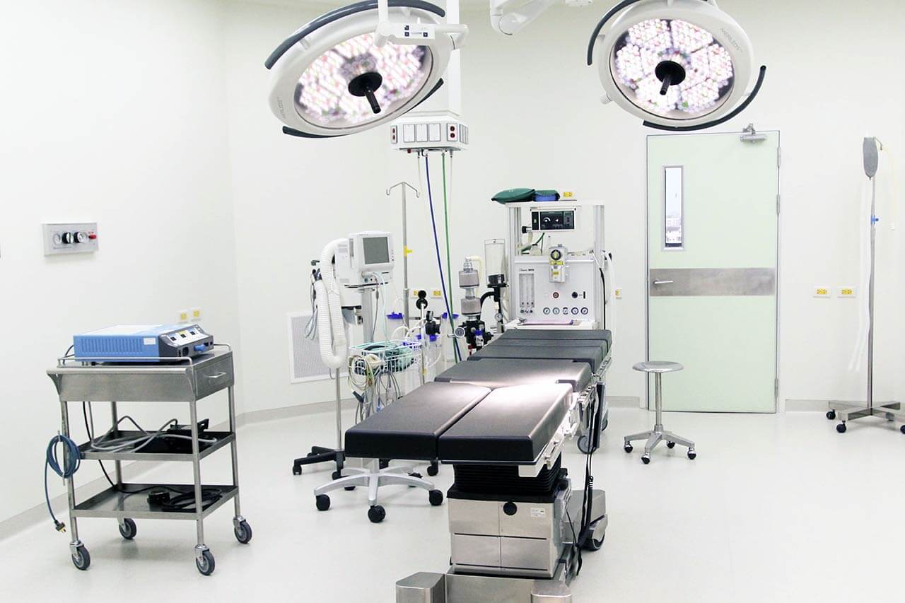
How program is carried out
During the first visit, the doctor will conduct a clinical examination and go through the results of the available diagnostic tests. After that, you will undergo the necessary additional examination, such as the assessment of liver and kidney function, ultrasound scan, CT scan and MRI. This will allow the doctor to determine which vessels are feeding the tumor and its metastases, as well as determine how well you will tolerate the procedure.
Chemoembolization begins with local anesthesia and catheterization of the femoral artery. The thin catheter is inserted through a few centimeters long incision of the blood vessel. The doctor gradually moves the catheter to the vessel feeding the primary tumor or its metastases. The procedure is carried out under visual control, an angiographic device is used for this. The vascular bed and the position of the catheter in it are displayed on the screen of the angiograph.
When the catheter reaches a suspected artery, a contrast agent is injected through it. Due to the introduction of the contrast agent, the doctor clearly sees the smallest vessels of the tumor and the surrounding healthy tissues on the screen of the angiograph. After that, he injects emboli into the tumor vessels through the same catheter.
Emboli are the spirals or the liquid microspheres. The type of embolus is selected individually, taking into account the diameter of the target vessel. When carrying out chemoembolization, a solution of a chemotherapy drug is additionally injected into the tumor vessel. Due to the subsequent closure of the vessel lumen with an embolus, the chemotherapy drug influences the tumor for a long time. In addition, the drug does not enter the systemic circulation, which allows doctors to use high doses of chemotherapeutic agents without the development of serious side effects. Chemoembolization leads to the destruction of the tumor or slowing down its progression.
After that, the catheter is removed from the artery. The doctor puts a vascular suture on the femoral artery and closes it with a sterile dressing. During chemoembolization, you will be awake. General anesthesia is not used, which significantly reduces the risks of the procedure and allows performing it on an outpatient basis, avoiding long hospital stay.
After the first procedure, you will stay under the supervision of an interventional oncologist and general practitioner. If necessary, you will receive symptomatic treatment. As a rule, a second chemoembolization procedure is performed in 3-5 days after the first one in order to consolidate the therapeutic effect. After that, you will receive recommendations for further follow-up and treatment.
Required documents
Medical records
MRI/CT scan (not older than 3 months)
Biopsy results (if available)
Service
Price from:
Type of program :
Price for 1 day:
Expected duration of the program:
The minimum duration of the program:
Select
You may also book:
Hospital directly:
Saving:
BookingHealth Price from:
About the department
Using modern diagnostic and therapeutic methods(digital X-ray, mammography, ultrasound, CT/MRI and angiography), the Department of Radiology provides visualization diagnostics and treatment. In particular, it treats diseases of blood vessels, liver - in particular tumors - and pain syndromes. Visualization allows gentle minimally invasive placement of the catheter and other instruments. The department unites two clinics - the Vivantes Auguste-Viktoria Hospital and the Wenckebach Hospital. Both hospitals cooperate with other specializations. The head of the department is Priv.-Doz. Dr. med. Andreas Koops.
One of the directions of the Department of Radiology and Interventional Therapy is treatment of liver tumors. Various methods are used to fight the tumor or prevent its growth. These include destruction of cancer tissues with high temperature (thermal ablation), radio frequencies (RFA), or microwaves (microwave ablation); treatment with medications that are injected into the blood vessels next to the tumor (transarterial chemoembolization of TACE); treatment with radioactive materials that are injected into the tumor (selective internal radiotherapy - SIRT, as well as transarterial radioembolization - TARE).
In addition, specialists of the department treat malignant tumors by outpatient, maximally sparing implantation of port systems in the shoulder area. In particular, chemotherapy can be received through this system. The Department of Radiology and Interventional Therapy at the Vivantes Auguste-Viktoria Hospital is the only center within Berlin where port systems in the shoulder area are installed quickly and without long waiting period under local anesthesia.
Photo of the doctor: (c) Vivantes - Netzwerk für Gesundheit GmbH
About hospital
The Vivantes Auguste-Victoria Hospital, located in Berlin's Tempelhof-Schöneberg district, is a hospital of specialized medical supplies with 692 beds in it. Annually 12 departments of the hospital receive about 70,000 patients - 47,000 in outpatient and 23,000 in inpatient settings. The hospital has special expertise and received international recognition in the field of Urology, especially in the treatment of prostate cancer and also Oncology, Neurology, Cardiology and other directions.
The possibilities of urological treatment include robot-assisted surgeries using the Da Vinci system, holmium laser enucleation (HoLEP), modern diagnostics, including fusion biopsy technique (MRI + ultrasound). Further, magnetic resonance imaging is also used for cardiac diagnostics. The clinic also stands out by its advanced diagnostic technologies of radiotherapy, diagnostic and therapeutic capabilities of nuclear medicine, including radiological synoviorthesis and joint therapy by methods of nuclear medicine.
In order to provide the best treatment and care, the hospital complies with strict quality criteria. By taking part in the "Quality Medicine Initiative" program, the entire medical team is constantly working to further improve the services offered.
Photo: (c) depositphotos
Accommodation in hospital
Patients rooms
Patients can use the accommodation services of the comfortable Vivantes Auguste-Victoria Hospital.
The spacious, bright rooms in modern style have at their disposal big bathrooms, electrically adjustable comfortable beds and free internet.
In addition, patients are also offered hotel services, including, for example, a bathrobe, towels, slippers.
Meals and Menus
Gastronomic assortment of the hospital includes:
Buffet breakfast and dinner in the restaurant
Vast choice of meals for lunch
Intermediate meals: yogurt and fruit
Bakery and confectionery
Hot and cold drinks, etc.
Further details
Standard rooms include:
Shower
Toilet
Wi-Fi
TV
Accompanying person
Subject to prior approval for an additional fee, an accompanying person may be accommodated in the patient's room.
Select your currency:
Your guarantee
One year of support after the treatment
Insurance to cover unforeseen expenses arising from complications during and 48 months after treatment (coverage up to 200,000 €)
Reduced costs by 40-70% (contracts with Hospitals)
In addition, we are the only TÜV-certified company with an ISO 9001:2015 certificate
thank you for reaching out! One of our medical advisors is currently reviewing your details. You can expect a call from us within one business day to discuss your request.
Note: The incoming call will appear as a German number or your local area code. This call is entirely free of charge.
Booking Health guarantees:
Expert Placement
We use rigorous statistical analysis to ensure you are treated at the best-rated facility for your specific diagnosis.
Total Cost Protection
No hidden fees or unexpected bills. We provide a fixed price guarantee, backed by insurance that covers any additional complications.
12-Month Extended Care
Your recovery is our priority. Benefit from a full year of direct medical support following your procedure.


 BookingHealth Price from:
