The program includes:
- Initial presentation in the clinic
- clinical history taking
- review of medical records
- physical examination
- laboratory tests:
- complete blood count
- general urine analysis
- biochemical analysis of blood
- TSH-basal, fT3, fT4
- tumor markers (AFP, CEA, СА-19-9)
- inflammation indicators (CRP, ESR)
- indicators of blood coagulation
- abdominal ultrasound scan
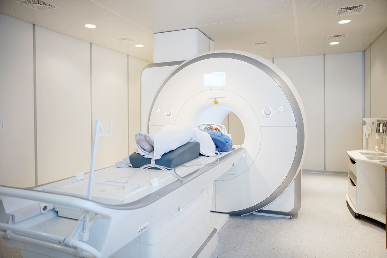
- CT/MRI of abdomen
- preoperative care
- percutaneous embolization (coiling) or chemoembolization
- symptomatic treatment
- cost of essential medicines
- nursing services
- elaboration of further recommendations
How program is carried out
During the first visit, the doctor will conduct a clinical examination and go through the results of the available diagnostic tests. After that, you will undergo the necessary additional examination, such as the assessment of liver and kidney function, ultrasound scan, CT scan and MRI. This will allow the doctor to determine which vessels are feeding the tumor and its metastases, as well as determine how well you will tolerate the procedure.
Chemoembolization begins with local anesthesia and catheterization of the femoral artery. The thin catheter is inserted through a few centimeters long incision of the blood vessel. The doctor gradually moves the catheter to the vessel feeding the primary tumor or its metastases. The procedure is carried out under visual control, an angiographic device is used for this. The vascular bed and the position of the catheter in it are displayed on the screen of the angiograph.
When the catheter reaches a suspected artery, a contrast agent is injected through it. Due to the introduction of the contrast agent, the doctor clearly sees the smallest vessels of the tumor and the surrounding healthy tissues on the screen of the angiograph. After that, he injects emboli into the tumor vessels through the same catheter.
Emboli are the spirals or the liquid microspheres. The type of embolus is selected individually, taking into account the diameter of the target vessel. When carrying out chemoembolization, a solution of a chemotherapy drug is additionally injected into the tumor vessel. Due to the subsequent closure of the vessel lumen with an embolus, the chemotherapy drug influences the tumor for a long time. In addition, the drug does not enter the systemic circulation, which allows doctors to use high doses of chemotherapeutic agents without the development of serious side effects. Chemoembolization leads to the destruction of the tumor or slowing down its progression.
After that, the catheter is removed from the artery. The doctor puts a vascular suture on the femoral artery and closes it with a sterile dressing. During chemoembolization, you will be awake. General anesthesia is not used, which significantly reduces the risks of the procedure and allows performing it on an outpatient basis, avoiding long hospital stay.
After the first procedure, you will stay under the supervision of an interventional oncologist and general practitioner. If necessary, you will receive symptomatic treatment. As a rule, a second chemoembolization procedure is performed in 3-5 days after the first one in order to consolidate the therapeutic effect. After that, you will receive recommendations for further follow-up and treatment.
Required documents
- Medical records
- MRI/CT scan (not older than 3 months)
- Biopsy results (if available)
Service
You may also book:
 BookingHealth Price from:
About the department
The Department of Interventional Radiology, Neuroradiology and Nuclear Medicine at the Hospital Cologne-Holweide provides the full range of services in the areas of its specialization. The department's team of physicians performs many image-guided diagnostic procedures and radioisotope examinations using state-of-the-art equipment. Effective minimally traumatic image-guided interventional procedures are also available here for the treatment of stroke, tumors, vascular stenosis, vascular obstructions, abscesses, etc. The department has advanced medical equipment, including a high-performance SOMATOM Definition Flash CT scanner, several 1.5 Tesla magnetic resonance imaging devices, and a dual-plane angiography machine. The medical facility has a picture archiving and communication system (PACS), which ensures fast processing and transmission of diagnostic results to different departments at the hospital. Doctors cooperate closely with specialists from other departments. Most diagnostic examinations and therapeutic procedures are performed on an outpatient basis. The doctors at the medical facility pay attention to comprehensive patient counseling, striving to provide complete information about the upcoming diagnostic procedures or treatment. The Head Physician of the department is Prof. Dr. med. Axel Gossmann.
The department's interventional radiologists specialize in performing image-guided invasive examinations and therapeutic procedures. Of particular interest in the field of diagnostics is a biopsy, which is biological material sampling for follow-up cytologic and histologic examinations. Biopsy procedures are performed under ultrasound or CT guidance. Doctors in this area also perform many effective therapeutic procedures under CT or angiography guidance. For example, the department often performs chemoembolization. This is a local treatment for malignant tumors that involves occluding the blood vessels that supply the neoplasm with emboli (microspheres) that contain chemotherapeutic agents. Procedures like balloon dilation and stent implantation are also performed under imaging guidance here. As a rule, these procedures are indicated in cases of vascular stenosis and obstruction of various localizations.
An integral part of the department's clinical practice is neuroradiology. Doctors in this specialization are responsible for image-guided diagnostics and CT- and MRI-guided endovascular treatment of brain and spinal cord diseases. A very demanded treatment method in the field of neuroradiology is aspiration thrombectomy. This procedure involves puncturing the blood vessel with a needle that is guided to the pathological focus through a specialized catheter, allowing for direct removal of the thrombus and thereby normalizing blood supply to the brain. The department's neuroradiologists also specialize in coiling for brain aneurysms and embolization for angiomas and brain and spinal malignancies.
A team of experts in nuclear medicine offers a broad range of radioisotope diagnostics. State-of-the-art equipment is available in the department for scintigraphy of the myocardium, kidneys, bones, lungs, brain, thyroid gland, and other organs. When conducting scintigraphy, doctors administer pharmaceuticals with radioactive markers, which accumulate in tissues or organs and emit specific radiation. This radiation is recorded by external detectors on the gamma camera, resulting in an image formation. Scintigraphy is a painless and completely safe diagnostic method that, in many cases, provides doctors with unique clinical data.
The department's clinical focuses include:
- Interventional radiology
- Image-guided interventional therapeutic procedures
- Balloon dilatation followed by stent implantation for vascular stenosis and obstruction
- Chemoembolization for malignant tumors
- Radiofrequency ablation for malignant tumors
- Puncture for abscesses and other pathological fluid accumulations
- Image-guided interventional therapeutic procedures
- Interventional neuroradiology
- Image-guided interventional therapeutic procedures
- Lysis therapy and aspiration thrombectomy for stroke
- Coiling for brain aneurysms
- Embolization for angiomas
- Embolization for brain and spinal tumors
- Image-guided interventional therapeutic procedures
- Nuclear medicine
- Radioisotope diagnostics
- Myocardial scintigraphy
- Skeletal scintigraphy
- Bone marrow scintigraphy
- Kidney scintigraphy
- Lung ventilation perfusion scintigraphy
- Brain scintigraphy
- Thyroid scintigraphy
- Parathyroid scintigraphy
- Adrenal scintigraphy
- Radioisotope diagnostics
- Other medical services
Curriculum vitae
Since 2008, Prof. Dr. med. Axel Gossmann has been the Head Physician of the Department of Interventional Radiology, Neuroradiology and Nuclear Medicine at the Hospital Cologne-Holweide. Since May 2013, he has held the position of Medical Director at the Hospital Cologne-Merheim. In September 2014, the specialist became the Head Physician of the Department of Diagnostic and Interventional Radiology at the University of Witten/Herdecke. Since April 2022, Prof. Axel Gossmann has also been Medical Director of the Cologne Hospital Network (Kliniken der Stadt Köln).
Photo of the doctor: (c) Krankenhaus Köln-Holweide
About hospital
According to the reputable Focus magazine, the Hospital Cologne-Holweide ranks among the top medical centers in North Rhine-Westphalia!
The medical complex is an academic hospital of the University of Cologne, providing patients with high-quality healthcare based on the latest achievements in university medicine. The hospital admits patients with various diseases, including cancer, diseases of the hematopoietic system, urinary system, gastrointestinal tract, ENT organs, etc. The hospital is known for having one of the largest Departments of Obstetrics in Germany, where more than 1,800 babies are born every year. The health facility also houses the specialized Breast Cancer Center and the Colon Cancer Center, whose treatment success rates are consistently high. The hospital has advanced technical resources and highly qualified staff to provide patients with effective medical care in the areas of its specialization.
The hospital has a total of 465 beds. The medical facility annually admits about 20,500 inpatients and more than 63,900 outpatients for diagnostics and treatment. The hospital provides comfortable conditions and modern infrastructure to meet the needs and wishes of patients. The medical team at the hospital prefers a multidisciplinary approach to medical care, which is particularly valuable in treating complex conditions such as cancer.
In 2009, the hospital was successfully certified by "High-Quality Management of Acute Pain Syndrome" TÜV Rheinland Group in gynecology, surgery, urology, otorhinolaryngology, and anesthesiology. In addition, the hospital became the first emergency medical facility for adults in Cologne to be KTQ® (Cooperation for Transparency and Quality in Healthcare) certified.
The specialists at the hospital always strive to provide each patient with high-quality medical care in a pleasant atmosphere. Doctors are open to dialog, gladly answer all questions that patients are interested in, and provide moral support throughout the therapeutic process.
Photo: (с) depositphotos
Accommodation in hospital
Patients rooms
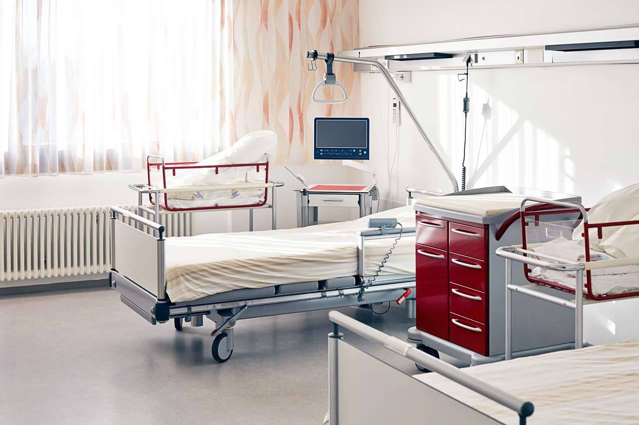
The patients of the Hospital Cologne-Holweide live in comfortable single and double rooms designed in light colors. Each patient room has an ensuite bathroom with a shower and a toilet. The furnishings of a standard patient room include an automatically adjustable bed with an orthopedic mattress, a bedside table, a wardrobe for personal belongings, a table and chairs for receiving visitors, and a TV with international channels. Wi-Fi is also available in the patient rooms.
The patients can stay in enhanced-comfort rooms, if desired. Such rooms are more spacious and additionally equipped with a safe, a mini-fridge, and upholstered furniture.
Meals and Menus
The patients are offered three tasty and healthy meals a day: breakfast, lunch, and dinner. Breakfast and dinner are served as buffets, and for lunch, there is a set of three menus to choose from.
If, for some reason, you do not eat all the foods, you will be offered an individual menu. Please inform the medical staff about your dietary preferences prior to treatment.
Further details
Standard rooms include:
Religion
A worship service is held in the chapel at the hospital every Sunday. There is also a prayer room at the hospital where one can pray in private.
The services of representatives of other religions are available upon request.
Accompanying person
Your accompanying person may stay with you in your patient room or at the hotel of your choice during the inpatient program.
Hotel
You may stay at the hotel of your choice during the outpatient program. Our managers will support you for selecting the best option.


