The program includes:
- Initial presentation in the clinic
- clinical history taking
- review of medical records
- physical examination
- laboratory tests:
- complete blood count
- general urine analysis
- biochemical analysis of blood
- inflammation indicators (CRP, ESR)
- indicators of blood coagulation
- neurological examination
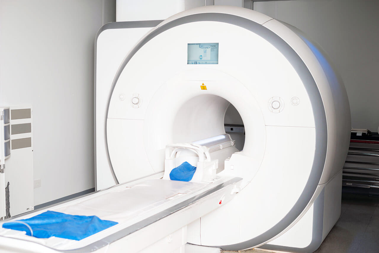
- CT/MRI scan
- neuropsychological tests (if indicated):
- ENMG (electroneuromyography)
- EEG (electroencephalography)
- SEPs (somatosensory evoked potentials)
- VEPs (visually evoked potentials)
- BAEP tests (brainstem auditory evoked potentials)
- preoperative care
- MRI-guided laser ablation of brain metastases
- postoperative MRI control
- symptomatic treatment
- control examinations
- the cost of essential medicines and materials
- nursing services
- full hospital accommodation
- developing of further guidance by specialists
in oncology and radiotherapy
How program is carried out
During the first visit, the physician will conduct a clinical examination and neurological examination, and go through the results of the available diagnostic tests. After that, you will undergo the necessary additional examination, such as the laboratory blood and urine tests, MRI scan with obtaining 3D images and creating volumetric images of the whole brain. Based on the results of an additional examination, the physician will clarify the localization of brain metastases. After that, the preparation according to the preoperative standard will begin.
Laser ablation of metastases begins with the formation of access to the brain. When using a catheter with a laser fiber, a hole with a diameter of 3.2 mm is sufficient for this. The miniature size and flexibility of the catheter help preserve healthy brain tissue.
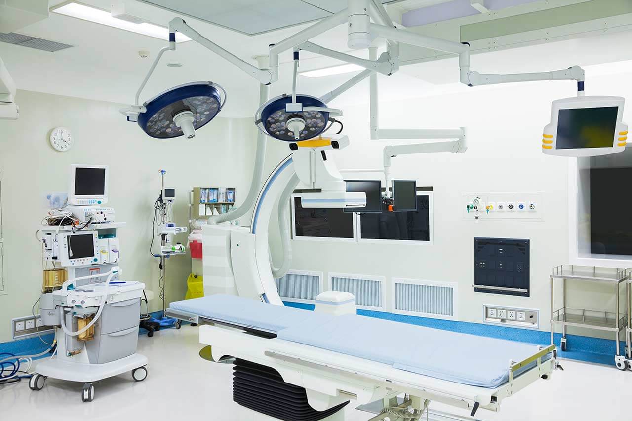
When the catheter reaches metastasis, energy is delivered through the laser fiber. Under the influence of high temperature, metastases are destroyed. The procedure is guided by real-time MRI. Surgeons also assess the temperature of healthy brain tissue using thermographic images to avoid overheating.
After the completion of the operation, you will be transferred to the ward, under the round-the-clock supervision of doctors and nurses. In several days, the attending physician will evaluate the results of the postoperative MRI scans, schedule the date of discharge from the hospital and give you detailed recommendations for further follow-up and treatment.
Required documents
- Medical records
- MRI/CT scan (not older than 3 months)
- Biopsy results (if available)
Service
You may also book:
 BookingHealth Price from:
About the department
The Department of Adult and Pediatric Neurosurgery, Spinal Surgery at the Nuremberg Hospital offers the full range of medical services in the fields of its competence. The department's team of doctors specializes in the surgical treatment of all diseases of the nervous system and also brilliantly performs operations for spinal diseases. The medical facility has an experienced team of pediatric neurosurgeons who perform surgery for brain tumors, hydrocephalus, vascular malformations of the brain, and other diseases. The department's specialists use state-of-the-art medical equipment to provide patients with the most effective treatment, in particular surgical microscopes, neuronavigation, laser devices, intraoperative ultrasound, equipment for stereotactic procedures, electrophysiological intraoperative monitoring, intraoperative fluorescence imaging, etc. A successful treatment outcome is facilitated by close interdisciplinary collaboration with experts in neurology, neuroradiology, otolaryngology, oral and maxillofacial surgery, and trauma surgery. Neurosurgeons thoroughly plan each operation and discuss the details of the upcoming surgical treatment with their patients to be able to meet all their needs and wishes. The Head Physician of the department is Prof. Dr. med. Karl-Michael Schebesch.
One of the department's key areas of specialization is the surgical treatment of brain tumors. The department's neurosurgeons have long experience treating gliomas, meningiomas, pituitary tumors, and brain metastases. Brain tumors at their early stages may not cause any symptoms, but as they grow, they compress vital nerve centers, leading to acute symptoms such as sensory and balance disorders, double vision or visual impairment, speech disorders, epileptic seizures, paralysis, etc. At the diagnostic stage, CT and/or MRI scans play a crucial role in detecting suspected brain tumors. During the elaboration of treatment tactics, the department’s doctors always take into account the type of tumor, its stage, size, and location, as well as the patient's age, general health condition, and the presence of comorbidities.
In most cases, a complete cure or long-term remission of brain cancer can only be achieved with the help of tumor resection surgery. At the same time, it is extremely important for the department's surgeons to perform the operation not only effectively but also safely for the patient, so special attention is paid to planning surgery. For example, the specialists often use navigated brain stimulation, which allows them to assess the proximity of the tumor to vital nerve centers and thereby minimize the risk of patient disability. State-of-the-art equipment, including computer-assisted navigation systems, intraoperative neurophysiological monitoring, surgical microscopes, intraoperative fluorescence imaging, and other devices, is also used directly during surgery. As a rule, brain tumor resection surgery is performed using minimally invasive or endoscopic techniques. During interdisciplinary tumor boards, doctors may also prescribe adjuvant conservative therapy, namely radiation therapy, chemotherapy, alternating electric field therapy, etc.
The department also successfully performs surgery for neurovascular diseases, including brain aneurysms, angiomas, cavernomas, and other pathologies. Clinical cases of patients with the above-mentioned pathologies are considered in close cooperation with neuroradiologists. The specialists cooperatively assess the patient's diagnostic data and determine whether open surgery or endovascular treatment (using an image-guided vascular approach) is required. If a decision is made to perform surgery, microsurgical techniques are most often used. The imaging of the cerebral vessels is provided by intraoperative infrared fluorescent video angiography.
The department's operating rooms are adapted for neurosurgical interventions in young patients. Of particular interest in the field of pediatric neurosurgery is the treatment of brain tumors, hydrocephalus, vascular malformations, cranial malformations, meningomyelocele, Arnold-Chiari malformations, etc. Whenever required, pediatricians, pediatric oncologists, pediatric surgeons, and other specialists may be involved in the therapeutic process. Most surgical interventions for diseases of the nervous system in children are performed using minimally invasive, microsurgical, and endoscopic techniques. The department's specialists widely use neuronavigation, neuromonitoring, intraoperative ultrasound imaging, and other technical options that ensure high accuracy and safety of surgical manipulations.
Spinal surgery is an integral part of the department's daily clinical practice. The department has gained impressive experience in the surgical treatment of spinal diseases, including herniated discs, spinal canal stenosis, vertebral fractures, compression syndrome, spondylolisthesis, and spinal and spinal cord tumors. Spinal surgery is performed in the medical facility using minimally traumatic surgical techniques under CT and intraoperative 3D imaging guidance. The risks for the patient in the postoperative period are therefore almost zero, and mobility is restored as soon as possible.
The department's key clinical focuses include:
- Adult neurosurgery
- Surgical treatment of brain tumors
- Gliomas
- Meningiomas
- Pituitary tumors
- Brain metastases
- Surgical treatment of neurovascular diseases
- Brain aneurysms
- Angiomas
- Cavernomas
- Surgical treatment of hydrocephalus
- Surgical treatment of chronic pain and movement disorders of neurological origin
- Sudeck's syndrome
- Pain syndrome caused by peripheral arterial disease of the lower limbs
- Pain syndrome caused by amputation
- Parkinson's disease
- Tremor
- Dystonia
- Spasticity
- Surgical treatment of brain tumors
- Pediatric neurosurgery
- Surgical treatment of brain tumors
- Surgical treatment of hydrocephalus
- Surgical treatment of vascular malformations
- Surgical treatment of cranial malformations
- Surgical treatment of meningomyelocele
- Surgical treatment of Arnold-Chiari malformation
- Spinal surgery
- Surgical treatment of herniated discs
- Surgical treatment of spinal stenosis
- Surgical treatment of vertebral fractures
- Surgical treatment of compression syndromes
- Surgical treatment of spondylolisthesis
- Surgical treatment of intervertebral joint osteoarthritis and cysts
- Surgical treatment of inflammatory lesions of the vertebral bodies and intervertebral discs
- Surgical treatment of spinal vascular malformations
- Surgical treatment of spinal and spinal cord tumors
- Other medical services
Curriculum vitae
Prof. Dr. med. Karl-Michael Schebesch heads the Department of Adult and Pediatric Neurosurgery, Spinal Surgery at the Nuremberg Hospital. The specialist graduated from the Faculty of Medicine at the University of Erlangen-Nuremberg. In June 2001, the doctor became a resident at the University Hospital Regensburg. In April 2008, Prof. Schebesch completed his clinical training and was board certified in Neurosurgery. In 2009, he became a Senior Physician, and in 2013, he held the position of a Managing Senior Physician in the Department of Neurosurgery. In 2012, he completed his habilitation procedures. In April 2017, Dr. Schebesch became the Professor for Neurosurgery, and in December 2019 he was appointed Deputy Head of the Department of Neurosurgery at the University Hospital Regensburg.
During his long clinical practice, the specialist has undergone multiple internships abroad, including in hospitals in Miami, Zurich, and Helsinki. Prof. Schebesch is the author of more than 90 publications in journals and is a reviewer for several international journals in Neurology and Neurosurgery.
Of particular interest to Dr. Schebesch are skull base surgery and neurovascular surgery.
Photo of the doctor: (c) Klinikum Nürnberg
About hospital
According to the reputable Focus magazine, the Nuremberg Hospital ranks among the top German medical facilities!
The hospital is one of the largest, highly specialized medical centers in Europe and positions itself as the maximum care hospital. The healthcare facility is an academic hospital of the Paracelsus Medical University in Nuremberg. It houses 42 departments, institutes, and highly specialized centers focusing on various medical fields. All the hospital's employees work hand in hand for the benefit of their patients. The specialists strive to provide top-class medical care for every patient. Moreover, the medical team always shows a humane attitude and understanding towards the patient's life situation, making every effort to support them during the entire therapeutic process.
The total number of beds in the hospital is 2,233. The medical team consists of more than 8,400 employees, including many world-famous doctors and professors who had their clinical training at the best medical facilities in Germany, other European countries, and the USA. The hospital admits more than 100,000 inpatients and more than 170,000 outpatients annually. The number of patients who come to the hospital steadily increases every year, which is the best confirmation of its high standards and outstanding treatment results.
The cornerstone of successful clinical practice is state-of-the-art technical infrastructure. The hospital offers its patients innovative technologies such as the Da Vinci surgical system, devices for stereotactic procedures, intraoperative radiation therapy, angiography, PET CT devices, high-intensity focused ultrasound, 64-slice CT scanners, and other advanced medical devices. The combination of cutting-edge technical facilities and the high competence of the physicians allows for the provision of effective treatment even in the most complex cases.
The Nuremberg Hospital is undoubtedly one of Germany's leading medical facilities, where patients benefit from modern infrastructure, precise diagnostics, effective treatment, and responsive care.
Photo: (с) depositphotos
Accommodation in hospital
Patients rooms
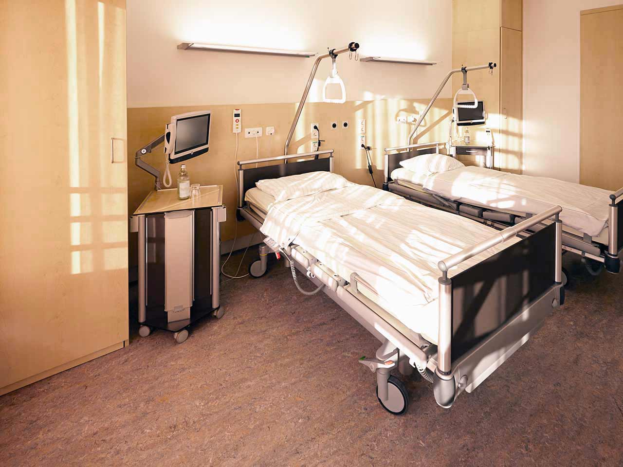
The patients at the Nuremberg Hospital live in comfortable single and double rooms. Each patient room is equipped with an ensuite bathroom with a shower and a toilet. Standard rooms include an automatically adjustable bed, a bedside table, a wardrobe for storing clothes, a table and chairs for receiving visitors, a TV, and a radio. The hospital also offers Wi-Fi.
If desired, patients can live in enhanced-comfort rooms. Such patient rooms are more spacious, and their furnishings correspond to the level of an upscale hotel.
Meals and Menus
The patients at the hospital are offered tasty and balanced meals three times a day: breakfast, lunch, and dinner. The patients have a daily choice of three dishes for lunch and dinner, while breakfast is served as a buffet.
If, for some reason, you do not eat all foods, you will be offered an individual menu. Please inform the medical staff about your food preferences prior to treatment.
Further details
Standard rooms include:
Accompanying person
Your accompanying person may stay with you in your patient room or at the hotel of your choice during the inpatient program.
Hotel
You may stay at the hotel of your choice during the outpatient program. Our managers will support you for selecting the best option.


