{"translation_price":"50","translation_doc_price":"40","child_coefficient":"1.1","transfer_price":"2.00","transfer_price_vip":"5.00","constant_transfer_price_vip":350,"constant_transfer_price":150,"constant_transfer_distanse":60,"type":"treatment","program_full_story":"<ul>\n\t<li>Initial presentation in the clinic<\/li>\n\t<li>clinical history taking<\/li>\n\t<li>review of medical records<\/li>\n\t<li>physical examination<\/li>\n\t<li>laboratory tests:\n\t<ul>\n\t\t<li>complete blood count<\/li>\n\t\t<li>general urine analysis<\/li>\n\t\t<li>biochemical analysis of blood<\/li>\n\t\t<li>TSH-basal, fT3, fT4<\/li>\n\t\t<li>tumor markers (AFP, CEA, \u0421\u0410-19-9)<\/li>\n\t\t<li>inflammation indicators (CRP, ESR)<\/li>\n\t\t<li>indicators of blood coagulation<\/li>\n\t<\/ul>\n\t<\/li>\n\t<li>abdominal ultrasound\u200b scan<\/li>\n\t<li>CT\/MRI of abdomen<\/li>\n\t<li>preoperative care<\/li>\n\t<li>percutaneous embolization (coiling) or chemoembolization<\/li>\n\t<li>symptomatic treatment<\/li>\n\t<li>cost of essential medicines<\/li>\n\t<li>nursing services<\/li>\n\t<li>elaboration of further recommendations<\/li>\n<\/ul>\n<div class=\"program_indications_for_surgery\"><h4>Indications<\/h4><ul>\n\t<li>Inoperable liver metastases<\/li>\n\t<li>Poor response to systemic chemotherapy<\/li>\n<\/ul>\n\n<p><strong>Treatment is not indicated<\/strong> in:<\/p>\n\n<ul>\n\t<li>Presence of extrahepatic metastases<\/li>\n\t<li>Affection of more than 70% of the liver<\/div><div class=\"program_how_program_going mt-4\"><h4>How program is carried out<\/h4><p style=\"text-align:justify\"><strong>During the first visit<\/strong>, the doctor will conduct a clinical examination and go through the results of the available diagnostic tests. After that, you will undergo the necessary additional examination, such as the assessment of liver and kidney function, ultrasound scan, CT scan and MRI. This will allow the doctor to determine which vessels are feeding the tumor and its metastases, as well as determine how well you will tolerate the procedure.<\/p>\n\n<p style=\"text-align:justify\"><strong>Chemoembolization <\/strong>begins with local anesthesia and catheterization of the femoral artery. The thin catheter is inserted through a few centimeters long incision of the blood vessel. The doctor gradually moves the catheter to the vessel feeding the primary tumor or its metastases. The procedure is carried out under visual control, an angiographic device is used for this. The vascular bed and the position of the catheter in it are displayed on the screen of the angiograph.<\/p>\n\n<p style=\"text-align:justify\">When the catheter reaches a suspected artery, a contrast agent is injected through it. Due to the introduction of the contrast agent, the doctor clearly sees the smallest vessels of the tumor and the surrounding healthy tissues on the screen of the angiograph. After that, he injects emboli into the tumor vessels through the same catheter.<\/p>\n\n<p style=\"text-align:justify\">Emboli are the spirals or the liquid microspheres. The type of embolus is selected individually, taking into account the diameter of the target vessel. When carrying out chemoembolization, a solution of a chemotherapy drug is additionally injected into the tumor vessel. Due to the subsequent closure of the vessel lumen with an embolus, the chemotherapy drug influences the tumor for a long time. In addition, the drug does not enter the systemic circulation, which allows doctors to use high doses of chemotherapeutic agents without the development of serious side effects. Chemoembolization leads to the destruction of the tumor or slowing down its progression.<\/p>\n\n<p style=\"text-align:justify\">After that, the catheter is removed from the artery. The doctor puts a vascular suture on the femoral artery and closes it with a sterile dressing. During chemoembolization, you will be awake. General anesthesia is not used, which significantly reduces the risks of the procedure and allows performing it on an outpatient basis, avoiding long hospital stay.<\/p>\n\n<p style=\"text-align:justify\"><strong>After the first procedure<\/strong>, you will stay under the supervision of an interventional oncologist and general practitioner. If necessary, you will receive symptomatic treatment. As a rule, a second chemoembolization procedure is performed in 3-5 days after the first one in order to consolidate the therapeutic effect. After that, you will receive recommendations for further follow-up and treatment.<\/p>\n<\/div><div class=\"program_required_documents mt-4\"><h4>Required documents<\/h4><ul>\n\t<li style=\"text-align: justify;\">Medical records<\/li>\n\t<li style=\"text-align: justify;\">Abdominal ultrasound (if available)<\/li>\n\t<li style=\"text-align: justify;\">MRI\/CT scan of the abdomen (if available)<\/li>\n\t<li style=\"text-align: justify;\">Biopsy results (if available)<\/li>\n<\/ul>\n<\/div>","program_full_story_crm":"<ul>\n\t<li>Initial presentation in the clinic<\/li>\n\t<li>clinical history taking<\/li>\n\t<li>review of medical records<\/li>\n\t<li>physical examination<\/li>\n\t<li>laboratory tests:\n\t<ul>\n\t\t<li>complete blood count<\/li>\n\t\t<li>general urine analysis<\/li>\n\t\t<li>biochemical analysis of blood<\/li>\n\t\t<li>TSH-basal, fT3, fT4<\/li>\n\t\t<li>tumor markers (AFP, CEA, \u0421\u0410-19-9)<\/li>\n\t\t<li>inflammation indicators (CRP, ESR)<\/li>\n\t\t<li>indicators of blood coagulation<\/li>\n\t<\/ul>\n\t<\/li>\n\t<li>abdominal ultrasound\u200b scan<\/li>\n\t<li>CT\/MRI of abdomen<\/li>\n\t<li>preoperative care<\/li>\n\t<li>percutaneous embolization (coiling) or chemoembolization<\/li>\n\t<li>symptomatic treatment<\/li>\n\t<li>cost of essential medicines<\/li>\n\t<li>nursing services<\/li>\n\t<li>elaboration of further recommendations<\/li>\n<\/ul>\n<div class=\"program_indications_for_surgery\"><h4>Indications<\/h4><ul>\n\t<li>Inoperable liver metastases<\/li>\n\t<li>Poor response to systemic chemotherapy<\/li>\n<\/ul>\n\n<p><strong>Treatment is not indicated<\/strong> in:<\/p>\n\n<ul>\n\t<li>Presence of extrahepatic metastases<\/li>\n\t<li>Affection of more than 70% of the liver<\/div><div class=\"program_how_program_going mt-4\"><h4>How program is carried out<\/h4><p style=\"text-align:justify\"><strong>During the first visit<\/strong>, the doctor will conduct a clinical examination and go through the results of the available diagnostic tests. After that, you will undergo the necessary additional examination, such as the assessment of liver and kidney function, ultrasound scan, CT scan and MRI. This will allow the doctor to determine which vessels are feeding the tumor and its metastases, as well as determine how well you will tolerate the procedure.<\/p>\n\n<p style=\"text-align:justify\"><strong>Chemoembolization <\/strong>begins with local anesthesia and catheterization of the femoral artery. The thin catheter is inserted through a few centimeters long incision of the blood vessel. The doctor gradually moves the catheter to the vessel feeding the primary tumor or its metastases. The procedure is carried out under visual control, an angiographic device is used for this. The vascular bed and the position of the catheter in it are displayed on the screen of the angiograph.<\/p>\n\n<p style=\"text-align:justify\">When the catheter reaches a suspected artery, a contrast agent is injected through it. Due to the introduction of the contrast agent, the doctor clearly sees the smallest vessels of the tumor and the surrounding healthy tissues on the screen of the angiograph. After that, he injects emboli into the tumor vessels through the same catheter.<\/p>\n\n<p style=\"text-align:justify\">Emboli are the spirals or the liquid microspheres. The type of embolus is selected individually, taking into account the diameter of the target vessel. When carrying out chemoembolization, a solution of a chemotherapy drug is additionally injected into the tumor vessel. Due to the subsequent closure of the vessel lumen with an embolus, the chemotherapy drug influences the tumor for a long time. In addition, the drug does not enter the systemic circulation, which allows doctors to use high doses of chemotherapeutic agents without the development of serious side effects. Chemoembolization leads to the destruction of the tumor or slowing down its progression.<\/p>\n\n<p style=\"text-align:justify\">After that, the catheter is removed from the artery. The doctor puts a vascular suture on the femoral artery and closes it with a sterile dressing. During chemoembolization, you will be awake. General anesthesia is not used, which significantly reduces the risks of the procedure and allows performing it on an outpatient basis, avoiding long hospital stay.<\/p>\n\n<p style=\"text-align:justify\"><strong>After the first procedure<\/strong>, you will stay under the supervision of an interventional oncologist and general practitioner. If necessary, you will receive symptomatic treatment. As a rule, a second chemoembolization procedure is performed in 3-5 days after the first one in order to consolidate the therapeutic effect. After that, you will receive recommendations for further follow-up and treatment.<\/p>\n<\/div>","is_ambulant":"1","bh_fee":"0","only_for_children":"0","no_service":"0","with_prepayment":"1","show_calculator":"1","paket_type":"1","btn_type":"2","clinic_icon":"5ff81b474b0d4.jpg","city":"Bonn","clinic_site":"https:\/\/www.wegeklinik.com\/","department_recommend":"3","country":"Germany","country_id":"1","clinic_name":"WEGE Clinic Bonn","cinic_name":"WEGE Clinic Bonn","department_id":"3870","duration":"5","direction":"Interventional radiology","min_duration":0,"clinic_id":"2596","paketPrice":3200,"paket":"<ul>\n <li> Interpreter up to 8 hours<\/li>\n <li>Translation of up to 5 pages<\/li>\n <li>Visa support<\/li>\n\n<\/ul>","title":"Treatment of liver metastases with percutaneous embolization (coiling) or chemoembolization","price":{"val":9806.43,"type":"val"},"price_surcharge":0,"price_surcharge_clear":0,"extra_service_clinic":[],"extra_service":[],"translation_hours":"0","translation_doc_count":null,"roads":[{"id":"7","distance":"27","airport_title":"Cologne-Bonn"},{"id":"4","distance":"87","airport_title":"Duesseldorf"},{"id":"25","distance":"127","airport_title":"Dortmund Airport"}],"pakets":[],"lang":{"day":"Day","days":"days","ambulatory":"Outpatient","stationaryProgram":"Inpatient"}}
Treatment of liver metastases with percutaneous embolization (coiling) or chemoembolization in WEGE Clinic Bonn
percutaneous embolization (coiling) or chemoembolization
symptomatic treatment
cost of essential medicines
nursing services
elaboration of further recommendations
Indications
Inoperable liver metastases
Poor response to systemic chemotherapy
Treatment is not indicated in:
Presence of extrahepatic metastases
Affection of more than 70% of the liver
How program is carried out
During the first visit, the doctor will conduct a clinical examination and go through the results of the available diagnostic tests. After that, you will undergo the necessary additional examination, such as the assessment of liver and kidney function, ultrasound scan, CT scan and MRI. This will allow the doctor to determine which vessels are feeding the tumor and its metastases, as well as determine how well you will tolerate the procedure.
Chemoembolization begins with local anesthesia and catheterization of the femoral artery. The thin catheter is inserted through a few centimeters long incision of the blood vessel. The doctor gradually moves the catheter to the vessel feeding the primary tumor or its metastases. The procedure is carried out under visual control, an angiographic device is used for this. The vascular bed and the position of the catheter in it are displayed on the screen of the angiograph.
When the catheter reaches a suspected artery, a contrast agent is injected through it. Due to the introduction of the contrast agent, the doctor clearly sees the smallest vessels of the tumor and the surrounding healthy tissues on the screen of the angiograph. After that, he injects emboli into the tumor vessels through the same catheter.
Emboli are the spirals or the liquid microspheres. The type of embolus is selected individually, taking into account the diameter of the target vessel. When carrying out chemoembolization, a solution of a chemotherapy drug is additionally injected into the tumor vessel. Due to the subsequent closure of the vessel lumen with an embolus, the chemotherapy drug influences the tumor for a long time. In addition, the drug does not enter the systemic circulation, which allows doctors to use high doses of chemotherapeutic agents without the development of serious side effects. Chemoembolization leads to the destruction of the tumor or slowing down its progression.
After that, the catheter is removed from the artery. The doctor puts a vascular suture on the femoral artery and closes it with a sterile dressing. During chemoembolization, you will be awake. General anesthesia is not used, which significantly reduces the risks of the procedure and allows performing it on an outpatient basis, avoiding long hospital stay.
After the first procedure, you will stay under the supervision of an interventional oncologist and general practitioner. If necessary, you will receive symptomatic treatment. As a rule, a second chemoembolization procedure is performed in 3-5 days after the first one in order to consolidate the therapeutic effect. After that, you will receive recommendations for further follow-up and treatment.
Required documents
Medical records
Abdominal ultrasound (if available)
MRI/CT scan of the abdomen (if available)
Biopsy results (if available)
Service
Price from:
Type of program :
Price for 1 day:
Expected duration of the program:
The minimum duration of the program:
Select
You may also book:
Hospital directly:
Saving:
BookingHealth Price from:
About the department
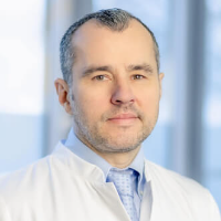
The Department of Interventional Radiology at the WEGE Clinic Bonn offers the full range of modern diagnostic procedures using ionizing radiation, image-guided tissue sampling (biopsy), and minimally invasive therapeutic procedures. Patients may be seen on an inpatient or outpatient basis. To achieve optimal results, the department works closely with specialists in radiation therapy, oncology, neurology, neurosurgery and neuro-oncology, as well as urology, orthopedics, pain management, and palliative care. This collaboration helps ensure an accurate diagnosis and the most effective treatment. The Head Physician of the department is Prof. Dr. med. Attila Kovács.
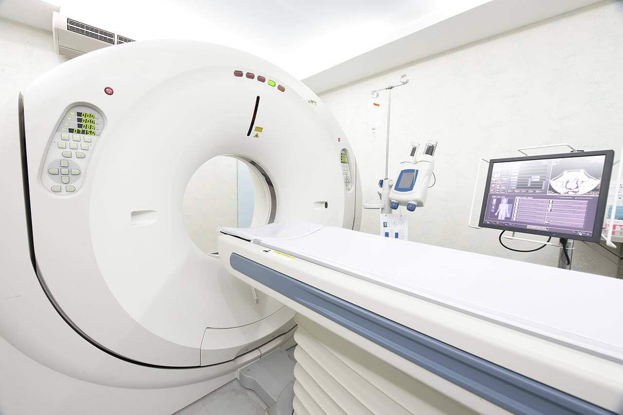
The department performs precise and safe diagnostics using ionizing radiation systems. Key diagnostic options include magnetic resonance imaging, computed tomography, radiography and digital subtraction angiography for vascular imaging. In addition, the department's experts perform all types of biopsies, including vacuum and punch biopsies. All tissue sampling procedures are performed under CT guidance (with the exception of prostate biopsies). Prostate tissue is obtained under MRI guidance. The department also performs systemic chemotherapy and immunotherapy sensitivity testing.
The department offers the widest range of image-guided interventional procedures. One of the most requested interventional procedures is transarterial chemoembolization (TACE). This is a local, minimally invasive therapy used to treat liver cancer and metastases to the kidneys or chest wall. In the early stages of liver cancer, ablation procedures can also achieve good results. However, when the tumor has reached a fairly large size or metastases have spread, the patient is prescribed transarterial chemoembolization. During this procedure, the arteries are embolized with small microspheres, which helps to cut off the supply of oxygen and nutrients to the tumor. At the same time, the microspheres deliver a high dose of a chemotherapy drug to the tumor and release it gradually. Such particles are called drug-coated granules. They allow physicians to target cancer cells more intensively. The department performs two types of transarterial chemoembolization – tumor selective and organ segment selective. Tumor-selective therapy is performed in the presence of a single metastasis in the liver, while segmental chemoembolization is performed in the presence of multiple liver lesions. The advantage of this therapy is the targeted destruction of the tumor and metastases while sparing healthy liver tissue.
The department also offers radiofrequency ablation, which uses heat to destroy the tumor. Variations of this technique include thermal ablation and microwave ablation. However, radiofrequency ablation is the most common method of thermal tumor destruction. It is used to treat malignant neoplasms and metastases in the liver, lungs, kidneys and adrenal glands, retroperitoneal lymph nodes, bones, and soft tissues. The procedure is performed under sedation (anesthesia is not required). Under CT guidance, a special probe is inserted into the body to increase the temperature of the tumor. Radiofrequency ablation allows the physician to heat the tumor cells to a temperature of 60 to 100°C. The heat affects the tumor until it is destroyed. The goal of radiofrequency ablation is to achieve the same result as classical tumor removal. The best results can be achieved with this technique for tumors up to 4 cm in size or liver tumors up to 5 cm in size.
It is worth mentioning that the doctors of the department also have excellent qualifications in kyphoplasty for the treatment of vertebral fractures (minimally invasive procedure). Kyphoplasty helps to relieve pain, and in many cases it can completely eliminate the pain syndrome. The patient lies on his stomach during the procedure. It takes about 20 minutes and is usually performed under general anesthesia. The essence of the therapeutic method is the introduction of a special cannula through the skin (at the level of the affected vertebra), which serves as a working channel for the introduction of the balloon. This balloon is filled with radiopaque fluid and inflated under constant x-ray control. It has two functions: to align the vertebra and to create a bed for the introduction of bone cement, which will help to rebuild the vertebra after the fracture. Kyphoplasty uses a special biocement that mimics real human bone tissue. Kyphoplasty has some risks (for example, damage to the nerve roots or the spinal cord itself), but the high professionalism of the department's doctors allows them to minimize these risks. To relieve symptoms and strengthen the back muscles after the therapeutic manipulation, additional drug therapy may be required.
The department specializes in the diagnostics and treatment of the following diseases:
Benign pathologies
Auditory nerve neuroma
Arteriovenous malformations
Systemic musculoskeletal pain syndromes
Erectile dysfunction
Focal cortical hyperplasia
Benign brain lesions
Benign prostatic hyperplasia
Benign uterine tumors
Varicocele
Testicular pain
Pituitary adenoma
Certain types of infertility
Focal liver formations
Meningiomas
Pathological vertebral fractures
Uterine fibroids
Malignant pathologies
Bowel cancer
Liver cancer, including metastatic lesions
Kidney cancer, including metastatic lesions
Skin cancer, including metastatic lesions
Breast cancer
Prostate cancer
Soft tissue metastasis
Other benign and malignant diseases
The department's range of medical services includes:
Diagnostic examinations
Biopsy (aspiration, punch biopsy)
Systemic chemotherapy and immunotherapy sensitivity testing
Computed tomography
Magnetic resonance imaging
Radiography
Digital subtraction angiography
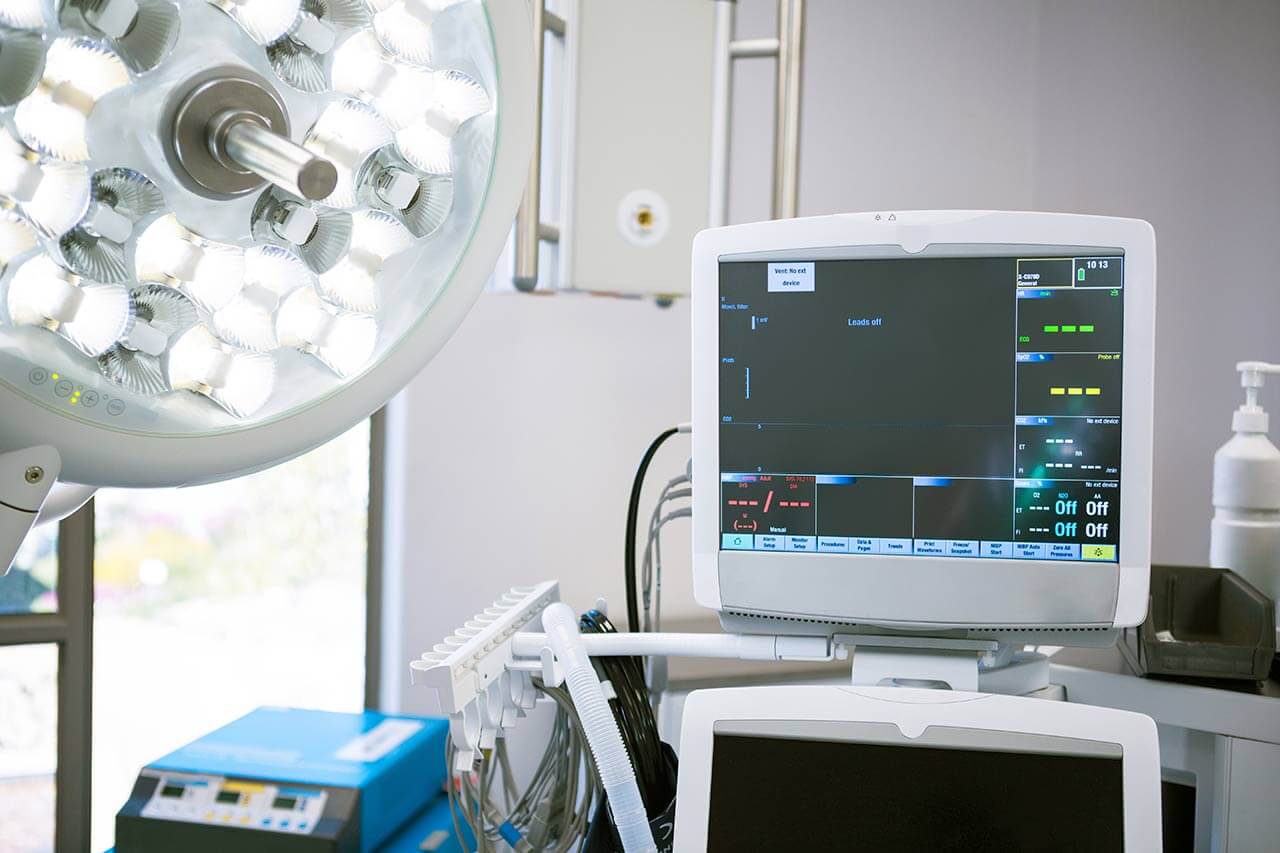
Interventional therapeutic procedures
Transarterial chemoembolization (TACE)
Thermal ablation
Radiofrequency ablation
Microwave ablation
Cryoablation
Electrochemotherapy
Prostatic artery embolization
Uterine artery embolization
Kyphoplasty
Other diagnostic and therapeutic options
Curriculum vitae
Higher Education
1989 - 1996 Medical studies in Kiel and Heidelberg.
Clinical and Scientific Career
1999 Doctoral thesis, Experimental Work on Biomechanical Support of the Heart, Department of Cardiac Surgery at the University Hospital Heidelberg. Subject: "Electron-microscopic examination of the musculus latissimus dorsi of the dog in chronic electrostimulation".
1997 - 2000 Work in the Department of Cardiac Surgery at the Ludwigshafen Clinic.
2000 - 2010 Work in the Department of Radiology at the University Hospital Bonn.
2005 Board certification in Diagnostic Radiology.
2009 Specialization in Neuroradiology.
2010 - 2012 Work in the Department of Radiology and Nuclear Medicine, University Hospital Schleswig-Holstein, Campus Luebeck.
2011 CODMAN Prize of the German Society of Neuroradiology.
2012 Habilitation. Subject: "Modern non-invasive diagnostics for the planning and monitoring of interventional and surgical therapy".
Since 12.2012 Head Physician, Department of Interventional Radiology, WEGE Clinic Bonn.
Qualifications
Board certification in Diagnostic Radiology.
Specialization in Neuroradiology.
Certified Instructor of the German Society of Interventional Radiology and Minimally Invasive Treatment (DeGIR) in Interventional Radiology (A, B, C, D modules).
Certified Q3 Instructor in Cardiac MRI and CT.
Permission for advanced training courses in Radiology, Medical Association of North Rhine-Westphalia.
Memberships in Professional Societies
German Society of Radiology (DRG).
German Society of Interventional Radiology and Minimally Invasive Treatment (DeGIR).
European Society of Radiology (ESR).
Cardiovascular and Interventional Radiological Society of Europe (CIRSE).
European Society of Minimally Invasive Neurological Therapy (ESMINT).
Radiological Society of North America (RSNA).
Photo of the doctor: (c) WEGE Klinik
About hospital
The WEGE Clinic Bonn is a progressive medical facility that offers all the latest diagnostic and treatment methods for the recovery of cancer patients. The clinic was founded by the renowned radiologist and surgeon Robert Janker. Today, its history has more than 80 years of successful clinical practice, which has brought worldwide fame and reputation to the medical center. The clinic specializes in medical oncology, radiation oncology, diagnostic and interventional radiology, and palliative care.
The work of the clinic's medical team is based on the use of sparing and highly specialized therapeutic procedures. Each clinical case is individual and requires a special approach. The specialists of the medical facility guarantee their patients not only the most effective treatment, but also the highest level of medical care, respectful attitude, and understanding of the needs and wishes of each patient.
Patients can be treated on an inpatient or outpatient basis. The clinic's physicians work closely with many specialized cancer centers in Germany, as well as with many private physicians. In addition to conventional cancer treatments, the clinic also offers individual counseling, psychological support, social care, and other related activities.
The outstanding quality of diagnostics and treatment is confirmed by certification according to DIN EN ISO 9001: 2015 standards. The clinic is also certified by the German Cancer Society. The clinic is also a certified cooperation partner of the Cancer Center Bonn-Rhein-Sieg.
Photo: (c) WEGE Klinik, (c) depositphotos
Accommodation in hospital
Patients rooms
The patients at the WEGE Clinic Bonn stay in modern and comfortable rooms. Each room is equipped with an ensuite bathroom with shower and toilet. The standard room includes a comfortable automatically adjustable bed, a bedside table, a table and chairs for receiving visitors, a wardrobe for storing clothes, a radio, a TV, and a telephone (additional charges apply for using a telephone and a TV). The patient rooms also have Wi-Fi.
The patients with disabilities are accommodated in specially equipped rooms. The clinic also provides single enhanced-comfort rooms of a five-star hotel level.
Meals and Menus
The patients of the clinic are offered a healthy and varied diet. The wishes and needs of the patients are always taken into consideration. Patients have a choice of several daily menus, including a full menu, a basic menu, and diet and vegetarian menus. Breakfast and lunch are served in the clinic's cafeteria, while dinner is served in the patient's room.
Patients staying in enhanced-comfort rooms are offered an individual menu. In addition to breakfast, lunch, and dinner, the patient is also provided with coffee and delicious cakes, fruit in the room.
Further details
Standard rooms include:
Toilet
Shower
Wi-Fi
TV
Religion
Religious services can be provided upon request.
Accompanying person
During the inpatient program, an accompanying person may stay with you in a patient room or hotel of your choice.
Hotel
During the outpatient program, you may stay in a hotel of your choice. Managers will help you choose the most suitable options.
Select your currency:
Your guarantee
One year of support after the treatment
Insurance to cover unforeseen expenses arising from complications during and 48 months after treatment (coverage up to 200,000 €)
Reduced costs by 40-70% (contracts with Hospitals)
In addition, we are the only TÜV-certified company with an ISO 9001:2015 certificate
thank you for reaching out! One of our medical advisors is currently reviewing your details. You can expect a call from us within one business day to discuss your request.
Note: The incoming call will appear as a German number or your local area code. This call is entirely free of charge.
Booking Health guarantees:
Expert Placement
We use rigorous statistical analysis to ensure you are treated at the best-rated facility for your specific diagnosis.
Total Cost Protection
No hidden fees or unexpected bills. We provide a fixed price guarantee, backed by insurance that covers any additional complications.
12-Month Extended Care
Your recovery is our priority. Benefit from a full year of direct medical support following your procedure.


 BookingHealth Price from:
