Arteriovenous Malformation AVM — Platinum Coils Embolization (coiling): treatment in the Best Hospitals in the World
Treatment prices are regulated by national law of the corresponding countries, but can also include additional hospital coefficients. In order to receive the individual cost calculation, please send us the request and medical records.

Department of Interventional Neuroradiology
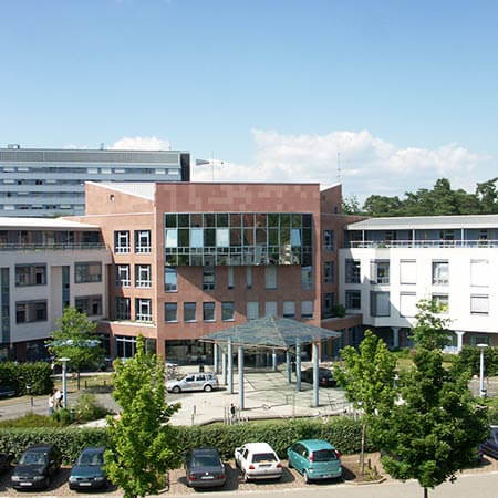
The Department of Interventional Neuroradiology offers the full range of services in the areas of its specialization. The medical facility provides imaging diagnostics and low-traumatic image-guided interventional treatment of nervous system diseases. The department's specialists have rich experience and exceptional professional skills in the field of interventional procedures for acute and chronic vascular diseases, such as ischemic strokes, brain hemorrhages, cerebral artery stenosis, brain aneurysms, and vascular malformations. The department's neuroradiologists cooperate closely with neurologists and neurosurgeons so that each patient receives an optimal treatment regimen based on the expert opinions of the specialists. The department's medical team has state-of-the-art computed tomography (CT), magnetic resonance imaging (MRI), and MR angiography systems that are actively used for diagnosing patients and therapeutic procedures. Medical care is provided in compliance with current clinical protocols. The department also offers many outpatient medical services, which is an advantage for many patients.


Department of Adult and Pediatric Diagnostic, Interventional Neuroradiology
The Department of Adult and Pediatric Diagnostic, Interventional Neuroradiology offers the widest range of services for the diagnostics and treatment of diseases of the brain, spinal cord and peripheral nerves using the advanced imaging systems. The department is equipped with modern technical devices, including 64-slice CT scanner, 1,5 and 3,0 Tesla MRI, biplane angiography system with 3D imaging of the blood vessels and bone structures, system for myelography of the entire spine and ultrasound scanners. All these devices are used not only for the diagnostics, but also for interventional procedures to treat obstructions, stenoses and other pathological vascular lesions in the head, neck and spine. The most popular interventional therapeutic procedures include clipping and coiling of cerebral vascular aneurysms, as well as cerebral vascular malformations. The department's doctors cooperate closely with specialists in the field of neurosurgery, neurology and pediatric neurology. Such an approach guarantees the patient a comprehensive assessment of the state of the nervous system and a favourable treatment outcome.


Department of Interventional Radiology
The Department of Interventional Radiology offers the full range of imaging examinations, as well as innovative image-guided minimally invasive techniques for the treatment of tumors, vascular diseases and internal pathologies (for example, CT, MRI, PET-CT, SPECT). The department's doctors have deep knowledge and colossal experience in the field of interventional radiological methods of treatment, which represent an excellent alternative to open surgical interventions. Despite the high level of technical equipment and the presence of advanced computerized systems, the focus is always on the person with his individual needs. Compliance with current clinical protocols and high professionalism of the department's specialists contribute to the successful clinical practice, as well as the reputability of the department among the best medical facilities of this kind in Germany.


Arteriovenous malformation (AVM) is an abnormal conglomerate of blood vessels in the brain or spinal cord. While blood vessels normally carry blood rich in oxygen and nutrients throughout the body, the vessels of arteriovenous malformations contain blood that is poor in these elements.
Overview
Arteriovenous malformation (AVM) is a disorder of the vessels that is characterized by one or more direct connections between arteries and veins. As arteries deliver oxygenated blood to healthy organs and tissues, they also deliver blood to malformations. For example, the arterial blood supply to a brain arteriovenous malformation is mainly derived from the internal carotid artery and vertebral basilar artery system.
However, when the brain arteriovenous malformation is adjacent to cerebral convexities or intracranial dural structures, the carotid and vertebral basilar arteries that supply the brain arteriovenous malformation may also originate from the arteries supplying the meninges (layers of protective brain membranes). Transdural blood supply to brain arteriovenous malformation may be from the external carotid artery or the meningeal branches of the internal carotid artery and the vertebral basilar artery systems.
Normally, blood enters tissues and cells through arteries, which branch into small capillaries. The blood then returns to the heart through the veins. In the case of arteriovenous malformation of the vessels, blood in the brain or spinal cord passes directly from the arteries to the veins, bypassing the capillaries, which forms a short closed cycle of blood flow, or shunt.
Most often, an AVM does not cause any severe symptoms. In many situations, the detection of arteriovenous malformation of the cerebral vessels is accidental, during the examination for other diseases. The symptoms of arteriovenous malformation (AVM) depend largely on the location of the pathological vascular formation. In rare cases, arteriovenous malformation causes epileptic seizures, neurological deficit, or pain.
About 0.2% of the population are carriers of arteriovenous malformations. As in an aneurysm, arteriovenous malformation can rupture and lead to cerebral hemorrhage (50% of all clinical manifestations). The course of the disease is less aggressive than with aneurysms. Epilepsy occupies second place in terms of manifestations of this disease.
Aneurysms (weakening of the artery wall) can occur in any artery. The most often aneurysms occur in the aorta – a large artery that carries blood from the heart to the rest of the body. Less common aneurysms are detected in the carotid artery.
Most popliteal and femoral artery aneurysms do not cause symptoms. However, blood clots can form inside the aneurysm. A loose blood clot is called an embolus. An embolus can travel along the bloodstream until he blocks an artery in the leg or foot. As a result, patients may experience an episode of sudden severe pain, numbness, coldness, and possibly paleness in the foot.
Arteriovenous congenital malformations are often connected with severe complications, such as stroke and neurological disorders. Arteriovenous malformation of cerebral vessels can be accompanied by headaches and epileptic seizures, as well as by other specific symptoms that usually depend on the location of the AVM.
Due to excessive blood accumulation in the area of AVM, patients may experience an increase in skin temperature, swelling, and redness. Sometimes patients experience a pulsating sensation within the affected area. Besides, the affected area is often inflamed and in some cases may become ulcerated. Extensive arteriovenous pathologies disrupt the blood supply and can even lead to the development of heart failure. In general, arteriovenous malformations manifest themselves with a variety of symptoms.
Diagnostics
Diagnosis of AVM usually begins with a thorough physical examination. Depending on the results of the examination and the suspicions that have arisen, the doctor may prescribe following instrumental examinations:
- Computed tomography (CT).
- Magnetic resonance imaging (MRI).
- MR-angiography.
- Cerebral arteriography. During the procedure, the surgeon inserts a thin tube (catheter) into the femoral artery and moves it under X-ray control until it reaches the carotid artery. After that, a contrast agent is injected into the vessel and a series of X-rays are taken.
Treatment
The leading method of the complex treatment of AVM is the surgical treatment. However, 23-60 % of AVMs are inoperable. This is due to the location of AVM in functionally significant areas of the brain or due to the large volume of malformations. Therefore, surgical treatments of the AVM often lead to fatal outcomes. In this regard, embolization treatments of AVM are widely used, as they cut off the feeding arteries of the AVM.
Doctors widely use coiling to treat arteriovenous malformations. Coiling is most commonly used in treatments of a cerebral aneurysm with the high risk of rupturing. Sometimes it is used to repair a ruptured aneurysm. There may be other clinical conditions in which doctors recommend a coiling procedure.
Embolization procedure is used as the first stage of treatments before microsurgical removal of large AVMs located in functionally important parts of the brain, or before radiosurgery. In 40% of cases, the embolization procedure allows achieving the destruction of the malformation and can be used as an independent treatment method.
Doctors also use endovascular embolization (endovascular coiling) to block blood flow into an aneurysm (the weakened area in the wall of an artery). If an aneurysm ruptures, it can cause life-threatening conditions. Limiting blood flow inside an aneurysm helps to prevent it from rupturing.
For endovascular coiling procedure, doctors use a catheter inserted into a groin artery. The catheter is inserted into the affected artery (with X-ray monitoring) where the coil is placed. The coils are made of soft platinum metal and are shaped like a spring. These coils are very small and thin.
Where can I undergo platinum coils embolization (coiling) abroad?
Health tourism is becoming more and more popular these days, as medicine abroad often ensures a much better quality of platinum coils embolization (coiling).
The following hospitals show the best success rates in platinum coils embolization (coiling):
- University Hospital Saarland Homburg, Germany
- HELIOS University Hospital Wuppertal, Germany
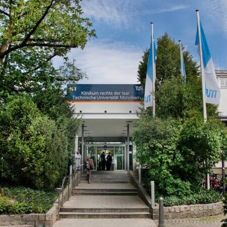
- University Hospital Rechts der Isar Munich, Germany
- Vivantes Neukölln Hospital Berlin, Germany
- University Hospital Ulm, Germany
You can find more information about the hospitals on the Booking Health website.
The cost of treatment abroad
The prices in hospitals listed on the Booking Health website are relatively low. With Booking Health, you can undergo platinum coils embolization (coiling) at an affordable price.
The cost of treatment varies, as the price depends on the hospital, the specifics of the disease, and the complexity of its treatment.
The cost of treatment with platinum coils embolization (coiling) in Germany is 17,215-34,111 EUR.
You might want to consider the cost of possible additional procedures and follow-up care. Therefore, the ultimate cost of treatment may differ from the initial price.
To make sure that the overall cost of treatment is suitable for you, contact us by leaving the request on the Booking Health website.
How can I undergo platinum coils embolization (coiling) abroad?
It is not easy to self-organize any treatment abroad. It requires certain knowledge and expertise. Thus, it is safer, easier, and less stressful to use the services of a medical tourism agency.
As the largest and most transparent medical tourism agency in the world, Booking Health has up-to-date information about platinum coils embolization (coiling) in the best hospitals. We will help you select the right clinic taking into account your wishes for treatment.
We want to help you and take on all the troubles. You can be free of unnecessary stress, while Booking Health takes care of all organizational issues regarding the treatment. Our services are aimed at undergoing platinum coils embolization (coiling) safely and successfully.
Medical tourism can be easy! All you need to do is to leave a request on the Booking Health website, and our manager will contact you shortly.

