Brain Aneurysm — Partial Resection and Coil Embolization: treatment in the Best Hospitals in the World
Treatment prices are regulated by national law of the corresponding countries, but can also include additional hospital coefficients. In order to receive the individual cost calculation, please send us the request and medical records.

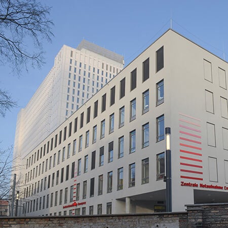
Department of Adult and Pediatric Neurosurgery, Spinal Surgery
The Department of Adult and Pediatric Neurosurgery, Spinal Surgery offers all the possibilities of modern surgical treatment for diseases of the central and peripheral nervous system in patients of all ages. More than 6,000 surgical procedures are performed annually in the department's high-tech operating rooms. Both planned and emergency neurosurgical procedures are performed here. The department's surgical team focuses on patients with cerebrovascular diseases, brain and skull base tumors, spine and spinal cord diseases, cerebrospinal fluid circulation disorders, and pathologies of the peripheral nervous system. The department's team of physicians also has extensive experience in functional neurosurgery: specialists perform deep brain stimulation for movement disorders, spinal cord stimulation for back pain, and vagus nerve stimulation for epilepsy. The department works closely with neurologists, radiologists, and nuclear medicine specialists to provide patients with the highest level of comprehensive medical care. The department is recognized as one of the top neurosurgical centers in Germany and beyond, as evidenced by consistently high treatment success rates and numerous quality certifications, including the German Cancer Society (DKG) Certificate, the German Spine Society (DWG) Certificate, and the Leading Medicine Guide Certificate.


Department of Neurosurgery and Spinal Surgery
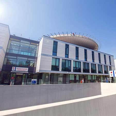
The Department of Neurosurgery and Spinal Surgery offers the full range of modern surgical procedures for patients with diseases of the brain, spinal cord, spine, and peripheral nervous system. The department has state-of-the-art equipment, including navigation systems, monitoring devices, surgical microscopes, intraoperative X-ray diagnostic systems, angiography systems, and laser equipment. In addition, a system for minimally invasive stereotactic interventions has been installed in the department's operating room. The department's neurosurgeons use the latest microsurgical and endoscopic techniques in their work. The medical facility is one of only three in Europe to successfully use the VISUALASE™ device for MRI-guided laser ablation to treat epilepsy. This minimally invasive procedure is highly effective and represents a real breakthrough in epilepsy surgery. The department's neurosurgeons always strive to provide the patient with optimal medical care. They talk in detail about each stage of treatment and are happy to answer the patient's questions. Neurologists and specialists in the field of neuroradiology are often involved in the therapeutic process, which guarantees a comprehensive approach to treatment.


Department of Adult and Pediatric Neurosurgery
The Department of Adult and Pediatric Neurosurgery offers the full range of surgical treatment of diseases of the brain, spine, spinal cord and nerves in adults and children. The department keeps pace with new trends in medicine, as well as contributes significantly to their development. Therefore, the most modern diagnostic and therapeutic methods are available here. An individual approach to each clinical case is crucial to ensure optimal treatment results with the preservation of all neurological functions.

A brain aneurysm (cerebral aneurysm, intracranial aneurysm) is a type of aneurysms in which one of the cerebral arteries is affected. The term "aneurysm" itself refers to the expansion of the arterial wall. The incidence of cerebral aneurysms among the general population is about 5%.
Content
- Overview
- Symptoms
- Diagnostics
- Treatment
- How the endovascular embolization is carried out?
- Where can I undergo partial resection and coil embolization treatment abroad?
- The cost of treatment abroad
- How can I undergo partial resection and coil embolization treatment abroad?
Overview
A brain aneurysm is a pathological local bulge of the artery wall.
Depending on the size of the aneurysm, there are few types of them:
- Regular-sized brain aneurysms (up to 15 mm)
- Large brain aneurysms (up to 25 mm)
- Gigantic brain aneurysms (more than 25 mm)
The larger the aneurysm is, the more it compresses the brain tissues and causes a more pronounced neurological picture. Protrusions can also be single or multiple, congenital, or acquired.
The disease often affects the vessels, inside which blood is flowing under high pressure. Such changes in the vessel wall lead to an increase in its size and abnormal pressure on the surrounding brain structures, which may disrupt the functions of the central nervous system.
However, a brain aneurysm is often detected only after an aneurysm rupture (in 50% of cases). If the aneurysm is unruptured, but simply increases in size, then it can manifest itself as a tumor (press on the surrounding brain tissues, causing certain neurological disorders). In terms of the outcomes of rupture of brain aneurysms, the statistics are quite harsh: 25% of patients die immediately or in a long-term period, 25% become severely disabled, 25% have a moderate disability, and only 25% do not have significant consequences for their health.
It is not clear why aneurysms occur in some people and not in others. In the first place among the causes of a cerebral aneurysm are congenital disorders of the structure of the vascular wall and atherosclerosis. Sometimes cerebral aneurysms occur as a result of inflammatory processes in the walls of an artery. This is usually due to blockage of the vessel lumen by an infected blood clot.
Brain aneurysms are dangerous for the patients due to the high risk of rupture of the aneurysm with hemorrhage into the brain tissue. Besides, the disease is often asymptomatic at the early stages of its development, which significantly complicates timely diagnosis making.
If a brain aneurysm is detected, it is necessary to immediately contact a neurosurgeon. The pathology is rather rare and specific, therefore not every neurosurgeon can provide a patient with the required help.
Symptoms
The danger of arterial aneurysms is in their asymptomatic and gradual development. At the early stages, the patient may not even be aware of a serious problem, since small aneurysms practically do not compress the brain tissue and do not cause discomfort.
The clinical picture manifests itself at the later stages of the disease. The first manifestations of a problem can include:
- Impaired vision, pain in the eyes and head
- Changes in the motor functions of the limbs – the patient can suddenly "forget" how to hold a spoon or his handwriting deteriorates
- Violation of tactile and pain sensitivity of the legs
- Seizures
- Numbness of the muscles of the face, inability to smile, sudden impairment of speech
These signs indicate compression of the brain tissue. A neurological deficit develops, which should become an alarm sign for seeking help from a neurologist.
As the disease progresses, the aneurysm can rupture. This condition is life threatening. The symptoms of rupture are:
- Intense headache that does not respond well to medications intake
- Nausea, vomiting
- Double vision (diplopia)
- Loss of consciousness
- Cramps
If a person experiences the described symptoms, an ambulance team should be called.
Identifying pathological conditions of the arteries of the brain is not always an easy task, especially if the patient does not feel any discomfort. In such cases, the diagnosis is suspected during preventive examinations.
Diagnostics
To identify an aneurysm, a neurologist may prescribe the following instrumental diagnostic procedures:
- Computed tomography (CT). The procedure is more often used to detect the already ruptured aneurysm with a formed hemorrhage in the brain tissue.
- Magnetic resonance imaging (MRI).
- Angiography of cerebral vessels. The method allows doctors to evaluate the size, localization, and structural features of the pathological protrusion.
If a ruptured aneurysm is suspected, the neurologist also performs a lumbar puncture to assess the condition of the cerebrospinal fluid.
Treatment
Surgery is the only effective treatment option for cerebral aneurysm. Having received the results of the patient's examination, the surgeon decides on the appropriateness of the surgical intervention or other kind of treatment. Since the consequences of a ruptured cerebral aneurysm, unlike an unruptured aneurysm, are lethal, surgical intervention can save the patient's life.
There are two methods of brain aneurysms treatments: surgical clipping of the aneurysm neck (surgery with opening the skull) or neuroradiological surgery that does not require opening the skull – endovascular embolization. Endovascular embolization includes filling the aneurysm with coils through a catheter, which is inserted through the femoral artery in the groin. The decision on the method of treatment is made by the medical council together with the patient. The council includes a neurosurgeon and an interventional radiologist. When choosing one of the two methods described above, the medical team takes into account many factors, such as the location of the aneurysm, its size, shape, width, and the conditions of adjacent vessels.
Endovascular treatment (performed by an interventional neuroradiologist) consists of diminishing the blood supply of the aneurysm by filling its lumen with special coils (coils are made of platinum). A cerebral aneurysm is approached with a very thin catheter, which is inserted through a small incision in the groin and then moved through the blood vessels. All this time, the location of the catheter is monitored using a special X-ray machine. This method is based on an advanced technique designed for minimally invasive closure of cerebral vascular defects, including aneurysms.
Endovascular embolization with coils is one of the most frequently performed in the world operations for the treatment of cerebral aneurysms. As with clipping, the purpose of the operation is to exclude an aneurysm from the general blood flow, which is necessary to prevent rupture.
Embolization of an aneurysm is one of the most common methods of treating disease. In most cases, endovascular embolization is the least invasive method aneurysms are treated with today. If it is impossible to carry out endovascular embolization or stenting, the usual surgical operation of vessel stenting or aneurysm clipping can be performed.
How the endovascular embolization is carried out?
Firstly, the surgeon and the patient discuss the possible risks and outcomes of treatment and choose the appropriate treatment method. The size of the aneurysm is extremely important for assessing prognosis and possible outcomes when deciding on a treatment scheme. The risk of rupture of an aneurysm with a diameter of up to 10 mm is estimated at 0-4% and, accordingly, is higher for bigger aneurysms. Also, it should be remembered that their size is not constant – aneurysms can increase, therefore, when choosing a conservative treatment, it is recommended to perform control imaging studies to assess the size of the aneurysm.
The operation is performed with access through the femoral artery – only skin puncture in the groin area is required. Along the blood vessels, catheters are sequentially moved into the vessels of the neck, cerebral arteries, and directly into the aneurysm cavity.
Embolization of an aneurysm results in the cessation of blood flow in the aneurysm cavity, maintaining normal blood flow in the cerebral artery. In the process of embolization, a catheter is inserted through the convenient vascular access (usually the inguinal one) under radiological control and moved up to the aneurysm. Then a thinner micro-catheter with a micro-wire coil inside it is inserted into the catheter and moved into the aneurysm cavity.
Through the catheter, platinum coils are inserted into the aneurysm. A coil fills the aneurysm cavity and blocks the flow of blood into it. A low voltage electric current is used to separate the micro spirals from the carrier conductor.
As soon as the tip of the catheter reaches the aneurysm cavity, coils are released from the catheter. A coil changes its shape and fills the aneurysm cavity. For large aneurysms, several coils may be needed. An aneurysm filled with a wire spiral is turned off from the systemic circulation and is gradually substituted with connective tissue, that excludes the risk of its rupture. As the blood flow in the aneurysm stops, the aneurysm is completely thrombosed, and it no longer poses a health threat.
The postoperative period after embolization is minimal. In the absence of hemorrhage or other undesirable conditions, a patient is discharged from the hospital in 2-3 days after the surgical intervention.
Where can I undergo partial resection and coil embolization treatment abroad?
Health tourism is becoming more and more popular these days, as medicine abroad often ensures a much better quality of partial resection and coil embolization treatment.
The following hospitals show the best success rates in partial resection and coil embolization treatment:
- Charite University Hospital Berlin, Germany
- Memorial Bahcelievler Hospital Istanbul, Turkey
- University Hospital HM Monteprincipe Madrid, Spain
- Beta Klinik Bonn, Germany
- University Hospital for Neurosurgery Salzburg, Austria
You can find more information about the hospitals on the Booking Health website.
The cost of treatment abroad
The prices in hospitals listed on the Booking Health website are relatively low. With Booking Health, you can undergo partial resection and coil embolization treatment at an affordable price.
The cost of treatment varies, as the price depends on the hospital, the specifics of the disease, and the complexity of its treatment.
The cost of treatment with partial resection and coil embolization in Germany is 22,087-38,216 EUR.
The cost of treatment with partial resection and coil embolization in Turkey is 16,096-19,867 EUR.
The cost of treatment with partial resection and coil embolization in Spain is approximately 13,285 EUR.
The cost of treatment with partial resection and coil embolization in Austria is approximately 28,087 EUR.
You might want to consider the cost of possible additional procedures and follow-up care. Therefore, the ultimate cost of treatment may differ from the initial price.
To make sure that the overall cost of treatment is suitable for you, contact us by leaving the request on the Booking Health website.
How can I undergo partial resection and coil embolization treatment abroad?
It is not easy to self-organize any treatment abroad. It requires certain knowledge and expertise. Thus, it is safer, easier, and less stressful to use the services of a medical tourism agency.
As the largest and most transparent medical tourism agency in the world, Booking Health has up-to-date information about partial resection and coil embolization treatment in the best hospitals. We will help you select the right clinic taking into account your wishes for treatment.
We want to help you and take on all the troubles. You can be free of unnecessary stress, while Booking Health takes care of all organizational issues regarding the treatment. Our services are aimed at undergoing partial resection and coil embolization treatment safely and successfully.
Medical tourism can be easy! All you need to do is to leave a request on the Booking Health website, and our manager will contact you shortly.
Authors:
The article was edited by medical experts, board certified doctors Dr. Vadim Zhiliuk and Dr. Sergey Pashchenko. For the treatment of the conditions referred to in the article, you must consult a doctor; the information in the article is not intended for self-medication!
Sources:

