{"translation_price":"50","translation_doc_price":"40","child_coefficient":"1.1","transfer_price":"2.00","transfer_price_vip":"5.00","constant_transfer_price_vip":350,"constant_transfer_price":150,"constant_transfer_distanse":60,"type":"treatment","program_full_story":"<ul>\n\t<li>Initial presentation in the hospital<\/li>\n\t<li>Clinical history taking<\/li>\n\t<li>Review of available medical records<\/li>\n\t<li>Physical examination<\/li>\n\t<li>Laboratory tests:\n\t<ul>\n\t\t<li>Complete blood count<\/li>\n\t\t<li>General urine analysis<\/li>\n\t\t<li>Biochemical analysis of blood<\/li>\n\t\t<li>Tumor markers<\/li>\n\t\t<li>Inflammation indicators (CRP, ESR)<\/li>\n\t\t<li>Coagulogram<\/li>\n\t<\/ul>\n\t<\/li>\n\t<li>Ultrasound\u200b scan<\/li>\n\t<li>CT scan \/ MRI<\/li>\n\t<li>Preoperative care<\/li>\n\t<li>Embolization or chemoembolization, 2 procedures<\/li>\n\t<li>Symptomatic treatment<\/li>\n\t<li>Cost of essential medicines<\/li>\n\t<li>Nursing services<\/li>\n\t<li>Elaboration of further recommendations<\/li>\n<\/ul>\n<div class=\"program_how_program_going mt-4\"><h4>How program is carried out<\/h4><p style=\"text-align: justify;\"><strong>During the first visit<\/strong>, the doctor will conduct a clinical examination and go through the results of the available diagnostic tests. After that, you will undergo the necessary additional examination, such as the assessment of liver and kidney function, ultrasound scan, CT scan and MRI. This will allow the doctor to determine which vessels are feeding the tumor and its metastases, as well as determine how well you will tolerate the procedure.<\/p>\n\n<p style=\"text-align: justify;\"><strong>Chemoembolization <\/strong>begins with local anesthesia and catheterization of the femoral artery. The thin catheter is inserted through a few centimeters long incision of the blood vessel. The doctor gradually moves the catheter to the vessel feeding the primary tumor or its metastases. The procedure is carried out under visual control, an angiographic device is used for this. The vascular bed and the position of the catheter in it are displayed on the screen of the angiograph.<\/p>\n\n<p style=\"text-align: justify;\">When the catheter reaches a suspected artery, a contrast agent is injected through it. Due to the introduction of the contrast agent, the doctor clearly sees the smallest vessels of the tumor and the surrounding healthy tissues on the screen of the angiograph. After that, he injects emboli into the tumor vessels through the same catheter.<\/p>\n\n<p style=\"text-align: justify;\">Emboli are the spirals or the liquid microspheres. The type of embolus is selected individually, taking into account the diameter of the target vessel. When carrying out chemoembolization, a solution of a chemotherapy drug is additionally injected into the tumor vessel. Due to the subsequent closure of the vessel lumen with an embolus, the chemotherapy drug influences the tumor for a long time. In addition, the drug does not enter the systemic circulation, which allows doctors to use high doses of chemotherapeutic agents without the development of serious side effects. Chemoembolization leads to the destruction of the tumor or slowing down its progression.<\/p>\n\n<p style=\"text-align: justify;\">After that, the catheter is removed from the artery. The doctor puts a vascular suture on the femoral artery and closes it with a sterile dressing. During chemoembolization, you will be awake. General anesthesia is not used, which significantly reduces the risks of the procedure and allows performing it on an outpatient basis, avoiding long hospital stay.<\/p>\n\n<p style=\"text-align: justify;\"><strong>After the first procedure<\/strong>, you will stay under the supervision of an interventional oncologist and general practitioner. If necessary, you will receive symptomatic treatment. As a rule, a second chemoembolization procedure is performed in 3-5 days after the first one in order to consolidate the therapeutic effect. After that, you will receive recommendations for further follow-up and treatment.<\/p>\n<\/div><div class=\"program_required_documents mt-4\"><h4>Required documents<\/h4><ul>\n\t<li style=\"text-align: justify;\">Medical records<\/li>\n\t<li style=\"text-align: justify;\">MRI\/CT scan (not older than 3 months)<\/li>\n\t<li style=\"text-align: justify;\">Biopsy results (if available)<\/li>\n<\/ul>\n<\/div>","program_full_story_crm":"<ul>\n\t<li>Initial presentation in the hospital<\/li>\n\t<li>Clinical history taking<\/li>\n\t<li>Review of available medical records<\/li>\n\t<li>Physical examination<\/li>\n\t<li>Laboratory tests:\n\t<ul>\n\t\t<li>Complete blood count<\/li>\n\t\t<li>General urine analysis<\/li>\n\t\t<li>Biochemical analysis of blood<\/li>\n\t\t<li>Tumor markers<\/li>\n\t\t<li>Inflammation indicators (CRP, ESR)<\/li>\n\t\t<li>Coagulogram<\/li>\n\t<\/ul>\n\t<\/li>\n\t<li>Ultrasound\u200b scan<\/li>\n\t<li>CT scan \/ MRI<\/li>\n\t<li>Preoperative care<\/li>\n\t<li>Embolization or chemoembolization, 2 procedures<\/li>\n\t<li>Symptomatic treatment<\/li>\n\t<li>Cost of essential medicines<\/li>\n\t<li>Nursing services<\/li>\n\t<li>Elaboration of further recommendations<\/li>\n<\/ul>\n<div class=\"program_how_program_going mt-4\"><h4>How program is carried out<\/h4><p style=\"text-align: justify;\"><strong>During the first visit<\/strong>, the doctor will conduct a clinical examination and go through the results of the available diagnostic tests. After that, you will undergo the necessary additional examination, such as the assessment of liver and kidney function, ultrasound scan, CT scan and MRI. This will allow the doctor to determine which vessels are feeding the tumor and its metastases, as well as determine how well you will tolerate the procedure.<\/p>\n\n<p style=\"text-align: justify;\"><strong>Chemoembolization <\/strong>begins with local anesthesia and catheterization of the femoral artery. The thin catheter is inserted through a few centimeters long incision of the blood vessel. The doctor gradually moves the catheter to the vessel feeding the primary tumor or its metastases. The procedure is carried out under visual control, an angiographic device is used for this. The vascular bed and the position of the catheter in it are displayed on the screen of the angiograph.<\/p>\n\n<p style=\"text-align: justify;\">When the catheter reaches a suspected artery, a contrast agent is injected through it. Due to the introduction of the contrast agent, the doctor clearly sees the smallest vessels of the tumor and the surrounding healthy tissues on the screen of the angiograph. After that, he injects emboli into the tumor vessels through the same catheter.<\/p>\n\n<p style=\"text-align: justify;\">Emboli are the spirals or the liquid microspheres. The type of embolus is selected individually, taking into account the diameter of the target vessel. When carrying out chemoembolization, a solution of a chemotherapy drug is additionally injected into the tumor vessel. Due to the subsequent closure of the vessel lumen with an embolus, the chemotherapy drug influences the tumor for a long time. In addition, the drug does not enter the systemic circulation, which allows doctors to use high doses of chemotherapeutic agents without the development of serious side effects. Chemoembolization leads to the destruction of the tumor or slowing down its progression.<\/p>\n\n<p style=\"text-align: justify;\">After that, the catheter is removed from the artery. The doctor puts a vascular suture on the femoral artery and closes it with a sterile dressing. During chemoembolization, you will be awake. General anesthesia is not used, which significantly reduces the risks of the procedure and allows performing it on an outpatient basis, avoiding long hospital stay.<\/p>\n\n<p style=\"text-align: justify;\"><strong>After the first procedure<\/strong>, you will stay under the supervision of an interventional oncologist and general practitioner. If necessary, you will receive symptomatic treatment. As a rule, a second chemoembolization procedure is performed in 3-5 days after the first one in order to consolidate the therapeutic effect. After that, you will receive recommendations for further follow-up and treatment.<\/p>\n<\/div>","is_ambulant":"1","bh_fee":"0","only_for_children":"0","no_service":"0","with_prepayment":"1","show_calculator":"1","paket_type":"1","btn_type":"0","clinic_icon":"6802a209368be.jpg","city":"Aachen","clinic_site":"http:\/\/www.ukaachen.de\/","department_recommend":"0","country":"Germany","country_id":"1","clinic_name":"University Hospital RWTH Aachen","cinic_name":"University Hospital RWTH Aachen","department_id":"2927","duration":"7","direction":"Interventional radiology","min_duration":0,"clinic_id":"252","paketPrice":7800,"paket":"<ul>\n <li>Interpreter up to 17 hours<\/li>\n <li>Translation up to 10 pages<\/li>\n <li>Visa support<\/li>\n\n \n<\/ul>","title":"Treatment of cholangiocarcinoma (Klatskin tumor) with embolization or chemoembolization","price":{"val":31354.73,"type":"val"},"price_surcharge":0,"price_surcharge_clear":0,"extra_service_clinic":[],"extra_service":[],"translation_hours":"0","translation_doc_count":null,"roads":[{"id":"7","distance":"88","airport_title":"Cologne-Bonn"},{"id":"4","distance":"101","airport_title":"Duesseldorf"},{"id":"6","distance":"250","airport_title":"Frankfurt a.M."}],"pakets":[],"lang":{"day":"Day","days":"days","ambulatory":"Outpatient","stationaryProgram":"Inpatient"}}
Treatment of cholangiocarcinoma (Klatskin tumor) with embolization or chemoembolization in University Hospital RWTH Aachen
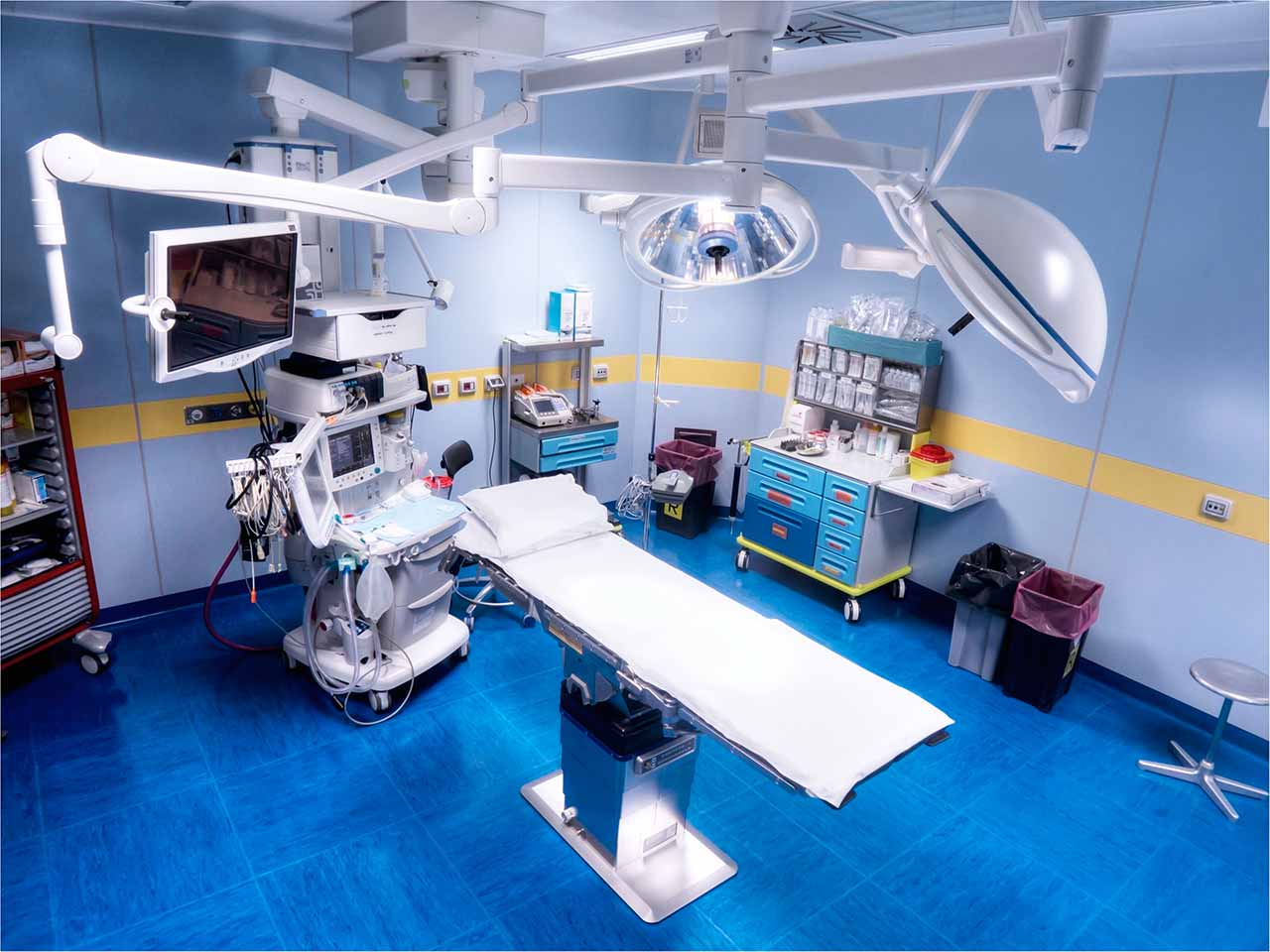
During the first visit, the doctor will conduct a clinical examination and go through the results of the available diagnostic tests. After that, you will undergo the necessary additional examination, such as the assessment of liver and kidney function, ultrasound scan, CT scan and MRI. This will allow the doctor to determine which vessels are feeding the tumor and its metastases, as well as determine how well you will tolerate the procedure.
Chemoembolization begins with local anesthesia and catheterization of the femoral artery. The thin catheter is inserted through a few centimeters long incision of the blood vessel. The doctor gradually moves the catheter to the vessel feeding the primary tumor or its metastases. The procedure is carried out under visual control, an angiographic device is used for this. The vascular bed and the position of the catheter in it are displayed on the screen of the angiograph.
When the catheter reaches a suspected artery, a contrast agent is injected through it. Due to the introduction of the contrast agent, the doctor clearly sees the smallest vessels of the tumor and the surrounding healthy tissues on the screen of the angiograph. After that, he injects emboli into the tumor vessels through the same catheter.
Emboli are the spirals or the liquid microspheres. The type of embolus is selected individually, taking into account the diameter of the target vessel. When carrying out chemoembolization, a solution of a chemotherapy drug is additionally injected into the tumor vessel. Due to the subsequent closure of the vessel lumen with an embolus, the chemotherapy drug influences the tumor for a long time. In addition, the drug does not enter the systemic circulation, which allows doctors to use high doses of chemotherapeutic agents without the development of serious side effects. Chemoembolization leads to the destruction of the tumor or slowing down its progression.
After that, the catheter is removed from the artery. The doctor puts a vascular suture on the femoral artery and closes it with a sterile dressing. During chemoembolization, you will be awake. General anesthesia is not used, which significantly reduces the risks of the procedure and allows performing it on an outpatient basis, avoiding long hospital stay.
After the first procedure, you will stay under the supervision of an interventional oncologist and general practitioner. If necessary, you will receive symptomatic treatment. As a rule, a second chemoembolization procedure is performed in 3-5 days after the first one in order to consolidate the therapeutic effect. After that, you will receive recommendations for further follow-up and treatment.
Required documents
Medical records
MRI/CT scan (not older than 3 months)
Biopsy results (if available)
Service
Price from:
Type of program :
Price for 1 day:
Expected duration of the program:
The minimum duration of the program:
Select
You may also book:
Hospital directly:
Saving:
BookingHealth Price from:
About the department
The Department of Adult and Pediatric Diagnostic, Interventional Radiology at the University Hospital RWTH Aachen offers the full range of services in these areas. Patient care is provided both on an outpatient and inpatient basis. In clinical practice, doctors apply the very latest methods of radiological diagnosis, as well as perform a variety of imaging-guided minimally invasive interventions. For these purposes, the department is equipped with state-of-the-art digital X-ray and ultrasound systems, CT and MRI scanners. The department is headed by Prof. Dr. med. Christiane Kuhl.
The service range of the department includes:
Diagnostic radiology
Conventional X-ray studies
Fluoroscopy
Digital subtraction angiography
Ultrasound examinations
Computed tomography (CT) and PET-CT
Magnetic resonance imaging (MRI)
Imaging-guided tissue sampling (biopsy) for histological examination
Installation of port systems and catheters for chemotherapy
Treatment of liver cancer and liver metastases
Transarterial chemoembolization
Selective internal radiation therapy
Radiofrequency ablation
Microwave ablation
Irreversible electroporation
Kidney cancer treatment
Radiofrequency ablation
Irreversible electroporation
Breast cancer treatment
Transarterial chemoembolization
Selective internal radiation therapy
Radiofrequency ablation
Irreversible electroporation for the treatment of liver metastases in breast cancer
Chemoperfusion for the treatment of inoperable breast cancer localized in the thorax
Treatment of lung cancer and lung metastases
Radiofrequency ablation
Microwave ablation
Treatment of benign diseases
Uterine artery embolization in uterine myomas
Radiofrequency ablation for the treatment of osteoid osteomas
Treatment of vascular tumors (arteriovenous malformations)
Treatment of vascular diseases
Treatment of peripheral arterial occlusive disease
Elimination of complications after the formation of a dialysis shunt, including stenosis of the venous vessels
Treatment of vascular pathologies of the abdominal organs and thorax
Embolization in acute bleeding
Varicocele treatment
Treatment of liver diseases
Treatment of portal hypertension and its complications (for example, ascites) with the use of transicular intrahepatic portosystemic shunts (TIPS)
Percutaneous transhepatic gallbladder drainage
Implantation of cava filters
Other minimally invasive procedures
Percutaneous endoscopic gastrostomy
Other imaging-guided interventional procedures
Curriculum vitae
1985 - 1991 Study of Human Medicine at the Rhenish Friedrich Wilhelm University of Bonn.
Professional Career
Since 2010, Head of the Department of Adult and Pediatric Diagnostic, Interventional Radiology, University Hospital RWTH Aachen.
2012 ‐ 2013 President of the Radiological Society of North Rhine-Westphalia.
2009 Invitation to the position of Full Professor and Head of the Department of Adult and Pediatric Diagnostic, Interventional Radiology, University Hospital RWTH Aachen.
2009 - 2010 Vice President, Rhenish Friedrich Wilhelm University of Bonn, International Affairs.
2004 - 2010 Deputy Head, Department of Radiology, University of Bonn.
2004 Full Professor of Radiology, Head in the Section for Diagnostic Imaging in Oncology and Interventional Oncology, University of Bonn, Department of Radiology.
1999 - 2004 Assistant Professor in Radiology, Department of Radiology, University of Bonn.
Clinical Training
2010 DEGIR certificate, stage 4, German Society of Interventional Radiology (license to conduct and teach the entire range of interventional procedures).
2002 Board certification in Neuroradiology.
2001 - 2002 Clinical training in Neuroradiological Interventions at the Department of Neuroradiology, Duisburg Hospital, Germany.
1999 Board certification in Radiology.
1996 - 1997 Residency in Neurosurgery, Department of Neurosurgery, University of Bonn.
1992 - 1999 Residency in Radiology, Department of Radiology, University of Bonn.
01.07.1993 Admission to medical practice.
01.01.1992 Temporary license for medical practice.
Membership in Professional Societies
German Society of Radiology (DRG).
Radiological Society of North America (RSNA).
American College of Radiology (ACR).
International Society of Magnetic Resonance Imaging in Medicine (ISMRM).
Society of Cardiovascular and Interventional Radiology (CIRSE).
German Society of Interventional Radiology (DEGIR).
European Society of Breast Imaging (EUSOBI).
American Society of Clinical Oncology (ASCO).
Cardiovascular and Interventional Radiology (CIRSE).
Society of Interventional Radiology (SIR).
World Conference on Interventional Oncology (WCIO).
Awards
2018 Impact Award of the National Consortium of Breast Centers (NCoBC), USA.
2015 RWTH Fellow.
2015 Gold Medal of the International Society of Magnetic Resonance Imaging in Medicine.
2008 Wachsmann Prize (Award for Educational Excellence) of the German Society of Radiology.
2007 Outstanding Teacher Award, Society for Magnetic Resonance Imaging in Medicine.
2004, 2006, 2008 "Best of ASCO", American Society of Clinical Oncology.
2006 Honorary Fellow, Breast Imaging Section, Royal College of Radiologist, UK.
2004 Award of the American College of Radiology Imaging Network (ACRIN).
2003 Award of the German Society of Radiology (Holthusen Ring).
2000 Award of the Ministery of Education and Research (BMBF).
2000 Award of the German Society of Radiology.
Peer Reviewer
RöFo.
European Radiology.
Journal of Magnetic Resonance Imaging (JMRI).
Magnetic Resonance Imaging (MRM).
The Breast.
Cancer.
Radiology.
Journal of the National Cancer Institute (JNCI).
Journal of Clinical Oncology (JCO).
NATURE (New Technology Section).
The Lancet.
Journal of the American Medical Association (JAMA).
Photo of the doctor: (c) Uniklinik RWTH Aachen
About hospital
According to the prestigious Focus magazine, the University Hospital RWTH Aachen ranks among the top German hospitals!
As a maximum care university medical facility, the hospital guarantees patients first-class medical services combined with a respectful and human attitude. The hospital integrates all the modern options for the accurate diagnostics, effective therapy and productive research activities within one specialized center.
The hospital has more than 60 departments, institutes and interdisciplinary centers. A competent team of professionals, consisting of more than 7,000 employees (more than 940 doctors, including about 95 professors), takes care of the patients' health. In addition, multidisciplinary teams of nurses, physiotherapists, as well as medical and technical staff are available here. The patients and respectful attitude to their social, cultural and religious affiliations are at the center of all employees' efforts.
The cornerstone of the successful clinical practice is the innovative technical base. The hospital offers the most advanced diagnostic and therapeutic equipment. Thus, the hospital has all the resources in order to provide top-class medical services.
Photo: (с) depositphotos
Accommodation in hospital
Patients rooms
The patients of the University Hospital RWTH Aachen live in comfortable single and double rooms. All patient rooms are designed with large windows, so one can enjoy a beautiful landscape view. Each patient room has an ensuite bathroom. The standard room furnishing includes an automatically adjustable bed, a bedside table, a spacious wardrobe, a TV, a radio and a telephone. Also, there is Wi-Fi access.
Meals and Menus
The patient and his accompanying person have a daily choice of three menus. If for any reason you do not eat all the foods, you will be offered an individual menu. Please inform the medical staff about your dietary preferences prior to the treatment.
Further details
Standard rooms include:
Toilet
Shower
Wi-Fi
TV
Religion
Religious services are available upon request.
Accompanying person
During the inpatient program, an accompanying person may stay with you in a patient room or at the hotel of your choice.
Hotel
During the outpatient program, you can live at a hotel of your choice. Managers will help you to choose the most suitable options.
The hospital offers a full range of laboratory tests (general, hormonal, tests for infections, antibodies, tumor markers, etc.), genetic tests, various modifications of ultrasound scans, CT scans, MRI and PET / CT, angiography, myelography, biopsy and other examinations. Treatment with medications, endoscopic and robotic operations, stereotaxic interventions is carried out here, modern types of radiation therapy are also used. The hospital offers patients all the necessary therapeutic techniques.
These are pathologies of the brain and spinal cord, benign and malignant tumors of various localizations, congenital and acquired heart defects, joint pathologies, stroke, neurodegenerative diseases, eye injuries, inflammatory skin diseases, allergies and other pathologies.
thank you for reaching out! One of our medical advisors is currently reviewing your details. You can expect a call from us within one business day to discuss your request.
Note: The incoming call will appear as a German number or your local area code. This call is entirely free of charge.
Booking Health guarantees:
Expert Placement
We use rigorous statistical analysis to ensure you are treated at the best-rated facility for your specific diagnosis.
Total Cost Protection
No hidden fees or unexpected bills. We provide a fixed price guarantee, backed by insurance that covers any additional complications.
12-Month Extended Care
Your recovery is our priority. Benefit from a full year of direct medical support following your procedure.


 BookingHealth Price from:
