{"translation_price":"50","translation_doc_price":"40","child_coefficient":"1.1","transfer_price":"2.00","transfer_price_vip":"5.00","constant_transfer_price_vip":350,"constant_transfer_price":150,"constant_transfer_distanse":60,"type":"treatment","program_full_story":"<ul>\n\t<li>Initial presentation in the hospital<\/li>\n\t<li>Clinical history taking<\/li>\n\t<li>Review of available medical records<\/li>\n\t<li>Physical examination<\/li>\n\t<li>Laboratory tests:\n\t<ul>\n\t\t<li>Complete blood count<\/li>\n\t\t<li>General urine analysis<\/li>\n\t\t<li>Biochemical analysis of blood<\/li>\n\t\t<li>Tumor markers<\/li>\n\t\t<li>Inflammation indicators (CRP, ESR)<\/li>\n\t\t<li>Coagulogram<\/li>\n\t<\/ul>\n\t<\/li>\n\t<li>Ultrasound\u200b scan<\/li>\n\t<li>CT scan \/ MRI<\/li>\n\t<li>Preoperative care<\/li>\n\t<li>Embolization or chemoembolization, 2 procedures<\/li>\n\t<li>Symptomatic treatment<\/li>\n\t<li>Cost of essential medicines<\/li>\n\t<li>Nursing services<\/li>\n\t<li>Elaboration of further recommendations<\/li>\n<\/ul>\n<div class=\"program_how_program_going mt-4\"><h4>How program is carried out<\/h4><p style=\"text-align:justify\"><strong>During the first visit<\/strong>, the doctor will conduct a clinical examination and go through the results of the available diagnostic tests. After that, you will undergo the necessary additional examination, such as the assessment of liver and kidney function, ultrasound scan, CT scan and MRI. This will allow the doctor to determine which vessels are feeding the tumor and its metastases, as well as determine how well you will tolerate the procedure.<\/p>\n\n<p style=\"text-align:justify\"><strong>Chemoembolization <\/strong>begins with local anesthesia and catheterization of the femoral artery. The thin catheter is inserted through a few centimeters long incision of the blood vessel. The doctor gradually moves the catheter to the vessel feeding the primary tumor or its metastases. The procedure is carried out under visual control, an angiographic device is used for this. The vascular bed and the position of the catheter in it are displayed on the screen of the angiograph.<\/p>\n\n<p style=\"text-align:justify\">When the catheter reaches a suspected artery, a contrast agent is injected through it. Due to the introduction of the contrast agent, the doctor clearly sees the smallest vessels of the tumor and the surrounding healthy tissues on the screen of the angiograph. After that, he injects emboli into the tumor vessels through the same catheter.<\/p>\n\n<p style=\"text-align:justify\">Emboli are the spirals or the liquid microspheres. The type of embolus is selected individually, taking into account the diameter of the target vessel. When carrying out chemoembolization, a solution of a chemotherapy drug is additionally injected into the tumor vessel. Due to the subsequent closure of the vessel lumen with an embolus, the chemotherapy drug influences the tumor for a long time. In addition, the drug does not enter the systemic circulation, which allows doctors to use high doses of chemotherapeutic agents without the development of serious side effects. Chemoembolization leads to the destruction of the tumor or slowing down its progression.<\/p>\n\n<p style=\"text-align:justify\">After that, the catheter is removed from the artery. The doctor puts a vascular suture on the femoral artery and closes it with a sterile dressing. During chemoembolization, you will be awake. General anesthesia is not used, which significantly reduces the risks of the procedure and allows performing it on an outpatient basis, avoiding long hospital stay.<\/p>\n\n<p style=\"text-align:justify\"><strong>After the first procedure<\/strong>, you will stay under the supervision of an interventional oncologist and general practitioner. If necessary, you will receive symptomatic treatment. As a rule, a second chemoembolization procedure is performed in 3-5 days after the first one in order to consolidate the therapeutic effect. After that, you will receive recommendations for further follow-up and treatment.<\/p>\n<\/div>","program_full_story_crm":"<ul>\n\t<li>Initial presentation in the hospital<\/li>\n\t<li>Clinical history taking<\/li>\n\t<li>Review of available medical records<\/li>\n\t<li>Physical examination<\/li>\n\t<li>Laboratory tests:\n\t<ul>\n\t\t<li>Complete blood count<\/li>\n\t\t<li>General urine analysis<\/li>\n\t\t<li>Biochemical analysis of blood<\/li>\n\t\t<li>Tumor markers<\/li>\n\t\t<li>Inflammation indicators (CRP, ESR)<\/li>\n\t\t<li>Coagulogram<\/li>\n\t<\/ul>\n\t<\/li>\n\t<li>Ultrasound\u200b scan<\/li>\n\t<li>CT scan \/ MRI<\/li>\n\t<li>Preoperative care<\/li>\n\t<li>Embolization or chemoembolization, 2 procedures<\/li>\n\t<li>Symptomatic treatment<\/li>\n\t<li>Cost of essential medicines<\/li>\n\t<li>Nursing services<\/li>\n\t<li>Elaboration of further recommendations<\/li>\n<\/ul>\n<div class=\"program_how_program_going mt-4\"><h4>How program is carried out<\/h4><p style=\"text-align:justify\"><strong>During the first visit<\/strong>, the doctor will conduct a clinical examination and go through the results of the available diagnostic tests. After that, you will undergo the necessary additional examination, such as the assessment of liver and kidney function, ultrasound scan, CT scan and MRI. This will allow the doctor to determine which vessels are feeding the tumor and its metastases, as well as determine how well you will tolerate the procedure.<\/p>\n\n<p style=\"text-align:justify\"><strong>Chemoembolization <\/strong>begins with local anesthesia and catheterization of the femoral artery. The thin catheter is inserted through a few centimeters long incision of the blood vessel. The doctor gradually moves the catheter to the vessel feeding the primary tumor or its metastases. The procedure is carried out under visual control, an angiographic device is used for this. The vascular bed and the position of the catheter in it are displayed on the screen of the angiograph.<\/p>\n\n<p style=\"text-align:justify\">When the catheter reaches a suspected artery, a contrast agent is injected through it. Due to the introduction of the contrast agent, the doctor clearly sees the smallest vessels of the tumor and the surrounding healthy tissues on the screen of the angiograph. After that, he injects emboli into the tumor vessels through the same catheter.<\/p>\n\n<p style=\"text-align:justify\">Emboli are the spirals or the liquid microspheres. The type of embolus is selected individually, taking into account the diameter of the target vessel. When carrying out chemoembolization, a solution of a chemotherapy drug is additionally injected into the tumor vessel. Due to the subsequent closure of the vessel lumen with an embolus, the chemotherapy drug influences the tumor for a long time. In addition, the drug does not enter the systemic circulation, which allows doctors to use high doses of chemotherapeutic agents without the development of serious side effects. Chemoembolization leads to the destruction of the tumor or slowing down its progression.<\/p>\n\n<p style=\"text-align:justify\">After that, the catheter is removed from the artery. The doctor puts a vascular suture on the femoral artery and closes it with a sterile dressing. During chemoembolization, you will be awake. General anesthesia is not used, which significantly reduces the risks of the procedure and allows performing it on an outpatient basis, avoiding long hospital stay.<\/p>\n\n<p style=\"text-align:justify\"><strong>After the first procedure<\/strong>, you will stay under the supervision of an interventional oncologist and general practitioner. If necessary, you will receive symptomatic treatment. As a rule, a second chemoembolization procedure is performed in 3-5 days after the first one in order to consolidate the therapeutic effect. After that, you will receive recommendations for further follow-up and treatment.<\/p>\n<\/div>","is_ambulant":"1","bh_fee":"0","only_for_children":"0","no_service":"0","with_prepayment":"1","show_calculator":"1","paket_type":"1","btn_type":"0","clinic_icon":"6000307acc78a.jpg","city":"Erlangen","clinic_site":"http:\/\/www.uk-erlangen.de\/","department_recommend":"1","country":"Germany","country_id":"1","clinic_name":"University Hospital Erlangen","cinic_name":"University Hospital Erlangen","department_id":"1535","duration":"7","direction":"Interventional radiology","min_duration":0,"clinic_id":"933","paketPrice":7800,"paket":"<ul>\n <li>Interpreter up to 17 hours<\/li>\n <li>Translation up to 10 pages<\/li>\n <li>Visa support<\/li>\n\n \n<\/ul>","title":"Treatment of bladder cancer with embolization or chemoembolization","price":{"val":29304.31,"type":"val"},"price_surcharge":0,"price_surcharge_clear":0,"extra_service_clinic":[],"extra_service":[],"translation_hours":"0","translation_doc_count":null,"roads":[{"id":"27","distance":"14","airport_title":"N\u00fcrnberg"},{"id":"2","distance":"183","airport_title":"Munich"},{"id":"6","distance":"209","airport_title":"Frankfurt a.M."}],"pakets":[],"lang":{"day":"Day","days":"days","ambulatory":"Outpatient","stationaryProgram":"Inpatient"}}
Treatment of bladder cancer with embolization or chemoembolization in University Hospital Erlangen
During the first visit, the doctor will conduct a clinical examination and go through the results of the available diagnostic tests. After that, you will undergo the necessary additional examination, such as the assessment of liver and kidney function, ultrasound scan, CT scan and MRI. This will allow the doctor to determine which vessels are feeding the tumor and its metastases, as well as determine how well you will tolerate the procedure.
Chemoembolization begins with local anesthesia and catheterization of the femoral artery. The thin catheter is inserted through a few centimeters long incision of the blood vessel. The doctor gradually moves the catheter to the vessel feeding the primary tumor or its metastases. The procedure is carried out under visual control, an angiographic device is used for this. The vascular bed and the position of the catheter in it are displayed on the screen of the angiograph.
When the catheter reaches a suspected artery, a contrast agent is injected through it. Due to the introduction of the contrast agent, the doctor clearly sees the smallest vessels of the tumor and the surrounding healthy tissues on the screen of the angiograph. After that, he injects emboli into the tumor vessels through the same catheter.
Emboli are the spirals or the liquid microspheres. The type of embolus is selected individually, taking into account the diameter of the target vessel. When carrying out chemoembolization, a solution of a chemotherapy drug is additionally injected into the tumor vessel. Due to the subsequent closure of the vessel lumen with an embolus, the chemotherapy drug influences the tumor for a long time. In addition, the drug does not enter the systemic circulation, which allows doctors to use high doses of chemotherapeutic agents without the development of serious side effects. Chemoembolization leads to the destruction of the tumor or slowing down its progression.
After that, the catheter is removed from the artery. The doctor puts a vascular suture on the femoral artery and closes it with a sterile dressing. During chemoembolization, you will be awake. General anesthesia is not used, which significantly reduces the risks of the procedure and allows performing it on an outpatient basis, avoiding long hospital stay.
After the first procedure, you will stay under the supervision of an interventional oncologist and general practitioner. If necessary, you will receive symptomatic treatment. As a rule, a second chemoembolization procedure is performed in 3-5 days after the first one in order to consolidate the therapeutic effect. After that, you will receive recommendations for further follow-up and treatment.
Service
Price from:
Type of program :
Price for 1 day:
Expected duration of the program:
The minimum duration of the program:
Select
You may also book:
Hospital directly:
Saving:
BookingHealth Price from:
About the department
According to the Focus magazine, the Department of Adult and Pediatric Urology, Andrology at the University Hospital Erlangen ranks among the best departments in Germany specializing in prostate cancer treatment!
The department offers the full range of diagnostic and therapeutic services in the fields of its expertise. The department's medical team specializes in the treatment of diseases of the urinary tract in women and men, as well as in the treatment of pathologies of the reproductive system in adult men and children, andrological diseases in men. The primary focus of the clinical practice of the department's doctors is medical care for patients with kidney, bladder, ureteral, urethral, prostate, testicular and penile cancers. The department's specialists keep pace with medical innovations, so most surgical interventions are performed using sparing laparoscopic techniques that do not require large incisions in the skin and soft tissues. To treat prostate cancer, doctors successfully use robot-assisted prostatectomy with the da Vinci surgical system. This intervention has many advantages over classical surgery and is available only in the best hospitals in the world. In addition, the department's doctors demonstrate excellent results in the treatment of kidney stone disease, urinary incontinence and pelvic organ prolapse. A specially trained team of doctors has been formed for the treatment of urological pathologies in children. Of particular interest is the correction of congenital genitourinary anomalies in young patients. The Chief Physician of the department is Prof. Dr. med. Bernd Wullich.
Since July 2012, the department has been using the innovative da Vinci surgical system, which allows performing sparing surgical treatment of urological pathologies. The main advance in this area is robot-assisted prostatectomy. Performing such an operation with the da Vinci surgical system provides the patient with many advantages, such as shorter hospital stay, earlier extraction of the urinary catheter and restoration of normal urination, less severe pain syndrome, lower risk of infection and complications, and minimal blood loss. The intervention is indicated for patients with prostate cancer.
The department's therapeutic offer in the field of urologic oncology includes not only prostate cancer treatment. The urologists of the medical facility also provide medical care to the patients suffering from bladder, kidney, ureteral, urethral, testicular and penile cancers. The treatment is carried out in collaboration with oncologists, radiation therapists, radiologists and other related specialists. The treatment regimen for each cancer patient is developed at an interdisciplinary tumor board. The first-line therapy is usually surgical resection of the malignant tumor, but in many patients this monotherapy is not sufficient. To destroy the remaining malignant cells in the body and achieve remission, doctors additionally use chemotherapy, radiation therapy, immunotherapy, ablative methods, targeted therapy and other types of treatment. Cancer patients also receive highly qualified psychological support.
The treatment of urinary incontinence and pelvic organ prolapse is also an important focus of the department's clinical activities. Preference is always given to conservative therapeutic methods. These include drug therapy, special therapeutic exercises to strengthen the pelvic floor muscles, behavioral therapy, biofeedback, etc. If conservative methods do not give the desired result, doctors resort to surgery. The most effective interventions today are TVT and TVT-O sling procedures.
The diagnostics and treatment of urological diseases in children occupies an important place in the department's work. To carry out comprehensive examinations, doctors cooperate closely with the Departments of Pediatric and Adolescent Medicine, Radiology and Nuclear Medicine. The department's pediatric urologists most often deal with the surgical treatment of congenital malformations of the kidneys and urinary tract (for example, ureteral stenosis, megaloureter), endoscopic and non-surgical treatment of vesicoureteral reflux, treatment of bladder exstrophy, neurogenic urinary disorders, as well as surgical treatment of diseases of the internal and external genitalia (epispadias, hypospadias). The therapeutic options include the treatment of kidney and bladder tumors and the formation of an artificial bladder. The service range is also complemented by therapy for urgent urological conditions. Since January 2019, the Section of Pediatric Urology has been certified as a Training Center for Pediatric Urology by the European Board of Paediatric Urology (EBPU).
In the field of andrology, the department admits men with infertility, erectile dysfunction, hormonal disorders (testosterone deficiency), penile pathologies (phimosis and paraphimosis, penile curvature), testicular pathologies (varicocele, hydrocele). As a rule, the treatment of these pathological conditions involves the use of conservative methods.
The department's main clinical focuses include:
Urology
Diagnostics and treatment of urological cancers
Prostate cancer
Testicular cancer
Penile cancer
Bladder cancer
Kidney cancer
Ureteral and urethral cancer
Diagnostics and treatment of benign urological diseases
Benign neoplasms of the kidneys, bladder, urethra, ureter
Benign prostatic hyperplasia
Urinary tract infections in men
Urinary and fecal incontinence, as well as functional disorders and diseases of the pelvic organs and pelvic floor in men and women
Kidney stone disease
Diagnostics and treatment of urological diseases in children
Congenital malformations of the kidneys and urinary tract (ureteral stricture, megaureter, bladder and urethral malformations)
Vesicoureteral reflux
Emergency conditions in children (testicular torsion, epididymitis, paraphimosis)
Kidney stone disease in children
Diseases of the external genitalia (cryptorchidism, phimosis, varicocele)
Hypospadias, epispadias, and intersex development of genitalia
Exstrophy-epispadias complex
Neurogenic urinary disorders (up to an enlarged bladder), for example, in spina bifida
Tumors of the genitourinary system (for example, Wilms' tumor, rhabdomyosarcoma) in collaboration with specialists in pediatric oncology and radiation therapy
Functional disorders of bladder emptying (enuresis, urinary incontinence in children)
Andrology
Diagnostics and treatment of andrological diseases in men
Erectile dysfunction
Male infertility
Hormonal disorders in men
Age-related changes in men
Penile diseases
Testicular disease
Other medical services
The department's range of surgical services includes:
Prostatectomy with the da Vinci surgical system
Bipolar electroresection for benign prostatic hyperplasia (TURP)
Surgical removal of bladder tumors through the urethra (TUR-B) and photodynamic diagnostics, if required
Minimally invasive stent implantation for urinary outflow disorders (for example, kidney stone disease)
Endourologic stone removal from the bladder, ureters and kidneys with possible preliminary destruction
Percutaneous removal of kidney stones with possible preliminary destruction
Extracorporeal shock wave lithotripsy
Conventional 3D laparoscopy
Sclerotherapy for varicocele (Tauber operation)
Surgical treatment of the foreskin narrowing (phimosis)
Surgical treatment of hydrocele
Surgery for suspected penile cancer (tissue biopsy for histological examination, plastic surgery, laser therapy)
Surgery for pathological changes in the testicles and epididymis
Other surgical options
Curriculum vitae
Higher Education and Professional Experience
1979 - 1985 Study of Medicine at the University of Freiburg.
1985 Doctoral thesis defense.
1987 - 1989 Education grant of the German Research Foundation (Germany and USA).
1995 Board certification in Human Genetics.
1998 Board certification in Urology.
1999 Venia legendi in Human Genetics, Adult and Pediatric Urology.
2003 Extraordinary Professorship.
2007 Invitation to the Chair of Urology at the Friedrich-Alexander University Erlangen-Nuremberg.
Memberships in Professional Societies
American Association for Cancer Research.
German Society of Urology.
German Cancer Society.
German Prostate Cancer Consortium.
European Society of Urology.
German Transplant Society.
Special Clinical Qualifications
Organ preservation surgery for kidney cancer (open and minimally invasive).
Nerve-preserving surgery for prostate tumors (open and minimally invasive).
Bladder replacement surgery.
Drug tumor therapy.
Urethral surgery.
Special Scientific Qualifications
Tumor genetics/epigenetics.
Biomarkers.
Evidence-based medicine.
Biobanking (storing of biological samples).
Permission to Hold Upgrade Training Courses
Permission to hold upgrade training courses in urology (for 5 years).
Permission to hold upgrade training courses in special urologic surgery.
Permission to hold upgrade training courses in X-ray diagnostics of the urinary tract.
Permission to hold upgrade training courses in drug tumor therapy.
Photo of the doctor: (c) Universitätsklinikum Erlangen
About hospital
According to the Focus magazine, University Hospital Erlangen ranks among the best medical facilities in Germany!
The hospital is one of the leading healthcare facilities in Bavaria and offers top-class medical care distinguished by the close intertwining of clinical activities with research and training of medical students. The hospital was founded in 1815 and today is proud of its rich traditions, numerous medical achievements and an excellent reputation not only in Germany, but also in the international arena. The hospital has 25 specialized departments, 7 institutes and 41 interdisciplinary centers, whose experts work tirelessly for the benefit of their patients.
The hospital has the status of a maximum care center, and therefore it represents almost all fields of modern medicine. Oncology, transplant medicine, and robot-assisted surgery are among the top priorities of the clinical activities of the medical complex. Oncology is represented by the Comprehensive Cancer Center Erlangen, which is one of 13 centers of excellence in Germany certified by the German Cancer Society. The university hospital has a high-tech center with high success rates for heart, liver, kidney, pancreas, cornea and bone marrow transplants. In addition, the hospital is a leader in the use of robot-assisted surgery. The medical facility has at its disposal innovative robotic technologies, in particular the da Vinci Surgical System, with the help of which surgeons perform many sparing interventions in various medical fields.
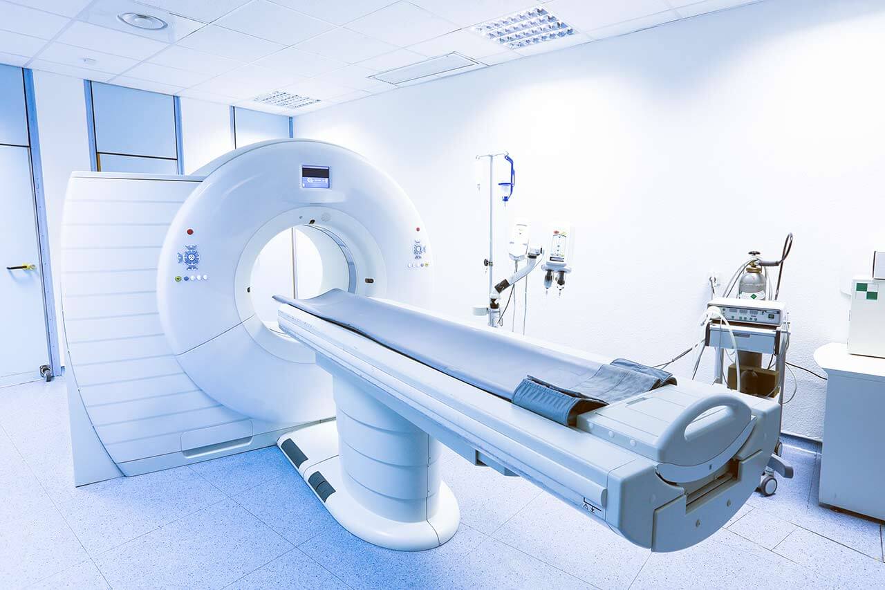
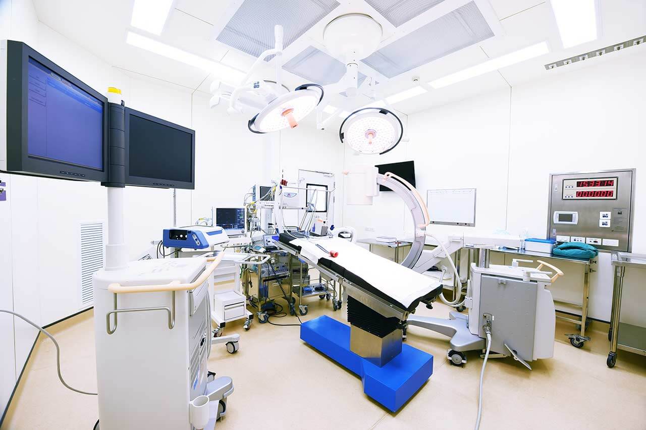
The medical team of the hospital consists of highly professional therapists, surgeons and nursing staff. The focus of their efforts is on the patient, his health and peace of mind, as well as comfort during treatment. The clinical practice of doctors is based on an individual approach to each case, which results in high treatment success rates. State-of-the-art technical equipment also plays an important role in the therapeutic process. The hospital is proud of the most advanced devices for imaging diagnostics (X-ray, ultrasound, CT, MRI, PET-CT, SPECT-CT, etc.), endoscopic examinations, laboratory tests, as well as specially equipped operating rooms for robot-assisted interventions, image-guided therapeutic manipulations, minimally invasive and classical surgeries of any complexity. Thus, the doctors of the university hospital have all the necessary resources to effectively treat the most severe pathologies and save lives.
The combination of high-tech equipment, experienced and highly qualified personnel, as well as strict adherence to the standards of modern medicine, form a solid foundation for the provision of the best medical care at the European level. An undeniable proof of the high prestige of the hospital is the constantly growing number of patients who come here from various regions of Germany and other countries of the world.
Photo: (с) depositphotos
Accommodation in hospital
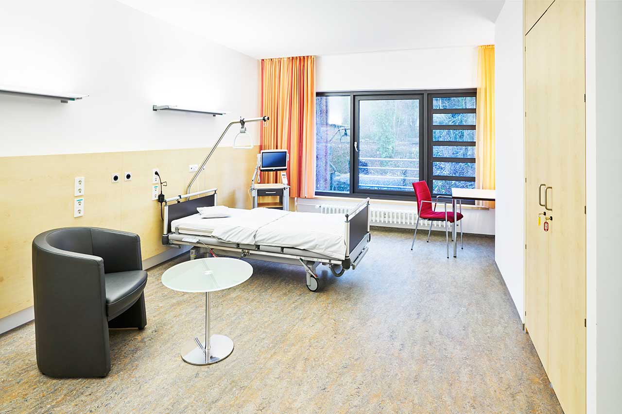
Patients rooms
The patients of the University Hospital Erlangen live in comfortable rooms with light colors and modern design. Each patient room has an ensuite bathroom with shower and toilet. The furnishing of the patient room includes an automatically adjustable bed with an orthopedic mattress, a bedside table, a wardrobe, a table and chairs for receiving visitors, a TV, a radio and a telephone. Wi-Fi can be provided upon request. The use of a mobile phone is prohibited in many rooms of the hospital.
Patients can also live in enhanced-comfort rooms with a more sophisticated design. The enhanced-comfort rooms additionally include upholstered furniture, a minifridge and a safe.
Meals and Menus
The hospital offers healthy and tasty food distinguished by many awards, including the 1st place in the prestigious ESSEN PRO GESUNDHEIT competition of the Bavarian State Ministry of the Environment and Consumer Protection.
The patient and the accompanying person have three meals a day. Breakfast is served buffet style: scrambled eggs, boiled eggs, sausage, cheese, bread and buns with butter and jam, cereals, etc. There are three set menus for lunch and dinner to choose from: a classic menu featuring local cuisine dishes, a Mediterranean menu and a vegetarian menu.
If for some reason you do not eat all the foods, you will be offered an individual menu. Please inform the medical staff about your dietary preferences prior to the treatment.
The hospital also houses many cafeterias, which will delight with a wide range of delicious dishes and drinks.
Further details
Standard rooms include:
Toilet
Shower
Wi-Fi
TV
Religion
The hospital regularly hosts catholic and evangelical devine services. The services of representatives of other religions are available upon request.
Accompanying person
During an inpatient program, an accompanying person can stay with you in the patient room or in a hotel of your choice.
Hotel
During an outpatient program, you can stay in a hotel of your choice. The managers will help you choose the most suitable options.
Select your currency:
Your guarantee
One year of support after the treatment
Insurance to cover unforeseen expenses arising from complications during and 48 months after treatment (coverage up to 200,000 €)
Reduced costs by 40-70% (contracts with Hospitals)
In addition, we are the only TÜV-certified company with an ISO 9001:2015 certificate
thank you for reaching out! One of our medical advisors is currently reviewing your details. You can expect a call from us within one business day to discuss your request.
Note: The incoming call will appear as a German number or your local area code. This call is entirely free of charge.
Booking Health guarantees:
Expert Placement
We use rigorous statistical analysis to ensure you are treated at the best-rated facility for your specific diagnosis.
Total Cost Protection
No hidden fees or unexpected bills. We provide a fixed price guarantee, backed by insurance that covers any additional complications.
12-Month Extended Care
Your recovery is our priority. Benefit from a full year of direct medical support following your procedure.

 BookingHealth Price from:
