The program includes:
- Initial presentation in the hospital
- Clinical history taking
- Evaluating the available medical reports
- General clinical examination
- Urological examination
- Laboratory tests:
- Complete blood count
- Biochemical analysis of blood
- Inflammation indicators (CRP, ESR)
- Indicators of blood coagulation
- Tumor markers, PSA
- Ultrasound scan of the urogenital system
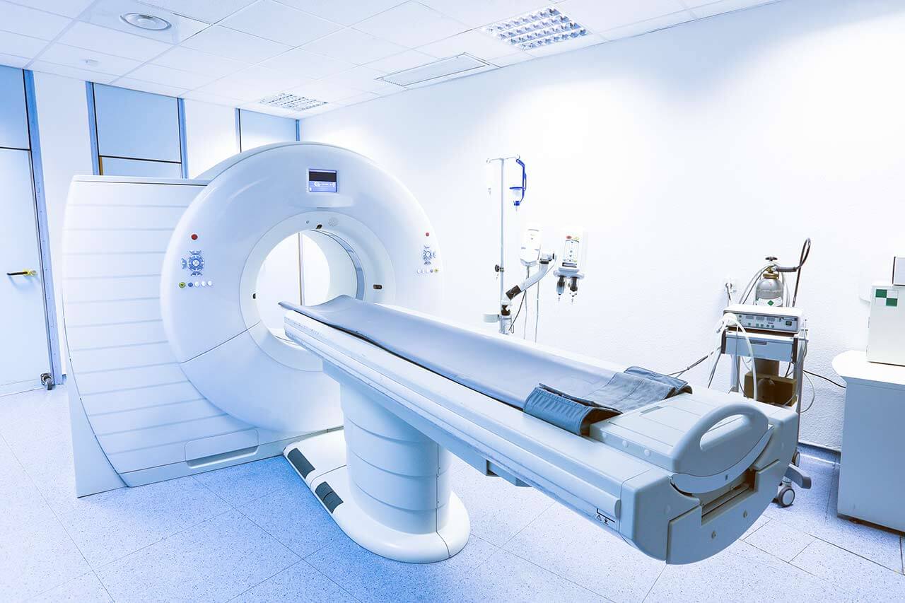
- CT scan/MRI of the abdomen and pelvis
- Preoperative care
- Removal of prostate tumor metastases with a gamma probe
- Consultations of related specialists
- Symptomatic treatment
- Cost of essential medicines
- Nursing services
- Control examinations
- Stay in the hospital with full board
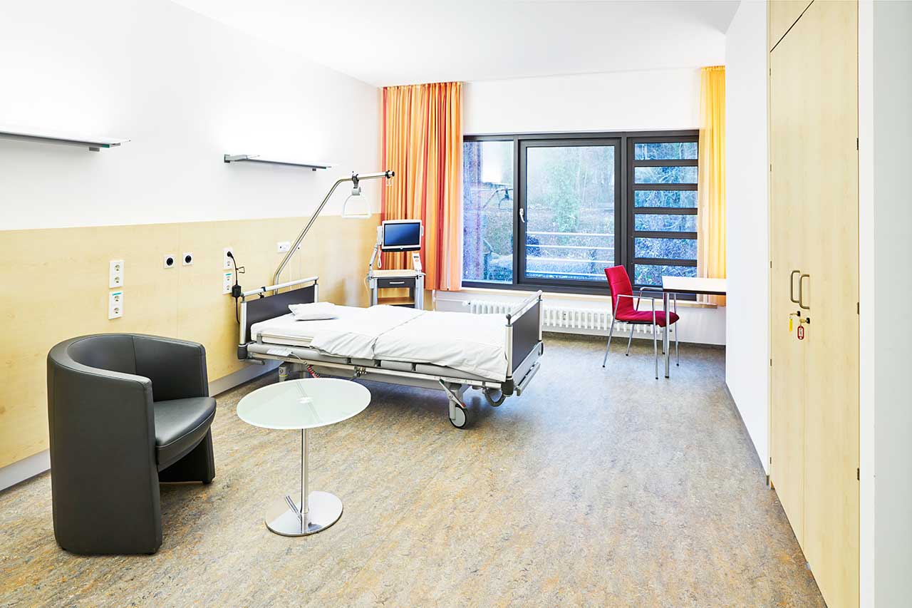
- Accommodation in 2-bedded ward
- Recommendations regarding further treatment
Indications
- Confirmed prostate cancer with suspected metastasizing
How program is carried out
During the first visit, the physician will conduct a clinical examination and go through the results of the available examinations. After that, you will undergo the necessary additional examination, such as the assessment of liver and kidney function, ultrasound scan of the abdominal and pelvic organs. Based on the results, the physician will determine the number and localization of metastases, and plan the upcoming intervention.
On the eve of the intervention, you will receive an injection of radioactively labeled PSMA. PSMA is the substance that binds to the smallest prostate cancer metastases in the lymph nodes and other tissues. The radioactive label will allow the surgeon to find all the metastases during the operation.
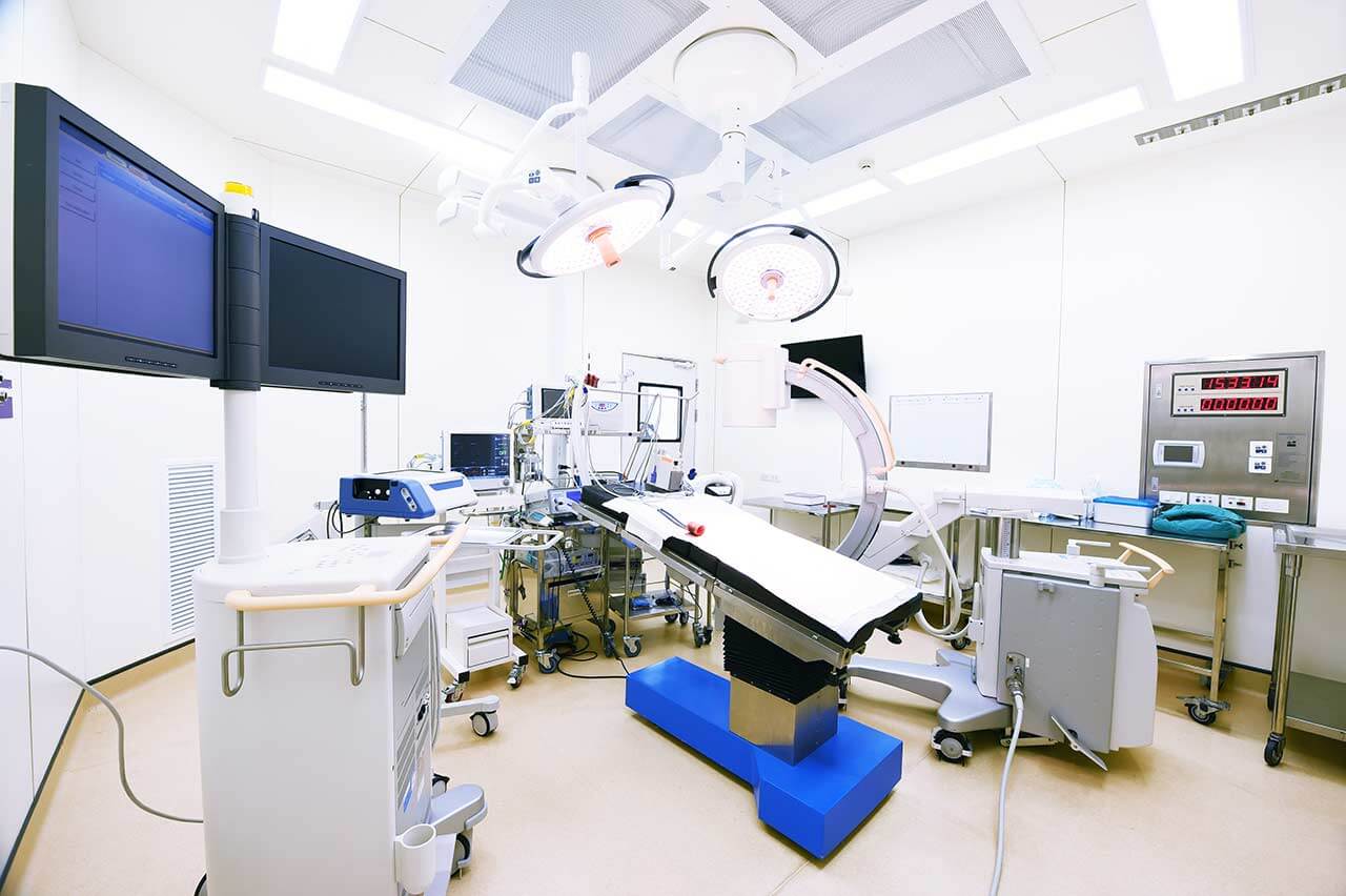
Removal of prostate tumor metastases with a gamma probe starts with general anesthesia. After anesthesia, the surgeon makes small incisions through which he inserts endoscopic instruments, a gamma probe and a video camera into the pelvic cavity and abdominal cavity. The gamma probe detects the lymph nodes affected by metastases by a radioactive label, and the surgeon removes them endoscopically. The video camera continuously transmits a three-dimensional image of the operating field in 12-fold magnification to the monitor.
As the surgeon sees the operating field in multiple magnification, he preserves the nerve endings and large blood vessels. This significantly reduces the surgical risks. The gamma probe, in turn, allows detecting and removing all metastases, which minimizes the risk of prostate cancer recurrence.
After the completion of the operation, you will be transferred back to the ward, under the supervision of the attending physician and nursing staff. Due to the minimal invasiveness of the operation and the short duration of general anesthesia, you will not need to stay in the intensive care unit for a long time.
Finally, the attending physician will evaluate the results of control examinations, schedule the date of discharge from the hospital and give you detailed recommendations for further follow-up and treatment.
Required documents
- Medical records
- PSA blood test
- MRI/CT scan (not older than 3 months)
- Bone scintigraphy (if available)
- Biopsy results (if available)
Service
You may also book:
 BookingHealth Price from:
About the department
The Department of Nuclear Medicine at the University Hospital Erlangen offers the full range of diagnostic and therapeutic services in the area of its specialization. Of particular interest are PET/CT, PET/MRI, SPECT/CT, as well as innovative therapeutic methods of nuclear medicine, such as peptide radioreceptor therapy for neuroendocrine tumors, liver tumor radioembolization, and treatment of prostate cancer using radioligands to prostate-specific membrane antigen (PSMA therapy). The department is headed by Prof. Dr. med. Torsten Kuwert.
The department's doctors most often admit patients suffering from cancers, diseases of the nervous and cardiovascular system. The patients receive medical care from the highly qualified internationally renowned specialists with profound clinical training and rich experience. The department's doctors use only reliable radioisotopes, which do not have a harmful effect on the human body and do not cause any side effects. The patients are admitted both on an inpatient and outpatient basis.
The most commonly used diagnostic methods in the department are positron emission tomography combined with computed tomography (PET/CT). The diagnostic test provides high-quality imaging and has a high resolution, thanks to which it helps the doctors to detect the slightest pathological changes that cannot be diagnosed with other procedures. PET/CT allows the doctors to diagnose cancer at early stages, as well as accurately determine the size of the detected malignant tumor. In addition, PET/CT is the only diagnostic method, which can detect Alzheimer's disease at a very early stage.
The department includes a specialized outpatient clinic for the diagnosis and treatment of benign and malignant thyroid diseases. The diagnostic options include ultrasound examination (there are three state-of-the-art high-resolution ultrasound systems), thyroid scintigraphy, and SPECT/CT, which is rarely required. The tests for determining hormone levels and antibodies to the thyroid gland are carried out in the department's in-house outpatient laboratory. Additional laboratory tests are carried out in the central laboratory of the university hospital. The doctors of the outpatient clinic are experts in radioiodine therapy. Prior to starting this therapy, they always inform patients about the details of the course of therapy and the indications for it. The clinical cases of patients with thyroid malignancies are reviewed at interdisciplinary medical boards, after which the optimal treatment tactics are selected. The essence of radioiodine therapy is to take radioactive iodine in the form of capsules or in liquid form. Radioactive iodine enters the bloodstream and is actively absorbed by the malignant cells of the thyroid gland, gradually destroying them. Before starting the course of treatment, the patient must follow a special low-iodine diet.
The department's medical team also successfully treats advanced prostate cancer using the innovative Lutetium-177 PSMA therapy. As a rule, such treatment is prescribed for men who have previously had radical prostatectomy and radiation therapy with poor results. The method is used only in progressive hospitals of the world's leading countries in terms of medicine and has already proven its high efficiency. The essence of Lutetium-177 PSMA therapy is to target cancer cells with beta-radiation of the isotope Lutetium-177, which is injected into the patient's body intravenously. Lutetium-177 binds to a molecule (ligand) that is similar to prostate-specific membrane antigen (PSMA), the content of which in prostate cancer cells is several times higher than in healthy prostate cells. Metastatic cells also actively express PSMA. The treatment with the isotope Lutetium-177 results in almost complete absorption of the entire injected dose by the target tissues, destroying the malignant cells and suppressing their further reproduction. The procedure is carried out under the PET-CT guidance, on an inpatient basis.
The department's range of services includes:
- Diagnostic options
- PET, PET/CT and PET/MRI
- Brain tumors (gliomas)
- Recurrent prostate cancer
- Neuroendocrine tumors
- Inflammatory processes
- Assessment of myocardial state
- Scintigraphy and SPECT/CT
- Endocrinology
- Thyroid scintigraphy with 99mTc or MIBI Tc-99m sodium pertechnetate
- Parathyroid scintigraphy
- Gastroenterology and pulmonology
- Lung perfusion scintigraphy
- Salivary gland scintigraphy
- Scintigraphy to assess the dynamics of gastric emptying
- Scintigraphy for suspected Meckel's diverticulum
- Detection of the source of hemorrhage using Tc-99m-labeled red blood cells
- Spleen scintigraphy
- Oncology
- Bone scintigraphy
- Scintigraphy to determine the expression of somatostatin receptors with Tc-99m-Tectreotyd
- I-123 MIBG scintigraphy
- Scintigraphy to determine the PSMA (prostate-specific membrane antigen) expression with Tc-99m MIP1404
- Cardiology
- Myocardial perfusion scintigraphy
- Assessment of myocardial viability
- Assessment of sympathetic innervation with the I-123 MIBG
- Nephrology and urology
- Dynamic kidney scintigraphy with TER-MAG-3 (Tc-99m-MAG-3)
- Static kidney scintigraphy (Tc-99m-DMSA)
- Neurology
- Brain perfusion scintigraphy
- Diagnostics of Parkinson's disease using FP-CIT-SPECT (dopamine transporter scintigraphy) and IBZM-SPECT (dopamine receptor-2 scintigraphy)
- Orthopedics and rheumatology
- Bone scintigraphy
- Diagnostics of inflammatory processes using anti-granulocyte scintigraphy
- Endocrinology
- PET, PET/CT and PET/MRI
- Therapeutic options
- Radioiodine therapy for benign and malignant thyroid diseases
- Peptide radioreceptor therapy (PRRT) for neuroendocrine tumors
- Lutetium-177 PSMA therapy for metastatic prostate cancer
- Selective internal radiation therapy for primary malignant liver tumors and liver metastases
- Xofigo® therapy (Radium-223-dichloride) for the treatment of bone metastases in prostate cancer
- Radiosynoviorthesis (RSO) for chronic inflammatory joint diseases
- Cevalin therapy
- Other diagnostic and therapeutic services
Curriculum vitae
Higher Education
- 1986 Master of Engineering, Rhine-Westphalian Technical Aachen University, Germany.
Professional Career
- 1986 - 1992 Assistant, Institute of Microelectronics/Children with Food Allergies, Juelich, Germany.
- 1992 - 1994 Associate Professor, Institute of Microelectronics/Children with Food Allergies, Juelich, Germany.
- 1994 - 1997 Associate Professor, Department of Nuclear Medicine, University of Muenster, Germany.
- 1997 Head and Professor, Department of Nuclear Medicine, Institute for Nuclear Medicine, Bad Oeynhausen, Germany.
- Head of the Department of Nuclear Medicine at the University Hospital Erlangen.
Photo of the doctor: (c) Universitätsklinikum Erlangen
About hospital
According to the Focus magazine, University Hospital Erlangen ranks among the best medical facilities in Germany!
The hospital is one of the leading healthcare facilities in Bavaria and offers top-class medical care distinguished by the close intertwining of clinical activities with research and training of medical students. The hospital was founded in 1815 and today is proud of its rich traditions, numerous medical achievements and an excellent reputation not only in Germany, but also in the international arena. The hospital has 25 specialized departments, 7 institutes and 41 interdisciplinary centers, whose experts work tirelessly for the benefit of their patients.
The hospital has the status of a maximum care center, and therefore it represents almost all fields of modern medicine. Oncology, transplant medicine, and robot-assisted surgery are among the top priorities of the clinical activities of the medical complex. Oncology is represented by the Comprehensive Cancer Center Erlangen, which is one of 13 centers of excellence in Germany certified by the German Cancer Society. The university hospital has a high-tech center with high success rates for heart, liver, kidney, pancreas, cornea and bone marrow transplants. In addition, the hospital is a leader in the use of robot-assisted surgery. The medical facility has at its disposal innovative robotic technologies, in particular the da Vinci Surgical System, with the help of which surgeons perform many sparing interventions in various medical fields.
The medical team of the hospital consists of highly professional therapists, surgeons and nursing staff. The focus of their efforts is on the patient, his health and peace of mind, as well as comfort during treatment. The clinical practice of doctors is based on an individual approach to each case, which results in high treatment success rates. State-of-the-art technical equipment also plays an important role in the therapeutic process. The hospital is proud of the most advanced devices for imaging diagnostics (X-ray, ultrasound, CT, MRI, PET-CT, SPECT-CT, etc.), endoscopic examinations, laboratory tests, as well as specially equipped operating rooms for robot-assisted interventions, image-guided therapeutic manipulations, minimally invasive and classical surgeries of any complexity. Thus, the doctors of the university hospital have all the necessary resources to effectively treat the most severe pathologies and save lives.
The combination of high-tech equipment, experienced and highly qualified personnel, as well as strict adherence to the standards of modern medicine, form a solid foundation for the provision of the best medical care at the European level. An undeniable proof of the high prestige of the hospital is the constantly growing number of patients who come here from various regions of Germany and other countries of the world.
Photo: (с) depositphotos
Accommodation in hospital
Patients rooms
The patients of the University Hospital Erlangen live in comfortable rooms with light colors and modern design. Each patient room has an ensuite bathroom with shower and toilet. The furnishing of the patient room includes an automatically adjustable bed with an orthopedic mattress, a bedside table, a wardrobe, a table and chairs for receiving visitors, a TV, a radio and a telephone. Wi-Fi can be provided upon request. The use of a mobile phone is prohibited in many rooms of the hospital.
Patients can also live in enhanced-comfort rooms with a more sophisticated design. The enhanced-comfort rooms additionally include upholstered furniture, a minifridge and a safe.
Meals and Menus
The hospital offers healthy and tasty food distinguished by many awards, including the 1st place in the prestigious ESSEN PRO GESUNDHEIT competition of the Bavarian State Ministry of the Environment and Consumer Protection.
The patient and the accompanying person have three meals a day. Breakfast is served buffet style: scrambled eggs, boiled eggs, sausage, cheese, bread and buns with butter and jam, cereals, etc. There are three set menus for lunch and dinner to choose from: a classic menu featuring local cuisine dishes, a Mediterranean menu and a vegetarian menu.
If for some reason you do not eat all the foods, you will be offered an individual menu. Please inform the medical staff about your dietary preferences prior to the treatment.
The hospital also houses many cafeterias, which will delight with a wide range of delicious dishes and drinks.
Further details
Standard rooms include:
Religion
The hospital regularly hosts catholic and evangelical devine services. The services of representatives of other religions are available upon request.
Accompanying person
During an inpatient program, an accompanying person can stay with you in the patient room or in a hotel of your choice.
Hotel
During an outpatient program, you can stay in a hotel of your choice. The managers will help you choose the most suitable options.


