{"translation_price":"50","translation_doc_price":"40","child_coefficient":"1.1","transfer_price":"2.00","transfer_price_vip":"5.00","constant_transfer_price_vip":350,"constant_transfer_price":150,"constant_transfer_distanse":60,"type":"treatment","program_full_story":"<ul>\n\t<li>Initial presentation in the clinic<\/li>\n\t<li>clinical history taking<\/li>\n\t<li>review of medical records<\/li>\n\t<li>physical examination<\/li>\n\t<li>laboratory tests:\n\t<ul style=\"list-style-type:circle\">\n\t\t<li>complete blood count<\/li>\n\t\t<li>general urine analysis<\/li>\n\t\t<li>biochemical analysis of blood<\/li>\n\t\t<li>inflammation indicators (CRP, ESR)<\/li>\n\t\t<li>indicators of blood coagulation<\/li>\n\t\t<li>tumor markers (CEA, CA19-9, CA125)<\/li>\n\t<\/ul>\n\t<\/li>\n\t<li>gynecological examination<\/li>\n\t<li>ultrasound scan: pelvis, abdomen<\/li>\n\t<li>preoperative care<\/li>\n\t<li>da Vinci myomectomy<\/li>\n\t<li>symptomatic treatment<\/li>\n\t<li>control examinations<\/li>\n\t<li>the cost of essential medicines and materials<\/li>\n\t<li>nursing services<\/li>\n\t<li>full hospital accommodation<\/li>\n\t<li>explanation of future recommendations<br \/>\n\t <\/li>\n<\/ul>\n<div class=\"program_how_program_going mt-4\"><h4>How program is carried out<\/h4><p style=\"text-align: justify;\"><strong>During the first visit<\/strong>, the physician will conduct a clinical examination and go through the results of the available diagnostic tests. After that, you will undergo the necessary additional examination, such as the assessment of liver and kidney function, ultrasound scan of the abdominal and pelvic organs, CT scan and MRI (if necessary). Based on the results of an additional examination, the physician will clarify the size and location of the myomatous nodes, choose the surgical technique and the type of anesthesia.<\/p>\n\n<p style=\"text-align: justify;\"><strong>Surgery with the da Vinci robot<\/strong> starts with general anesthesia. After anesthesia, the surgeon makes small incisions through which he inserts the manipulators of the da Vinci robot and a video camera into the pelvic cavity. Using manipulators, the doctor removes myomatous nodes, and the video camera continuously transmits a three-dimensional image of the operating field in 12-fold magnification to the monitor.<\/p>\n\n<p style=\"text-align: justify;\">Due to the precise movements of the da Vinci robotic arms, the surgeon can successfully remove multiple and large myomatous nodes on any wall of the uterus. After that, the wall of the uterus is sutured with a multilayer suture. This ensures the consistency of the scar on the uterus in the future and contributes to the preservation of the reproductive function.<\/p>\n\n<p style=\"text-align: justify;\">Excessive body mass, which usually complicates endoscopic surgery, is also not a direct contraindication to robotic intervention.<\/p>\n\n<p style=\"text-align: justify;\"><strong>After the completion of the operation<\/strong>, you will be transferred back to the ward, under the supervision of the attending physician and nursing staff. Due to the minimal invasiveness of the operation and the short duration of general anesthesia, you will not need to stay in the intensive care unit for a long time.<\/p>\n\n<p style=\"text-align: justify;\">Finally, the attending physician will evaluate the results of <strong>control examinations<\/strong>, schedule the date of discharge from the hospital and give you detailed recommendations for further follow-up and treatment.<\/p>\n<\/div><div class=\"program_required_documents mt-4\"><h4>Required documents<\/h4><ul>\n\t<li style=\"text-align: justify;\">Medical records<\/li>\n\t<li style=\"text-align: justify;\">Ultrasound scan (if available)<\/li>\n<\/ul>\n<\/div>","program_full_story_crm":"<ul>\n\t<li>Initial presentation in the clinic<\/li>\n\t<li>clinical history taking<\/li>\n\t<li>review of medical records<\/li>\n\t<li>physical examination<\/li>\n\t<li>laboratory tests:\n\t<ul style=\"list-style-type:circle\">\n\t\t<li>complete blood count<\/li>\n\t\t<li>general urine analysis<\/li>\n\t\t<li>biochemical analysis of blood<\/li>\n\t\t<li>inflammation indicators (CRP, ESR)<\/li>\n\t\t<li>indicators of blood coagulation<\/li>\n\t\t<li>tumor markers (CEA, CA19-9, CA125)<\/li>\n\t<\/ul>\n\t<\/li>\n\t<li>gynecological examination<\/li>\n\t<li>ultrasound scan: pelvis, abdomen<\/li>\n\t<li>preoperative care<\/li>\n\t<li>da Vinci myomectomy<\/li>\n\t<li>symptomatic treatment<\/li>\n\t<li>control examinations<\/li>\n\t<li>the cost of essential medicines and materials<\/li>\n\t<li>nursing services<\/li>\n\t<li>full hospital accommodation<\/li>\n\t<li>explanation of future recommendations<br \/>\n\t <\/li>\n<\/ul>\n<div class=\"program_how_program_going mt-4\"><h4>How program is carried out<\/h4><p style=\"text-align: justify;\"><strong>During the first visit<\/strong>, the physician will conduct a clinical examination and go through the results of the available diagnostic tests. After that, you will undergo the necessary additional examination, such as the assessment of liver and kidney function, ultrasound scan of the abdominal and pelvic organs, CT scan and MRI (if necessary). Based on the results of an additional examination, the physician will clarify the size and location of the myomatous nodes, choose the surgical technique and the type of anesthesia.<\/p>\n\n<p style=\"text-align: justify;\"><strong>Surgery with the da Vinci robot<\/strong> starts with general anesthesia. After anesthesia, the surgeon makes small incisions through which he inserts the manipulators of the da Vinci robot and a video camera into the pelvic cavity. Using manipulators, the doctor removes myomatous nodes, and the video camera continuously transmits a three-dimensional image of the operating field in 12-fold magnification to the monitor.<\/p>\n\n<p style=\"text-align: justify;\">Due to the precise movements of the da Vinci robotic arms, the surgeon can successfully remove multiple and large myomatous nodes on any wall of the uterus. After that, the wall of the uterus is sutured with a multilayer suture. This ensures the consistency of the scar on the uterus in the future and contributes to the preservation of the reproductive function.<\/p>\n\n<p style=\"text-align: justify;\">Excessive body mass, which usually complicates endoscopic surgery, is also not a direct contraindication to robotic intervention.<\/p>\n\n<p style=\"text-align: justify;\"><strong>After the completion of the operation<\/strong>, you will be transferred back to the ward, under the supervision of the attending physician and nursing staff. Due to the minimal invasiveness of the operation and the short duration of general anesthesia, you will not need to stay in the intensive care unit for a long time.<\/p>\n\n<p style=\"text-align: justify;\">Finally, the attending physician will evaluate the results of <strong>control examinations<\/strong>, schedule the date of discharge from the hospital and give you detailed recommendations for further follow-up and treatment.<\/p>\n<\/div>","is_ambulant":"0","bh_fee":"0","only_for_children":"0","no_service":"0","with_prepayment":"1","show_calculator":"1","paket_type":"1","btn_type":"2","clinic_icon":"60018b76e5d59.jpg","city":"Frankfurt am Main","clinic_site":"http:\/\/www.kgu.de","department_recommend":"1","country":"Germany","country_id":"1","clinic_name":"University Hospital Frankfurt am Main","cinic_name":"University Hospital Frankfurt am Main","department_id":"495","duration":"5","direction":"Gynecology","min_duration":0,"clinic_id":"256","paketPrice":3900,"paket":"<ul>\n <li>Interpreter up to 10 hours\n <\/li>\n <li>Translation up to 5 pages<\/li>\n <li>Visa support\n <\/li>\n \n<\/ul>","title":"Da Vinci myomectomy in uterine fibroids (myoma uteri)","price":{"val":10951.18,"type":"val"},"price_surcharge":0,"price_surcharge_clear":0,"extra_service_clinic":[{"id":"need_to_use_head_doctor","title":"Treatment by leading experts","type":"val","val":"2893.38","req":0},{"id":"1_person_room","title":" Single room (whole period)","parent":"duration","type":"count_val","val":"130.00"},{"id":"2_person_room","title":" double room (whole period)","parent":"duration","type":"count_val","val":"90.00"},{"id":"attendant_place","title":"Accommodation for the accompanying person ","parent":"duration","type":"count_val","val":"90.00"}],"extra_service":[],"translation_hours":"0","translation_doc_count":null,"roads":[{"id":"6","distance":"10","airport_title":"Frankfurt a.M."},{"id":"19","distance":"218","airport_title":"Stuttgart"},{"id":"4","distance":"430","airport_title":"Duesseldorf"}],"pakets":[],"lang":{"day":"Day","days":"days","ambulatory":"Outpatient","stationaryProgram":"Inpatient"}}
Da Vinci myomectomy in uterine fibroids (myoma uteri) in University Hospital Frankfurt am Main
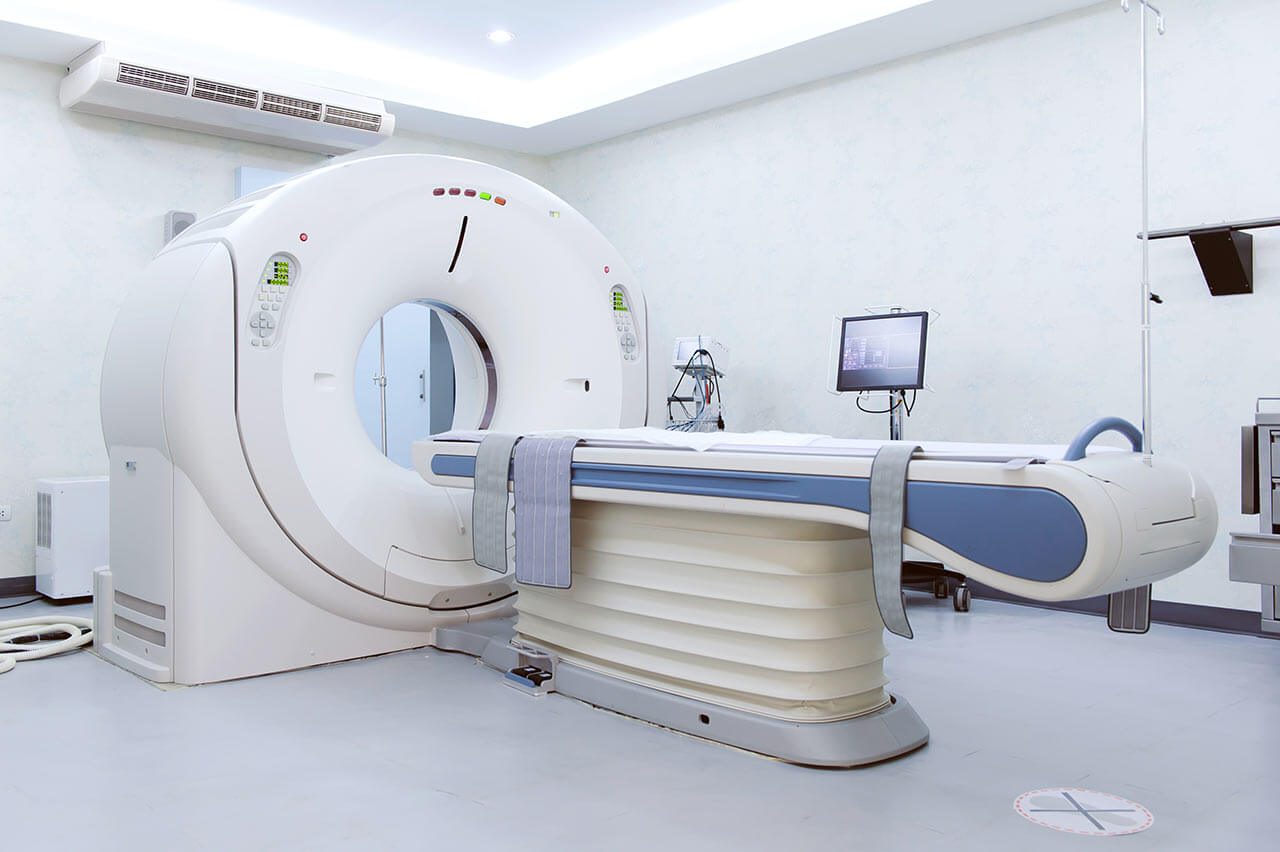
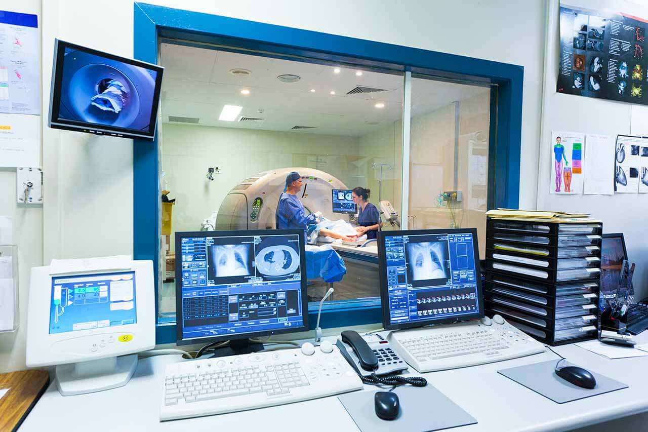
During the first visit, the physician will conduct a clinical examination and go through the results of the available diagnostic tests. After that, you will undergo the necessary additional examination, such as the assessment of liver and kidney function, ultrasound scan of the abdominal and pelvic organs, CT scan and MRI (if necessary). Based on the results of an additional examination, the physician will clarify the size and location of the myomatous nodes, choose the surgical technique and the type of anesthesia.
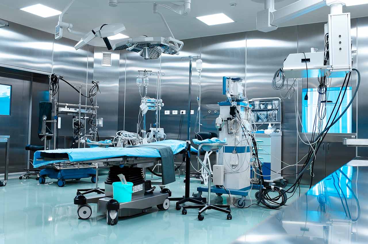
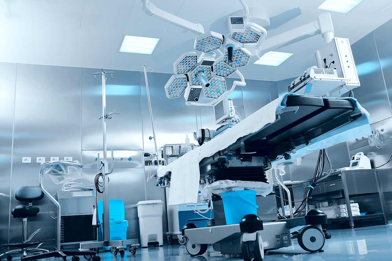
Surgery with the da Vinci robot starts with general anesthesia. After anesthesia, the surgeon makes small incisions through which he inserts the manipulators of the da Vinci robot and a video camera into the pelvic cavity. Using manipulators, the doctor removes myomatous nodes, and the video camera continuously transmits a three-dimensional image of the operating field in 12-fold magnification to the monitor.
Due to the precise movements of the da Vinci robotic arms, the surgeon can successfully remove multiple and large myomatous nodes on any wall of the uterus. After that, the wall of the uterus is sutured with a multilayer suture. This ensures the consistency of the scar on the uterus in the future and contributes to the preservation of the reproductive function.
Excessive body mass, which usually complicates endoscopic surgery, is also not a direct contraindication to robotic intervention.
After the completion of the operation, you will be transferred back to the ward, under the supervision of the attending physician and nursing staff. Due to the minimal invasiveness of the operation and the short duration of general anesthesia, you will not need to stay in the intensive care unit for a long time.
Finally, the attending physician will evaluate the results of control examinations, schedule the date of discharge from the hospital and give you detailed recommendations for further follow-up and treatment.
Required documents
Medical records
Ultrasound scan (if available)
Service
Price from:
Type of program :
Price for 1 day:
Expected duration of the program:
The minimum duration of the program:
Select
You may also book:
Hospital directly:
Saving:
BookingHealth Price from:
About the department
According to the Focus magazine, the Department of Adult and Pediatric Gynecology, Obstetrics and Mammology at the University Hospital Frankfurt am Main ranks among the top German departments specializing in the provision of obstetric services and breast cancer treatment!
The department offers all diagnostic and therapeutic options for patients of all ages with diseases of the female reproductive system, mammary gland, as well as provides the comprehensive pregnancy management, childbirth and postpartum care in accordance with the highest standards of modern obstetrics. One of the priority focuses of the department’s work is the treatment of oncopathology in women. All specialists have excellent qualifications and tremendous experience, and therefore they achieve successful results even in extremely difficult clinical cases.
The department is headed by Prof. Dr. med. Sven Becker, who has been treating gynecologic and breast diseases, as well as providing obstetric services, for more than 20 years. Prof. Becker studied medicine in Germany, France, Spain, and Japan, and had his clinical training at the Johns Hopkins Hospital at Johns Hopkins University, where he completed his board certification in gynecology and obstetrics according to American standards. During his professional career, the doctor has published more than 200 publications in various journals. He is actively involved in professional societies, including the German Society of Obstetrics and Gynecology (DGGG), the American College of Obstetricians and Gynecologists (ACOG), and the European Society of Gynecological Oncology (ESGO).
In the field of gynecology, the patients are provided with the treatment of general gynecological diseases, oncological pathologies of the female reproductive organs, endocrine diseases of gynecological genesis, therapy of urinary incontinence, pelvic prolapse, etc. A specially trained team of doctors is available to serve young patients with gynecological problems. They specialize in the treatment of infectious diseases, inflammatory processes of the genital organs in girls, correction of malformations and puberty disorders.
The department's mammologists deal with the diagnostics and treatment of breast cancer and benign breast diseases. The department is certified by the German Cancer Society and also has DIN EN ISO 9001: 2008 certification. The doctors have at their command the very latest therapy methods and surgical techniques, which allow them to fight against breast diseases as efficiently as possible. At the advanced stages of cancer, when the patient requires complete mammary gland resection, the doctors perform reconstructive interventions to ensure a good aesthetic result.
Another important focus of the department’s work is obstetrics. All specialists in this field make every efforts to ensure the best possible safety and make labor painless and as comfortable as possible for any woman. the department has modern ultrasound equipment for all types of ultrasound examinations in each trimester. The spectrum is complemented by chromosome analysis and fetal therapy (cordocentesis, drug treatment of cardiac arrhythmias in fetus (intrauterine administration of drugs)). The department's obstetricians have vast experience in managing high-risk pregnancies, multiple pregnancies. The patient can have a water birth, If necessary, a caesarean section can be performed as well. To relieve pain, the patients are offered peridural anesthesia.
The service range of the department includes:
General gynecology
Diagnostics and treatment of uterine fibroids
Hormone therapy
Placement of the hormonal coil Mirena®
Endometrial removal
Fibroid enucleation (organ-sparing surgery)
Hysterectomy (removal of the uterus)
Arterial embolization
Diagnostics and treatment of endometriosis
Hormone therapy
Placement of progestogen-containing intrauterine coil Mirena®
Surgical treatment (at the advanced stage of the disease)
Electrocoagulation
Diagnostics and treatment of cervical, vaginal and vulvar dysplasia
Cervical conization
Laser therapy using CO2 laser
Diagnostics and treatment of menstrual disorders
Gynecologic oncology
Ovarian cancer
Cervical cancer
Endometrial cancer
Vulvar cancer
Vaginal cancer
Borderline ovarian tumors
Cervical and vulvar dysplasia (precancerous conditions)
Rare ovarian tumors
Sarcomas of the uterus and reproductive organs
Urogynecology
Diagnostics and treatment of urinary incontinence
Conservative therapy (for example, hormone therapy, pelvic muscle strengthening, Biofeedback, drug therapy, etc.)
Surgical treatment (for example, TVT sling procedure, transobturator sling, colposuspension)
Diagnostics and treatment of pelvic prolapse
Pediatric gynecology
Preventive measures (tips on intimate hygiene, sex education, contraception and infection prevention)
Diagnostics and treatment of inflammatory processes of the reproductive organs
Diagnostics and correction of genital malformations in girls (for example, closure or absence of the vagina)
Diagnostics and treatment of developmental disorders in girls (for example, delayed mammary gland development, absence of menstruation)
Gynecological endocrinology and reproductive medicine
Reproductive problems (also in case of endometriosis, uterine fibroids, polycystic ovary syndrome)
Intrauterine insemination
In vitro fertilization
Intracytoplasmic sperm injection (ICSI)
Testicular sperm extraction
Microsurgical epididymal sperm aspiration
Preservation of fertility during chemotherapy, radiation therapy
Surgical treatment (organ-sparing interventions, plastic reconstructive surgery and aesthetic breast surgery)
Sentinel lymph node biopsy (including its removal)
Implantation of venous port systems for chemotherapy
Obstetrics
Integrated management of pregnancy, including high-risk pregnancy
Ultrasound screening in all trimesters
Fetal therapy
Chromosome analysis
Сhildbirth (including water birth)
Cesarean section
Postpartum care for mother and baby (Neonatal Intensive Care Unit)
Other diagnostic and therapeutic options
Curriculum vitae
Education
1987 - 1994 Study of Medicine at the Johannes Gutenberg University of Mainz.
1989 - 1990 Study at the Université René Descartes Paris.
1991 Study at the Autonomous University of Madrid.
1992 - 1993 Study at the Nihon University in Tokyo.
1994 Third Medical State Exam in Germany and the US State Exam (USMLE I+II).
Doctoral Thesis
1999 Doctoral thesis defense (magna cum laude) at the Institute for Physiological Chemistry, Johannes Gutenberg University of Mainz: "In vitro testing of potential antiviral agents against HIV in the MTT assay" (supervisor: Prof. Dr. med. Werner E.G. Müller).
Habilitation
2006 University Hospital Tuebingen: "Tumor cell dissemination and tumor cell persistence in breast cancer – the effect of systemic treatment and characteristics of cells" (supervisor: Prof. Dr. med. Diethelm Wallwiener).
Professional Career
1995 - 1996 Academic Teaching Hospital of the University of Frankfurt am Main, Department of Gynecology of the St. Joseph Hospital in Wiesbaden.
1996 - 1997 Institute of Pathology at the Technical University of Munich, University Hospital Rechts der Isar Munich.
1997 - 2001 Residency in Gynecology and Obstetrics at the Johns Hopkins Hospital of the Johns Hopkins University in Baltimore (Prof. Harold Fox).
2001 Board certification for Gynecology and Obstetrics by the American College of Obstetricians and Gynecologists (ACOG).
2001 Board certification for Gynecology and Obstetrics, Germany.
2002 - 2004 Head of the Section for Clinical Obstetrics/Delivery Unit, University Hospital Tuebingen
Since 2004 Operative Senior Physician, Head of the Section for Operative Gynecology, Department of Gynecology, University Hospital Tuebingen.
Since 2008 Leading Senior Physician, Department of Gynecology, University Hospital Tuebingen.
2008 Associate Professor, Department of Gynecology, University Hospital Tuebingen.
2010 Extraordinary Professor, University of Tuebingen.
Since 07.2012 Head of the Department of Adult and Pediatric Gynecology, Obstetrics and Mammology at the University Hospital Frankfurt am Main.
Membership in Professional Societies
German Society of Obstetrics and Gynecology (DGGG).
Professional Association of Gynecologists (BVF).
German Cancer Society (DKG).
German Senological Society (DSG).
American College of Gynecology and Obstetrics (ACOG).
Working Group on Gynecological Oncology (AGO).
Working Group on Gynecologic Endoscopy (AGE).
German Society for Perinatal Medicine (DGPM).
European Society for Obstetrics and Gynecology.
German Society of Ultrasound in Medicine (DEGUM).
European Society of Gynecological Oncology (ESGO).
European Society of Gynecologic Endoscopy (ESGE).
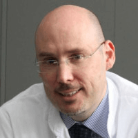
Photo of the doctor: (c) Universitätsklinikum Frankfurt
FOCUS, an internationally respected magazine, has consistently ranked the University Hospital Frankfurt am Main among the best hospitals in Germany!
The foundation of the hospital dates back to 1914. Since then, it has developed into a well-known German medical facility, whose clinical activities are based on the combination of rich traditions in medicine and scientific innovations. Like other leading German university hospitals, the University Hospital Frankfurt am Main is committed to providing world-class medical care through the implementation of state-of-the-art advancements for the benefit of patients, including those from abroad. The hospital offers many unique and highly effective therapies, which allow it to achieve remarkable treatment results and offer hope to patients even in the most complex cases.
The hospital runs 33 specialized departments and 23 research institutes, all equipped with the necessary technology and staffed by qualified specialists to provide the most effective medical care for every patient. The hospital has 1,593 beds for comfortable inpatient care. The University Hospital Frankfurt am Main annually provides its medical services to more than 50,000 inpatients and about 500,000 outpatients with conditions of varying complexity. These figures alone speak volumes about the medical facility's exceptional reputation among patients.
The University Hospital Frankfurt am Main leads in many medical fields, with neurology, neurosurgery, oncology, and cardiology being its top priorities. This medical facility also achieves outstanding results in organ and bone marrow transplantation, which are life-saving treatments for many patients with severe diseases.
Overall, this medical facility is a strong player in both German and European healthcare. The hospital's medical staff make every effort to help patients recover using the most advanced therapies currently available, and they excel at their task. The University Hospital Frankfurt am Main offers not only first-rate medical care, but also patient-centered service in a comfortable environment.
Photo: (c) depositphotos
Accommodation in hospital
Patients rooms
The patients of the University Hospital Frankfurt am Main live in comfortable rooms made in modern design and meeting the highest standards of European medicine. Each room is equipped with an ensuite bathroom with a toilet and a shower. The standard room includes a comfortable, automatically adjustable bed, a bedside table, a wardrobe, a table and chairs for receiving visitors and a TV. If desired, patients can use Wi-Fi. The patients can also stay in the enhanced-comfort rooms.
Meals and Menus
The patient and his accompanying person have a daily choice of three menus. If for any reason you do not eat all the food, you will be offered an individual menu. Please inform the medical staff about your dietary preferences prior to the treatment.
Further details
Standard rooms include:
Toilet
Shower
Wi-Fi
TV
Religion
Religious services are available upon request.
Accompanying person
During the inpatient program, an accompanying person may stay with you in a patient room or in a hotel of your choice.
Hotel
During the outpatient program, you may stay in a hotel of your choice. Managers will help you choose the most suitable options.
Select your currency:
Your guarantee
We are the only TÜV-certified company with an ISO 9001:2015 certificate
thank you for reaching out! One of our medical advisors is currently reviewing your details. You can expect a call from us within one business day to discuss your request.
Note: The incoming call will appear as a German number or your local area code. This call is entirely free of charge.
Booking Health guarantees:
Expert Placement
We use rigorous statistical analysis to ensure you are treated at the best-rated facility for your specific diagnosis.
Total Cost Protection
No hidden fees or unexpected bills. We provide a fixed price guarantee, backed by insurance that covers any additional complications.
12-Month Extended Care
Your recovery is our priority. Benefit from a full year of direct medical support following your procedure.


 BookingHealth Price from: