{"translation_price":"50","translation_doc_price":"40","child_coefficient":"1.1","transfer_price":"2.00","transfer_price_vip":"5.00","constant_transfer_price_vip":350,"constant_transfer_price":150,"constant_transfer_distanse":60,"type":"treatment","program_full_story":"<ul>\n\t<li>Initial presentation in the hospital<\/li>\n\t<li>Clinical history taking<\/li>\n\t<li>Review of available medical records<\/li>\n\t<li>Physical examination<\/li>\n\t<li>Ultrasound\u200b scan<\/li>\n\t<li>CT scan \/ MRI<\/li>\n\t<li>Preoperative care<\/li>\n\t<li>Embolization or chemoembolization, 2 procedures<\/li>\n\t<li>Symptomatic treatment<\/li>\n\t<li>Cost of essential medicines<\/li>\n\t<li>Nursing services<\/li>\n\t<li>Elaboration of further recommendations<\/li>\n<\/ul>\n<div class=\"program_how_program_going mt-4\"><h4>How program is carried out<\/h4><p style=\"text-align:justify\"><strong>During the first visit<\/strong>, the doctor will conduct a clinical examination and go through the results of the available diagnostic tests. After that, you will undergo the necessary additional examination, such as the assessment of liver and kidney function, ultrasound scan, CT scan and MRI. This will allow the doctor to determine which vessels are feeding the tumor and its metastases, as well as determine how well you will tolerate the procedure.<\/p>\n\n<p style=\"text-align:justify\"><strong>Chemoembolization <\/strong>begins with local anesthesia and catheterization of the femoral artery. The thin catheter is inserted through a few centimeters long incision of the blood vessel. The doctor gradually moves the catheter to the vessel feeding the primary tumor or its metastases. The procedure is carried out under visual control, an angiographic device is used for this. The vascular bed and the position of the catheter in it are displayed on the screen of the angiograph.<\/p>\n\n<p style=\"text-align:justify\">When the catheter reaches a suspected artery, a contrast agent is injected through it. Due to the introduction of the contrast agent, the doctor clearly sees the smallest vessels of the tumor and the surrounding healthy tissues on the screen of the angiograph. After that, he injects emboli into the tumor vessels through the same catheter.<\/p>\n\n<p style=\"text-align:justify\">Emboli are the spirals or the liquid microspheres. The type of embolus is selected individually, taking into account the diameter of the target vessel. When carrying out chemoembolization, a solution of a chemotherapy drug is additionally injected into the tumor vessel. Due to the subsequent closure of the vessel lumen with an embolus, the chemotherapy drug influences the tumor for a long time. In addition, the drug does not enter the systemic circulation, which allows doctors to use high doses of chemotherapeutic agents without the development of serious side effects. Chemoembolization leads to the destruction of the tumor or slowing down its progression.<\/p>\n\n<p style=\"text-align:justify\">After that, the catheter is removed from the artery. The doctor puts a vascular suture on the femoral artery and closes it with a sterile dressing. During chemoembolization, you will be awake. General anesthesia is not used, which significantly reduces the risks of the procedure and allows performing it on an outpatient basis, avoiding long hospital stay.<\/p>\n\n<p style=\"text-align:justify\"><strong>After the first procedure<\/strong>, you will stay under the supervision of an interventional oncologist and general practitioner. If necessary, you will receive symptomatic treatment. As a rule, a second chemoembolization procedure is performed in 3-5 days after the first one in order to consolidate the therapeutic effect. After that, you will receive recommendations for further follow-up and treatment.<\/p>\n<\/div><div class=\"program_required_documents mt-4\"><h4>Required documents<\/h4><ul>\n\t<li style=\"text-align: justify;\">Medical records<\/li>\n\t<li style=\"text-align: justify;\">MRI\/CT scan (not older than 3 months)<\/li>\n\t<li style=\"text-align: justify;\">Biopsy results (if available)<\/li>\n<\/ul>\n<\/div>","program_full_story_crm":"<ul>\n\t<li>Initial presentation in the hospital<\/li>\n\t<li>Clinical history taking<\/li>\n\t<li>Review of available medical records<\/li>\n\t<li>Physical examination<\/li>\n\t<li>Ultrasound\u200b scan<\/li>\n\t<li>CT scan \/ MRI<\/li>\n\t<li>Preoperative care<\/li>\n\t<li>Embolization or chemoembolization, 2 procedures<\/li>\n\t<li>Symptomatic treatment<\/li>\n\t<li>Cost of essential medicines<\/li>\n\t<li>Nursing services<\/li>\n\t<li>Elaboration of further recommendations<\/li>\n<\/ul>\n<div class=\"program_how_program_going mt-4\"><h4>How program is carried out<\/h4><p style=\"text-align:justify\"><strong>During the first visit<\/strong>, the doctor will conduct a clinical examination and go through the results of the available diagnostic tests. After that, you will undergo the necessary additional examination, such as the assessment of liver and kidney function, ultrasound scan, CT scan and MRI. This will allow the doctor to determine which vessels are feeding the tumor and its metastases, as well as determine how well you will tolerate the procedure.<\/p>\n\n<p style=\"text-align:justify\"><strong>Chemoembolization <\/strong>begins with local anesthesia and catheterization of the femoral artery. The thin catheter is inserted through a few centimeters long incision of the blood vessel. The doctor gradually moves the catheter to the vessel feeding the primary tumor or its metastases. The procedure is carried out under visual control, an angiographic device is used for this. The vascular bed and the position of the catheter in it are displayed on the screen of the angiograph.<\/p>\n\n<p style=\"text-align:justify\">When the catheter reaches a suspected artery, a contrast agent is injected through it. Due to the introduction of the contrast agent, the doctor clearly sees the smallest vessels of the tumor and the surrounding healthy tissues on the screen of the angiograph. After that, he injects emboli into the tumor vessels through the same catheter.<\/p>\n\n<p style=\"text-align:justify\">Emboli are the spirals or the liquid microspheres. The type of embolus is selected individually, taking into account the diameter of the target vessel. When carrying out chemoembolization, a solution of a chemotherapy drug is additionally injected into the tumor vessel. Due to the subsequent closure of the vessel lumen with an embolus, the chemotherapy drug influences the tumor for a long time. In addition, the drug does not enter the systemic circulation, which allows doctors to use high doses of chemotherapeutic agents without the development of serious side effects. Chemoembolization leads to the destruction of the tumor or slowing down its progression.<\/p>\n\n<p style=\"text-align:justify\">After that, the catheter is removed from the artery. The doctor puts a vascular suture on the femoral artery and closes it with a sterile dressing. During chemoembolization, you will be awake. General anesthesia is not used, which significantly reduces the risks of the procedure and allows performing it on an outpatient basis, avoiding long hospital stay.<\/p>\n\n<p style=\"text-align:justify\"><strong>After the first procedure<\/strong>, you will stay under the supervision of an interventional oncologist and general practitioner. If necessary, you will receive symptomatic treatment. As a rule, a second chemoembolization procedure is performed in 3-5 days after the first one in order to consolidate the therapeutic effect. After that, you will receive recommendations for further follow-up and treatment.<\/p>\n<\/div>","is_ambulant":"1","bh_fee":"0","only_for_children":"0","no_service":"0","with_prepayment":"1","show_calculator":"1","paket_type":"1","btn_type":"0","clinic_icon":"60018b76e5d59.jpg","city":"Frankfurt am Main","clinic_site":"http:\/\/www.kgu.de","department_recommend":"0","country":"Germany","country_id":"1","clinic_name":"University Hospital Frankfurt am Main","cinic_name":"University Hospital Frankfurt am Main","department_id":"2771","duration":"7","direction":"Interventional radiology","min_duration":0,"clinic_id":"256","paketPrice":7800,"paket":"<ul>\n <li>Interpreter up to 17 hours<\/li>\n <li>Translation up to 10 pages<\/li>\n <li>Visa support<\/li>\n\n \n<\/ul>","title":"Treatment of neuroendocrine tumor (NET) of the pancreas with embolization or chemoembolization","price":{"val":27640.18,"type":"val"},"price_surcharge":0,"price_surcharge_clear":0,"extra_service_clinic":[],"extra_service":[],"translation_hours":"0","translation_doc_count":null,"roads":[{"id":"6","distance":"10","airport_title":"Frankfurt a.M."},{"id":"19","distance":"218","airport_title":"Stuttgart"},{"id":"4","distance":"430","airport_title":"Duesseldorf"}],"pakets":[],"lang":{"day":"Day","days":"days","ambulatory":"Outpatient","stationaryProgram":"Inpatient"}}
Treatment of neuroendocrine tumor (NET) of the pancreas with embolization or chemoembolization in University Hospital Frankfurt am Main
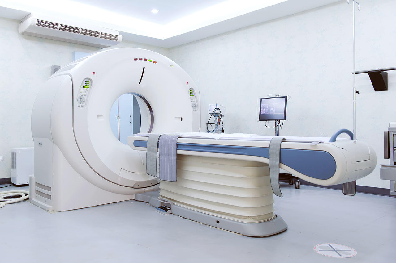
During the first visit, the doctor will conduct a clinical examination and go through the results of the available diagnostic tests. After that, you will undergo the necessary additional examination, such as the assessment of liver and kidney function, ultrasound scan, CT scan and MRI. This will allow the doctor to determine which vessels are feeding the tumor and its metastases, as well as determine how well you will tolerate the procedure.
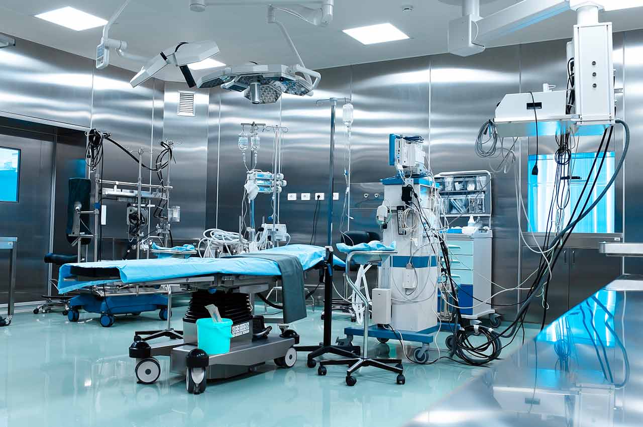
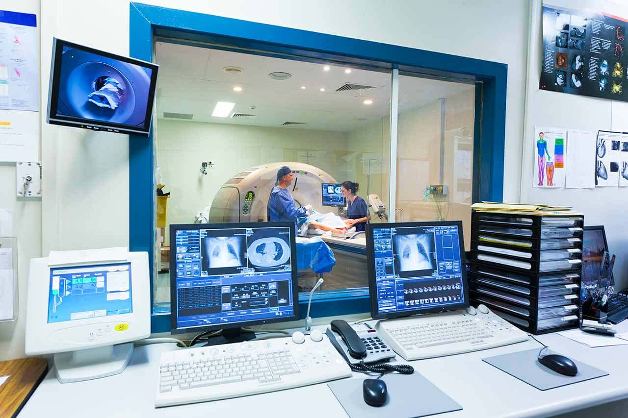
Chemoembolization begins with local anesthesia and catheterization of the femoral artery. The thin catheter is inserted through a few centimeters long incision of the blood vessel. The doctor gradually moves the catheter to the vessel feeding the primary tumor or its metastases. The procedure is carried out under visual control, an angiographic device is used for this. The vascular bed and the position of the catheter in it are displayed on the screen of the angiograph.
When the catheter reaches a suspected artery, a contrast agent is injected through it. Due to the introduction of the contrast agent, the doctor clearly sees the smallest vessels of the tumor and the surrounding healthy tissues on the screen of the angiograph. After that, he injects emboli into the tumor vessels through the same catheter.
Emboli are the spirals or the liquid microspheres. The type of embolus is selected individually, taking into account the diameter of the target vessel. When carrying out chemoembolization, a solution of a chemotherapy drug is additionally injected into the tumor vessel. Due to the subsequent closure of the vessel lumen with an embolus, the chemotherapy drug influences the tumor for a long time. In addition, the drug does not enter the systemic circulation, which allows doctors to use high doses of chemotherapeutic agents without the development of serious side effects. Chemoembolization leads to the destruction of the tumor or slowing down its progression.
After that, the catheter is removed from the artery. The doctor puts a vascular suture on the femoral artery and closes it with a sterile dressing. During chemoembolization, you will be awake. General anesthesia is not used, which significantly reduces the risks of the procedure and allows performing it on an outpatient basis, avoiding long hospital stay.
After the first procedure, you will stay under the supervision of an interventional oncologist and general practitioner. If necessary, you will receive symptomatic treatment. As a rule, a second chemoembolization procedure is performed in 3-5 days after the first one in order to consolidate the therapeutic effect. After that, you will receive recommendations for further follow-up and treatment.
Required documents
Medical records
MRI/CT scan (not older than 3 months)
Biopsy results (if available)
Service
Price from:
Type of program :
Price for 1 day:
Expected duration of the program:
The minimum duration of the program:
Select
You may also book:
Hospital directly:
Saving:
BookingHealth Price from:
About the department
The Department of Adult and Pediatric Diagnostic, Interventional Radiology at the University Hospital Frankfurt am Main provides the full range of services in these fields and specializes in conducting radiologic examinations, high-intensity focused ultrasound (HIFU) therapy, vascular, oncological, orthopedic and urologic interventions. The department includes specialized Sections for Gynecologic, Pediatric and Orthopedic Radiology. The Chief Physician of the department is Prof. Dr. med. Thomas Vogl.
In the field of diagnostics, the department offers the whole spectrum of modern diagnostic radiology, including CT and MRI diagnostics, X-ray diagnostics and ultrasound. To provide the fastest and most accurate diagnostics, the department has in its arsenal the very latest medical developments, for example, (128-slice) SOMATOM Definition AS scanner, SOMATOM Force Dual Source scanner, Siemens Somatom Definition AS Sliding mobile tomograph. These tomographs allow obtaining images of the highest quality with minimal radiation exposure to the patient.
In the field of interventional radiology, the department specializes in vascular, oncological, orthopedic interventions, as well as in prostate biopsy with an accuracy the nearest millimeter.
The department offers the full range of cancer interventions:
Laser-induced thermotherapy (LITT), which is a special minimally invasive thermotherapy for liver and lung tumors
Uterine fibroid embolization, which is a minimally invasive therapy for uterine myoma
Radiofrequency tumor ablation, which is a minimally invasive high-frequency thermoablation for liver and lung tumors
Selective internal radiation therapy with the use of SIR spheres, which is a minimally invasive radio-oncological treatment of liver tumors
Transpulmonary chemoembolization (TPCE), which is a minimally invasive and selective chemotherapy for lung tumors
Transarterial chemoperfusion (TACP), which is a minimally invasive percutaneous chemoperfusion for liver tumors
Prostate embolization in benign prostatic hyperplasia, which is a special minimally invasive therapy for benign prostatic hyperplasia
Other interventions
In addition, the department offers a wide range of minimally invasive vascular interventions. For example, excimer laser (ELA) angioplasty is successfully used in the treatment of peripheral arterial occlusive disease. It shows high rates of treatment effectiveness.
Also, the department offers special orthopedic interventions to treat herniated discs, osteoporosis and bone tumors. It uses a new minimally invasive method of treating herniated discs – ozone therapy. Its advantages include improving blood circulation and reducing inflammation, improving microcirculation and oxygen supply, etc. Another effective method is vertebroplasty/osteoplasty, which is used to reduce pain and stabilize the fractured vertebrae.
Another focus of the department is high-intensity focused ultrasound (HIFU) therapy. The department has one of the very first HIFU devices in Germany, which allows not only for tumor treatment, but also for its imaging. Focused ultrasound can be successfully used in the treatment of various benign and malignant tumors.
Indications for high intensity-focused ultrasound (HIFU) therapy:
Pancreatic tumors
Liver tumors
Kidney tumors
Breast fibroadenomas
Bone tumors
Uterine fibroids
Adenomyosis
Breast cancer
The main diagnostic and therapeutic focuses of the department are:
Vascular interventions (for example, excimer laser angioplasty)
Cancer interventions
Orthopedic interventions
MRI-guided prostate biopsy
Cancer treatment
Interdisciplinary Center for Liver Diseases
Other diagnostic and therapeutic options
Curriculum vitae
1976 - 1982 Study of Medicine at Ludwig Maximilian University of Munich.
1980 Training at the Higher School of Medicine of the Hebrew University, Hadassah Hospital, Jerusalem.
1980 - 1981 Assistant in the Department of Anatomy at the University of Munich.
1982 Admission to medical practice.
Since 1983, Assistant Physician in the Department of Radiology, University of Munich.
Professional Experience
1988 Internship at the Massachusetts General Hospital in Boston.
1988 - 1990 Additional qualification in Nuclear Medicine.
1989 Certified Radiologist specializing in Radiation Therapy.
1990 Senior Physician of the Department of Radiology, University of Munich. Focuses: conventional X-ray diagnostics, ultrasound, CT, interventional radiology, establishment and management of the Department of MRI Diagnostics and Spectroscopy.
1992 Specialized in Nuclear Medicine.
1992 Specialized in In-Vitro Nuclear Diagnostics.
1992 Appointment to the post of the Managing Senior Physician of the Department of Radiation Therapy at the Free University in Berlin, University Hospital Virchow.
1992 (C3) University Professor at the Free University of Berlin, General Radiology.
Certified Neuroradiologist.
1998 Invitation to hold a post of (C4) Professor in X-ray diagnostics, Frankfurt University.
1998 Invitation accepted.
Since 1998, Chief Physician of the Department of Adult and Pediatric Diagnostic, Interventional Radiology at the University Hospital Frankfurt am Main.
2005 Collaboration with the Friedrichsheim Orthopedic Clinic of the University of Frankfurt: Head of the Department of Radiology.
Main Clinical Focuses
Conventional X-ray diagnostics.
Traumatology.
Gastrointestinal radiology.
Interventional radiology.
Chemoembolization.
Angioplasty.
MRI diagnostics.
Main Research Focuses
MR-angiography.
In-vitro spectroscopy.
Development of contrast agents.
Interstitial laser therapy.
Hepatobiliary diagnostics.
Photo of the doctor: (c) Universitätsklinikum Frankfurt
About hospital
According to the reputable Focus magazine, the University Hospital Frankfurt am Main ranks among the top German medical facilities!
The hospital was founded in 1914 and today is a well-known German medical facility, which combines rich traditions and scientific innovations. A medical team of more than 6,500 employees cares about the health of patients around the clock, ensuring them with the highest standards of medical care and best possible safety.
The hospital has 32 specialized departments and more than 20 research institutes, which have all the necessary resources for the provision of the most effective care for any patient. The hospital has 1,488 beds for inpatient medical care. The medical facility diagnoses and treats more than 51,000 inpatients and about 44,800 outpatients every year. Due to the demonstration of outstanding treatment results, the number of patients seeking medical care here increases significantly annually.
The hospital presents all areas of modern medicine, whereas its special competence lies in neuroscience, oncology, cardiovascular medicine, cardiac surgery and other fields. Many treatment methods available here are unique not only in Europe, but also internationally.
Photo: (c) depositphotos
Accommodation in hospital
Patients rooms
The patients of the University Hospital Frankfurt am Main live in comfortable rooms made in modern design and meeting the highest standards of European medicine. Each room is equipped with an ensuite bathroom with a toilet and a shower. The standard room includes a comfortable, automatically adjustable bed, a bedside table, a wardrobe, a table and chairs for receiving visitors and a TV. If desired, patients can use Wi-Fi. The patients can also stay in the enhanced-comfort rooms.
Meals and Menus
The patient and his accompanying person have a daily choice of three menus. If for any reason you do not eat all the food, you will be offered an individual menu. Please inform the medical staff about your dietary preferences prior to the treatment.
Further details
Standard rooms include:
Toilet
Shower
Wi-Fi
TV
Religion
Religious services are available upon request.
Accompanying person
During the inpatient program, an accompanying person may stay with you in a patient room or in a hotel of your choice.
Hotel
During the outpatient program, you may stay in a hotel of your choice. Managers will help you choose the most suitable options.
Select your currency:
Your guarantee
We are the only TÜV-certified company with an ISO 9001:2015 certificate
thank you for reaching out! One of our medical advisors is currently reviewing your details. You can expect a call from us within one business day to discuss your request.
Note: The incoming call will appear as a German number or your local area code. This call is entirely free of charge.
Booking Health guarantees:
Expert Placement
We use rigorous statistical analysis to ensure you are treated at the best-rated facility for your specific diagnosis.
Total Cost Protection
No hidden fees or unexpected bills. We provide a fixed price guarantee, backed by insurance that covers any additional complications.
12-Month Extended Care
Your recovery is our priority. Benefit from a full year of direct medical support following your procedure.

 BookingHealth Price from:
