Astrocytoma treatment with MRI-guided laser ablation in University Hospital Goettingen
University Hospital Goettingen
Goettingen, Germany
During the first visit, the physician will conduct a clinical examination and neurological examination, and go through the results of the available diagnostic tests. After that, you will undergo the necessary additional examination, such as the laboratory blood and urine tests, MRI scan with obtaining 3D images and creating volumetric images of the whole brain. Based on the results of an additional examination, the physician will clarify the localization of brain metastases. After that, the preparation according to the preoperative standard will begin.
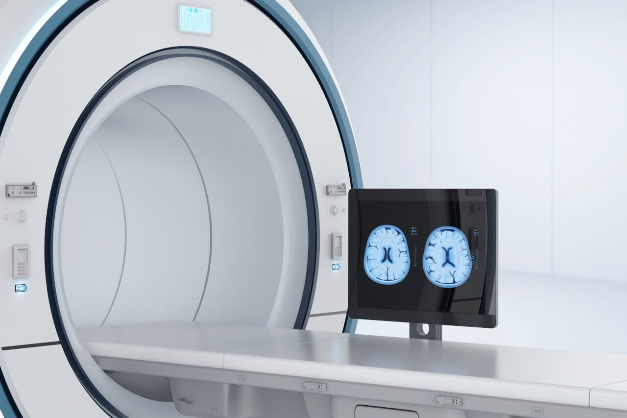
Laser ablation of metastases begins with the formation of access to the brain. When using a catheter with a laser fiber, a hole with a diameter of 3.2 mm is sufficient for this. The miniature size and flexibility of the catheter help preserve healthy brain tissue.
When the catheter reaches metastasis, energy is delivered through the laser fiber. Under the influence of high temperature, metastases are destroyed. The procedure is guided by real-time MRI. Surgeons also assess the temperature of healthy brain tissue using thermographic images to avoid overheating.
After the completion of the operation, you will be transferred to the ward, under the round-the-clock supervision of doctors and nurses. In several days, the attending physician will evaluate the results of the postoperative MRI scans, schedule the date of discharge from the hospital and give you detailed recommendations for further follow-up and treatment.
 BookingHealth Price from:
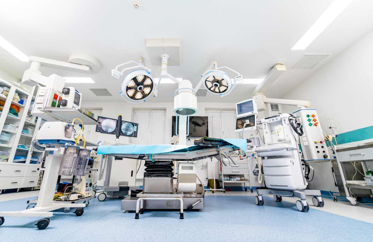
The Department of Adult and Pediatric Neurosurgery, Spinal Surgery at the University Hospital Goettingen offers the full range of surgical treatment for patients of all ages with diseases of the central and peripheral nervous system. The department is the largest Neurosurgery Center in Germany with certification by the German Cancer Society (DKG) in the treatment of neuro-oncological diseases and certification by the German Society for Orthopedics and Trauma Surgery (DGOU) and the German Society of Neurosurgery (DGNC) in the treatment of spinal pathologies. The department's medical team specializes in the surgical treatment of neurovascular diseases, movement disorders, with a special emphasis on the treatment of Parkinson's disease using deep brain stimulation, benign and malignant tumors of the brain and spinal cord, and neurosurgical diseases in children. A separate focus of the daily clinical practice of specialists is spinal surgery. The neurosurgeons of the department brilliantly perform microsurgical interventions for herniated intervertebral discs, spinal stenosis, and spinal instability. Surgical treatment focuses not only on achieving a good therapeutic result, but also on preserving the functions of the brain and spinal cord. The department's specialists use minimally invasive, endoscopic, and microsurgical techniques. The department has 3 high-tech operating rooms where more than 3,000 surgical procedures are performed annually. The department treats over 7,000 patients every year, about 2,000 of them in inpatient settings. The department has 62 inpatient beds. The Head Physician of the department is Prof. Dr. med. Veit Rohde.
The treatment of brain tumors is one of the primary clinical tasks of the department's team of neurosurgeons. The specialists treat patients with both benign and malignant brain tumors. For the treatment of benign tumors, surgical resection is sufficient, but when a brain malignancy is diagnosed, surgery is only the first step in the therapeutic process, usually followed by chemotherapy and/or radiation therapy. The goal of the department's neurosurgeons when treating a brain tumor is its complete removal because, in this case, the patient has a high chance of being cured or achieving a long-term and stable remission. When performing brain tumor surgery, the neurosurgeons in the department use modern surgical microscopes, 5-aminolevulinic acid fluorescent navigation, computer-assisted navigation, and intraoperative monitoring. All of these innovative technical capabilities allow for the most effective resection of a brain tumor with a high degree of safety. Each surgical procedure is carefully planned based on CT and MRI scans. The neurosurgeons in the department also specialize in surgery for brain tumors in children. This pathology is the second most common among young patients after leukemia. In addition, the specialists regularly perform operations to remove brain metastases due to skin cancer, breast cancer, lung cancer, and other oncological diseases. A standard treatment regimen for brain metastases includes a microsurgical resection followed by irradiation of the pathological focus. Chemotherapy may also be required, if clinically indicated.
The department successfully performs surgical treatment of neurovascular diseases such as brain aneurysms, angiomas, cavernomas, dural arteriovenous fistulas, and strokes. A brain aneurysm is an abnormal enlargement of a cerebral artery that may potentially cause life-threatening bleeding in the brain. In many cases, an aneurysm is characterized by an asymptomatic course, so it is diagnosed accidentally during an examination for other diseases. As for the treatment of this pathology, the neurosurgeons in the department offer coiling (a minimally traumatic endovascular intervention under imaging guidance) and classical operations. For the treatment of arteriovenous malformations of the brain and spinal cord, such as cavernomas, angiomas, and dural arteriovenous fistulas, the department performs mainly endovascular interventions, with open surgery being required only in complex clinical cases.
The department's specialists have excellent professional skills in the treatment of spinal pathologies. Neurosurgeons focus on patients with herniated discs, spinal stenosis, and spinal instability. These pathologies are accompanied by severe pain, which can often be relieved with drug therapy and physiotherapeutic treatment. In some cases, conservative treatment may not give the desired result, so doctors resort to surgical techniques. Surgery for spinal diseases is performed in the department through a minimally invasive approach under the guidance of a surgical microscope. It should be noted that spinal stabilization surgery is performed using an innovative robot-assisted system, which is available only in four university hospitals in Germany, including the University Hospital Goettingen. Thanks to the robotic device, specialists can percutaneously implant screw systems to stabilize the spine through skin incisions no longer than 2 cm. In addition, the use of the robot ensures maximum accuracy in positioning the screw system in the spine, which contributes to the best outcome of the surgical intervention and eliminates the risk of developing neurological deficits.
The department's range of medical services includes the following:
Since October 1, 2005, Prof. Dr. med. Veith Rohde has been the Head Physician of the Department of Adult and Pediatric Neurosurgery, Spinal Surgery at the University Hospital Goettingen. The specialist graduated from Johannes Gutenberg University Mainz, after which he received admission to medical practice in 1988, and in 1989 he received his doctorate. After this, Dr. Veith Rohde worked for two years as an Assistant Physician in the Department of Neurosurgery at the University Hospital Tuebingen. Over the next three years, he held the position of Assistant Physician in the Department of Neurosurgery at the Municipal Hospital Duisburg. In 1994, Prof. Veith took up a position as a Physician in the Department of Neurosurgery at the University Hospital RWTH Aachen, where, after one year, he completed his board certification in neurosurgery and became a Senior Physician. In 1999, Prof. Veith had his habilitation at the University Hospital RWTH Aachen. His scientific work focused on the use of transcranial magnetic stimulation before and during neurosurgical procedures. Since 1999, he has worked as a Senior Physician and Deputy Head Physician in the Department of Neurosurgery at the University Hospital RWTH Aachen. In 2003, Dr. Veith Rohde received additional qualifications in the field of special intensive care after neurosurgical procedures, and in 2005 he was appointed Extraordinary Professor at the Faculty of Medicine of the RWTH Aachen University.
Prof. Dr. med. Veith Rohde is a Member of the German Society of Neurosurgery (DGNC), the German Society of Neuroradiology (DGNR), and the German Society for Computer- and Robot-Assisted Surgery (CURAC).
Photo of the doctor: (c) Universitätsmedizin Göttingen
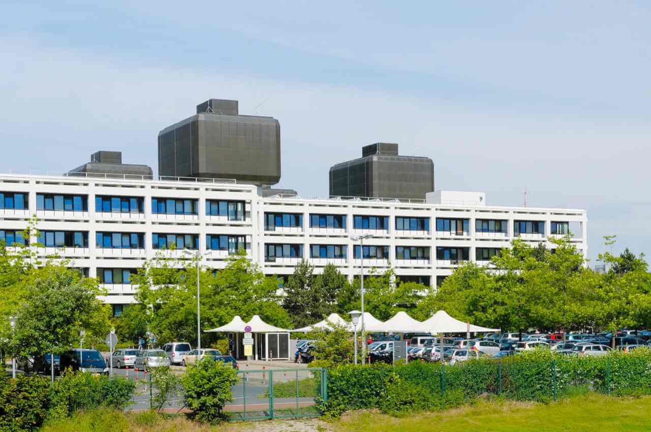
The University Hospital Goettingen positions itself as the largest multidisciplinary medical complex with a first-class level of service and rich traditions. Founded in 1737, it is one of the oldest healthcare facilities in Germany and Europe. The hospital has earned its place among the leading medical centers in Europe through a combination of successful clinical practice and fruitful research activities, resulting in effective diagnostic and treatment methods that save lives.
The hospital prides itself on its state-of-the-art equipment, the availability of which largely determines the success of treatment. The health of patients is in the safe hands of highly qualified specialists with impressive clinical experience and deep knowledge in their field of expertise. It is worth noting that the hospital employs many renowned professors with impeccable reputations who have been recognized by the national and international medical communities. Personalized medicine is a priority here. Each patient and their clinical case are unique, and the optimal set of therapeutic measures is selected on an individual basis.
The University Hospital Goettingen has 60 highly specialized departments and institutes representing almost all areas of modern medicine: general and abdominal surgery, cardiothoracic surgery, neurosurgery, oral and maxillofacial surgery, gastroenterology, endocrinology, oncology, hematology, nephrology, cardiology, pulmonology, neurology, gynecology, urology, ophthalmology, dermatology, pediatric and adolescent medicine, nuclear medicine, and radiation oncology. The hospital's bed capacity is 1,600, with many medical services provided on an outpatient basis. The medical complex treats more than 60,000 inpatients and more than 220,000 outpatients every year. The desire of such a huge number of people to undergo treatment at the hospital testifies to its excellent reputation and good therapeutic results.
By choosing treatment at the University Hospital Goettingen, patients can expect highly accurate diagnosis and effective treatment in a comfortable environment in compliance with strict international medical standards. The care and concern for the patient is at a very high level: the patient and his or her health are the main value of the medical team at the hospital. During the therapeutic process, the patient feels care, understanding, and empathy.
Photo: (с) depositphotos
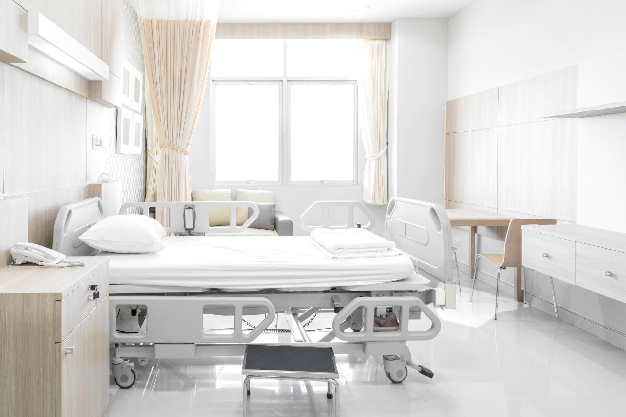
The patients of the University Hospital Goettingen stay in single and double rooms with all necessary amenities. The patient rooms are equipped with an automatically adjustable bed with an orthopedic mattress, a bedside table, a wardrobe, a table, chairs, a telephone and a TV. Patient rooms have access to Wi-Fi. For maximum patient comfort, each patient room has an ensuite bathroom with a shower and a toilet.
The hospital offers varied and tasty meals three times a day: breakfast, lunch, and dinner. If, for some reason, you do not eat all the foods, you will be offered an individual menu. Please inform the medical staff about your dietary preferences prior to your treatment.
The hospital also has a cozy restaurant, bistro, and cafeteria where you can have a tasty snack and enjoy a cup of hot tea or aromatic coffee with dessert.
Standard rooms include:
![]() Toilet
Toilet
![]() Shower
Shower
![]() Wi-Fi
Wi-Fi
![]() TV
TV
Your accompanying person may stay with you in your patient room or at the hotel of your choice during the inpatient program.
You may stay at the hotel of your choice during the outpatient program. Our managers will support you for selecting the best option.
