Ventricular septal defect belongs to the most common congenital heart defects. The ventricular septal defect is "aggressive" with regard to the development of such a dangerous complication as pulmonary hypertension. Because of this complication, some patients with large defects become inoperable and conducting surgery for them is impossible at this point. The main task of the physician is to refer the patient for surgery in time. Thus, it is vital to unravel what is particularly important to know about pathology and its management.
Content
- What is the ventricular septal defect?
- Why does it occur?
- Circulatory features of the ventricular septal defect
- Manifestation of VSD
- Treatment principles
- When is the surgery needed?
- Diagnostic examination before surgery
- Endovascular surgery specifics
- Rehabilitation
- Undergoing treatment in German hospitals
- The cost of treatment of the ventricular septal defect
- Why undergo treatment in Germany with Booking Health?
What is the ventricular septal defect?
The ventricular septum is a powerful muscular barrier that forms the inner walls of both the right and left ventricles, and in each, making up about 1/3 of their total area. It is as involved in the contraction and relaxation of the heart during each cycle as the rest of the ventricular walls.
At the onset of the development of the defect, both atria represent a single cavity. The interatrial septum is created during the heart formation, that is, in the intrauterine period. Between the atria, the so-called primary septum grows, closing the gap between the heart chambers. Then, a secondary orifice appears in position above the primary septum. After that, a secondary septum begins to grow and close the secondary orifice and extends to the bottom of the heart, where it is interrupted by the oval window. The ultimate "structure" allows the correct blood flow.
In the fetus, it is formed of three constituent parts. At the 4-5th week of pregnancy, all these parts should exactly match and connect, like a Lego set. If this is not happening, a hole remains in the septum. Soon after birth, when the normal blood flow in both circuits is established, there is a significant difference in the ventricular pressure. And then blood from the left ventricle goes simultaneously into the aorta (where it should be), and into the right ventricle (where it should not be). Thus, the right ventricle functions with increased load to provide the lungs with extra volume of already oxidized blood.
There are healthy folks who have a small gap in the atria. This gap is noticed in the fetus in the womb and it usually closes in the first months of life. This is not thought of as malformation and does not cause the development of those symptoms typical for septum defects. In adults, such defects can rarely lead to the development of sudden cerebral thromboembolism (stroke). Only in this case, the defect requires treatment.
However, in people with the significant defect, the blood overloaded right ventricle and right atrium increase in size, which leads to heart malfunction, heart failure, and various types of arrhythmias. The defect is manifested by shortness of breath, constant fatigue, and in advanced cases, edema and heart palpitations. Blood overload of the pulmonary artery leads to the development of frequent bronchopulmonary diseases, and, in neglected cases, to irreversible changes in the pulmonary vessels. In such cases, the closure of the ventricular septal defect is contraindicated.
Why does it occur?
Many heart defects, including VSD, most often occur in the first trimesters of pregnancy under the unfavorable influence. It is during this period that the cardiovascular system begins to form. A disruption of the normal process of formation and division of cells can lead to serious consequences.
The ventricular septal defect can be initiated by such circumstances as:
- Acute or chronic infectious disease in the pregnant woman at the beginning of pregnancy
- Effects of toxins, alcohol, nicotine on the pregnant woman's body.
- Hereditary factors.
In the case of VSD, the root cause of the pathology is very often not detected at all. Although, several diagnostic procedures can detect a ventricular septal defect and its causes. Most often, the necessary information about the health of the fetal heart can be obtained from the ultrasound examination.
Circulatory features of the ventricular septal defect
An abnormal blood flow through the ventricular septal defect causes a sharp overload.
Elevated pulmonary artery pressure leads to the compaction and backflow of blood, which causes insufficiently oxygenated blood to enter the organs, and ultimately starve them in patients with ventricular septal defects.
Manifestation of VSD
A minor pathology in newborns has little impact on their quality of life but is identified by a pronounced heart murmur. However, the blood passing through the narrow opening makes many sounds clearly audible to the doctor.
Cardiologists know that the more dangerous the defect, the less sound it makes. Children with large foramen magnum in the first hours after birth make themselves heard. When listening to the heart, the systolic murmur may not be very intense, but it is constant.
In the defect of the ventricular septum of the heart, circulatory disturbances associated with abnormal blood in circulation may cause bluish color of the skin of the face and body.
Subsequently, rapid fatigue, high incidence of infectious respiratory processes, development of heart failure are added to the clinical picture.
Treatment principles
With the help of drug therapy, the symptoms may go away or decrease. But if nothing changes, if the size of the heart increases and the size of the defect on ultrasound remains the same, it is necessary to change the treatment tactics.
In the first few months of life, ventricular septal defects, even large ones, may shrink or close on their own. If the child is not getting better, it is not recommended to wait, because the situation may aggravate, and it will be too late for surgical treatment.
The best results of surgery are achieved when large ventricular septal defects are removed before the period of two or two and a half years when the child does not develop heart failure. At that time, all processes are reversible. The heart goes back to normal volume and blood circulation normalizes. After surgery, patients are practically healthy. They can do anything and live with no negative consequences after surgery, and only the scar on the chest will be a reminder of treatment.
An intervention to repair a ventricular septal defect traditionally is open surgery, performed using artificial circulation. The ventricular septal defects are surgically corrected by suturing the hole (i.e., just a few stitches) or, by using a patch of synthetic (or specially treated biological) material that is quickly covered by the heart's own tissue. Conventional surgery for the treatment of ventricular septal defects is one of the most widespread and perfected procedures in the treatment of congenital heart disease, which gives excellent results. So there is no need to doubt it if patients have indications.
As of today, radiosurgical methods of ventricular septal defect closure are widely used. However, it is not always possible to perform such an operation. In this case, an important role is played by the anatomical localization of the defects, and the qualifications of the radiological surgeon.
Radical operations are not always possible as well. With a significant defect in a child who is too low in weight, with signs of heart failure, which cannot be managed conservatively, the very fact of a major surgery with artificial circulation can be dangerous, especially when it comes to children in the first months of life. And then there is a way out: split the surgical treatment into two stages. The first surgical procedure aims to improve the heart condition, while the second one serves to finally close the defect.
The discharge from the left ventricle can be reduced by increasing the resistance to ejection from the right ventricle. This will achieve, first of all, reduction of pressure below the artificially created obstacle, equalize pressure in the ventricles above the obstacle and, the volume of the discharge itself is going to decrease. And this is achieved through a cuff placed on the pulmonary artery above its valves, which narrows the artery. The blood flow obstruction created artificially gives the desired hemodynamic result. The second operation is performed after a few months. And in no case, it is recommended to postpone the treatment for several years, although the child's condition may not be a cause for concern.
The operating surgeon will tell you more precisely what the case is about and what treatment tactics are suitable. Technical difficulties (for example, the need to treat more than one defect) can make the surgery more difficult and time-consuming. But in an unusual, rare situation, the surgeon will certainly explain everything to you.
When is the surgery needed?
Currently, no medication can cause the defect to close completely. The defect can only be sutured or closed with surgical treatment.
Sometimes, minor defects disappear without treatment, eliminating the need for intervention. Tiny septal holes without significant blood flow abnormalities allow a wait-and-see approach for up to 6 to 36 months. However, significant defects are an absolute indication for surgery. Depending on the size of the ventricular septal defects, surgery can be performed soon after birth or postponed for some time.
In ventricular septal defect, the operation can be performed using conventional "open" techniques or with the use of minimally traumatic endovascular equipment.
Large defects are sutured after dissection of the thorax and heart chamber. The operation is performed by a large team of experienced surgeons with the help of a device for artificial circulation. Due to the modern equipment and the high skills of the operating cardiac surgeons, the risk of complications is reduced to a statistical minimum.
With small septal holes detected, endovascular surgery is preferred. The advantages are the absence of incisions and the lower probability of undesirable consequences.
Diagnostic examination before surgery
Preoperative preparation begins with a consultation appointment with a cardiovascular surgeon.
At admission to the hospital, a consultation with an anesthesiologist is mandatory, as this specialist will be present during the operation and will monitor the condition of vital body indicators.
Urine and blood laboratory diagnostics are also required; a total of several tests, showing general indexes, biochemical parameters, coagulation, group, and rhesus factor, and presence of infections are indicated.
During the preoperative period, the doctor may prescribe a course of drugs to regulate the heart rhythm and maintain normal blood pressure and myocardial function.
Endovascular surgery specifics
Local anesthesia is used during the surgery, but drug sedation can also be used.
The intervention is performed in a specially equipped radiology operating room by percutaneous (endovascular) access without cavitary incisions, cardiac arrest, and the use of artificial circulation devices.
The operating physician sees what is happening in the operating field in high magnification on the monitor screen. A safe contrast agent is injected into the bloodstream for good imaging.
Through a puncture in the skin, a flexible probe is introduced into the femoral vein, inside which a special occluder is placed in a folded form, which is supposed to eliminate the opening hole. The doctor advances the catheter to the site of the focus. Once it is at the desired point, the occluder automatically unfolds and blocks the hole. Over time, complete isolation of the blood flow is achieved.
At the surgery is completed, the surgeon performs a control radiographic review of the chest organs. If the patch is inserted with a displacement, it is possible to return it to the catheter and repeat the manipulation. This rules out the inaccurate insertion.
Percutaneous access surgery for the defect closure allows excluding the pathology without direct open surgery on the heart, minimizes the chances of complications and returns to the patients a quality of life unavailable before.
Rehabilitation
For some time, patients may feel discomfort in the throat area because of the insertion of the transesophageal transducer. Patients are prescribed medication to prohibit thrombosis for half a year after the conducted intervention. Patients are advised to limit physical activity for at least a month. In six months, the occluder is fully covered by the heart's own cells. Until then, patients should refrain from routine vaccinations. After 6 months, patients are, as a rule, completely healthy.
Undergoing treatment in German hospitals
Heart surgery in Germany is a specialty of many German hospitals. That is why more and more foreign patients rely on German doctors because heart surgery implies a high degree of professional experience.
On average, more than 100,000 cardiac surgical procedures are performed in German hospitals each year. Since there are around 80 cardiac surgery centers and cardiac surgery departments throughout Germany, operations are performed within a short time frame with a minimal waiting time.
The experience of the heart surgeons, as well as the use of different new methods for heart treatment in Germany justifies an excellent reputation that extends far beyond Germany. To give patients with ventricular septal defect the best possible treatment, doctors have access to modern surgical techniques. This makes it possible to perform sparing minimally invasive interventions.
The leading hospitals for treatment in Germany are:
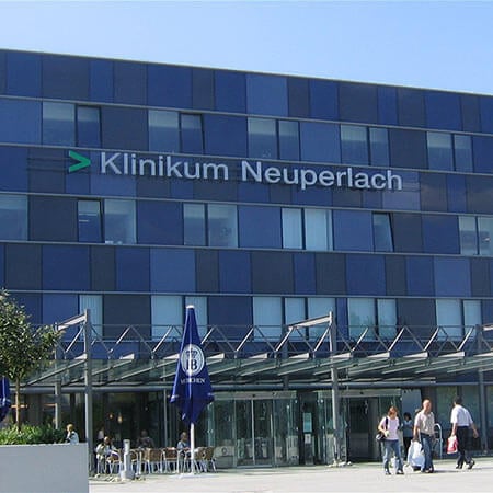
- Hospital Neuperlach Munich.
- University Hospital Ulm.
- University Hospital Heidelberg.
- Charite University Hospital Berlin.
- University Hospital RWTH Aachen.
Visit the hospital section of the Booking Health website to know more about the mentioned German hospitals.
The cost of treatment of the ventricular septal defect
The cost of treatment for ventricular septal defect depends on the required extent of surgery and the overall treatment plan, which is prescribed by the doctor for each patient, depending on the result of the medical examination.
- The average cost of treatment in Germany with surgical repair starts at 9,041 EUR.
- The average cost of treatment with a catheter procedure starts at 5, 849 EUR.
- The average prices for the establishment of the diagnosis start at 477 EUR.
- The average prices for rehabilitation start at 566 EUR.
Booking Health can consult you regarding the prices for medical services required in your case.
Why undergo treatment in Germany with Booking Health?
The best cardiology clinics in Germany are at your disposal to help with the treatment of ventricular septal defects and complex surgical procedures.
It is much safer to trust the organization of ventricular septal defect treatment in Germany to a company that has vast experience in the process. In doing so, you will not worry about anything and can focus on your recovery.
If you have any questions, do not hesitate to contact Booking Health for any information you need.
Authors: Dr. Vadim Zhiliuk, Dr. Sergey Pashchenko
